

Abstract
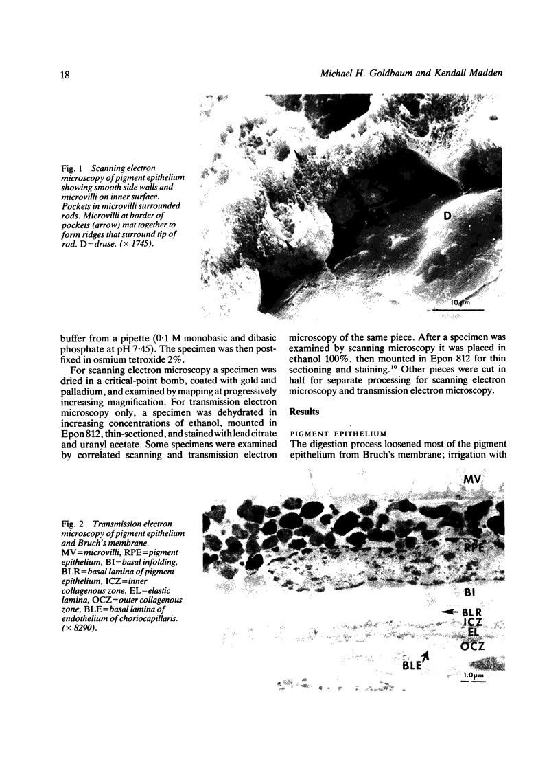
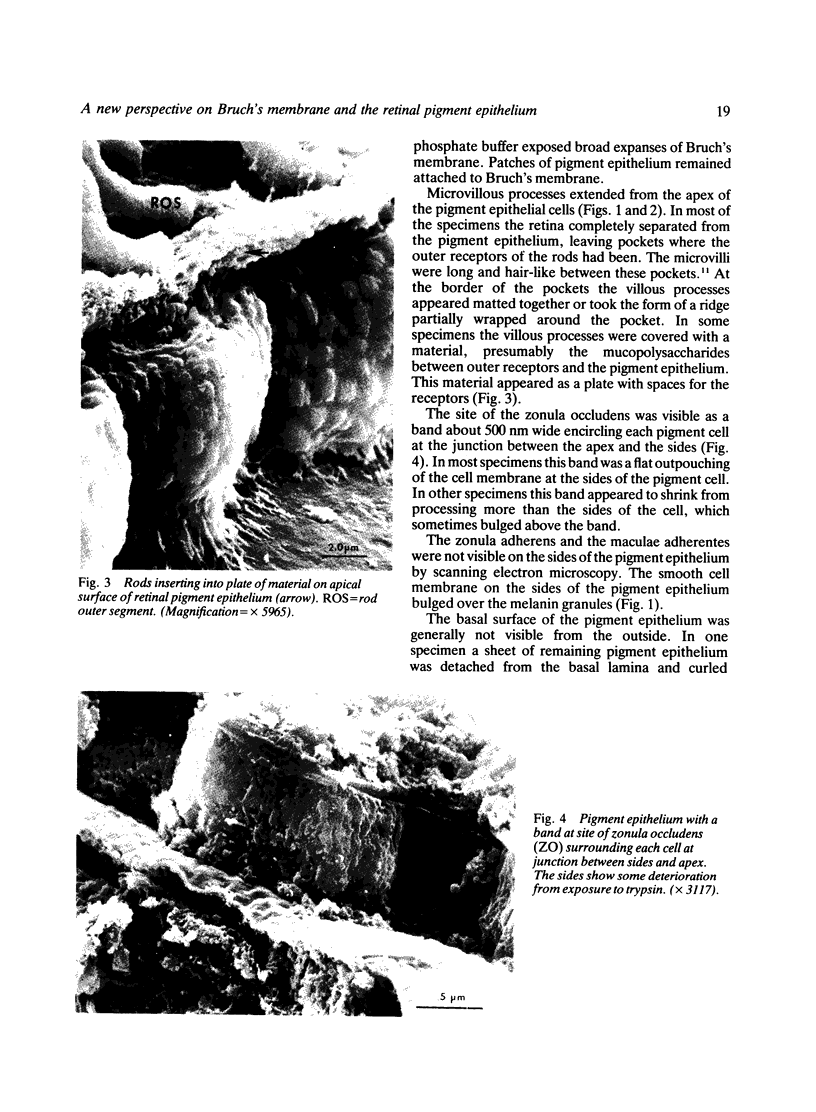
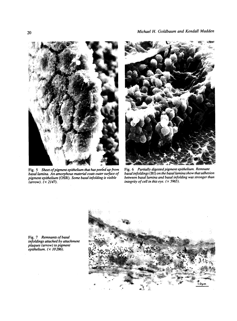
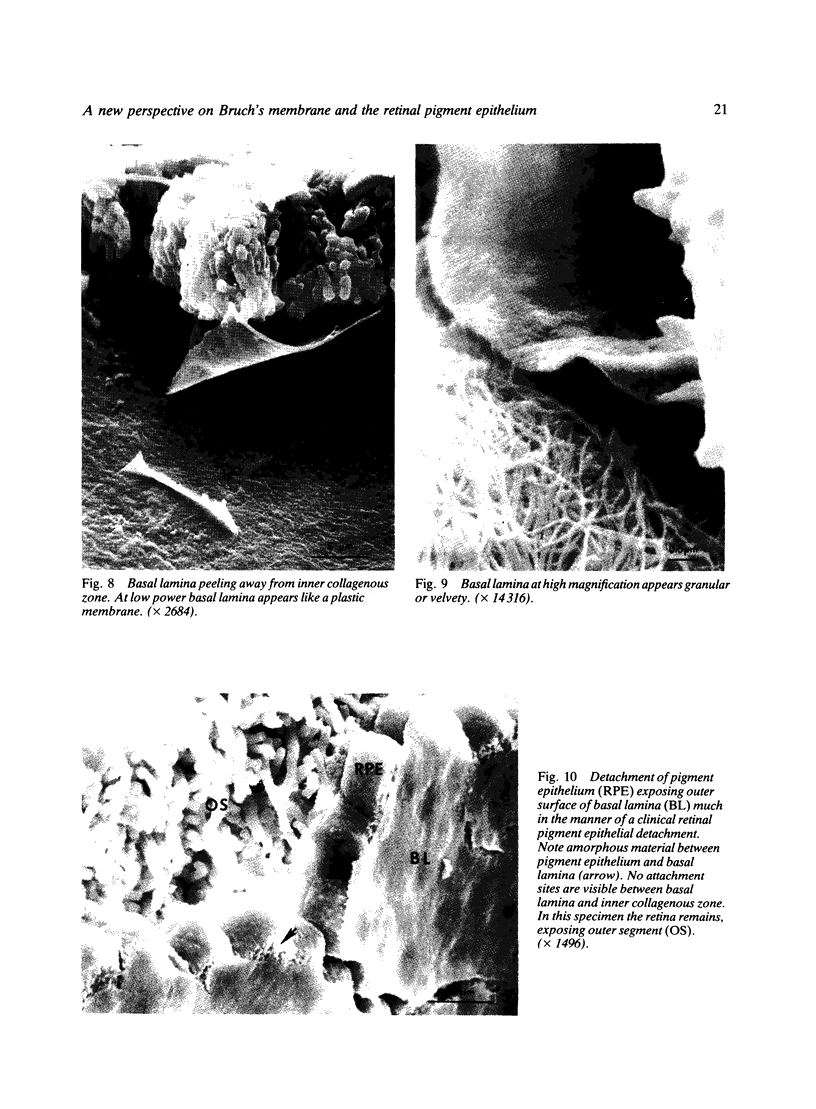
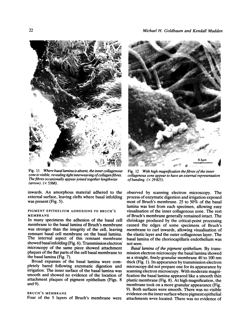
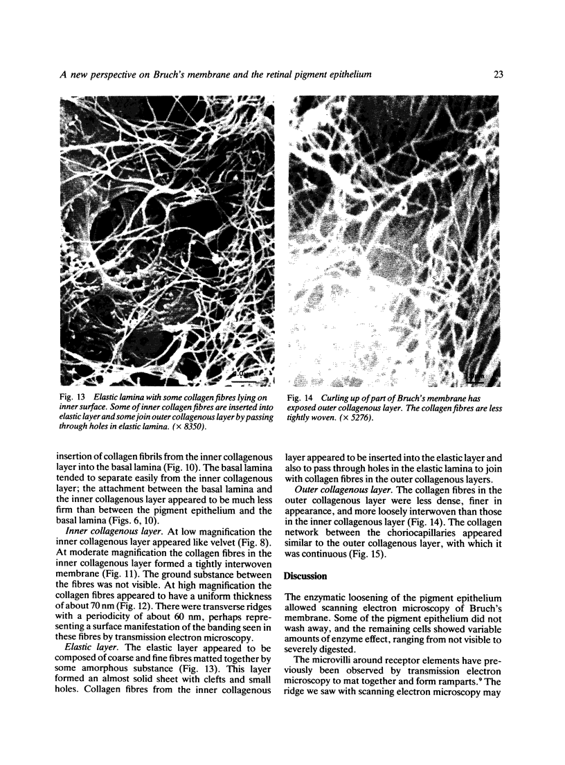
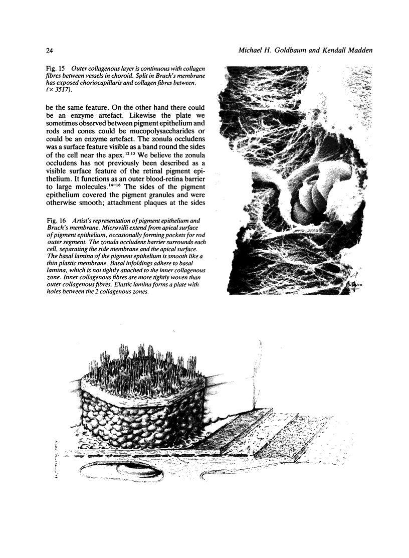
Trypsin digestion of retinal pigment epithelium is a technique that bares Bruch's membrane, to allow topographical examination by scanning electron microscopy. Twenty-five human eyes were examined. The zonula occludens of the pigment epithelium was clearly seen as a surface feature, but attachment plaques at the sides and base were not visible. The adhesion between the pigment epithelium and the basal lamina was stronger than between the basal lamina and the rest of Bruch's membrane. Surface features of the basal lamina, inner collagenous zone, elastic layer, and outer collagenous zone were seen in a way that previously required an artist's representation constructed from microscopic sections.
Full text
PDF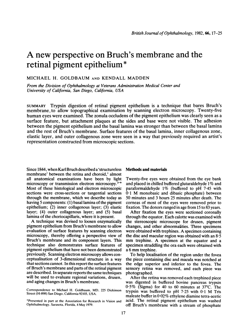
Images in this article

Selected References
These references are in PubMed. This may not be the complete list of references from this article.
- FARQUHAR M. G., PALADE G. E. Junctional complexes in various epithelia. J Cell Biol. 1963 May;17:375–412. doi: 10.1083/jcb.17.2.375. [DOI] [PMC free article] [PubMed] [Google Scholar]
- HOGAN M. J. MACULAR DISEASES: PATHOGENESIS. ELECTRON MICROSCOPY OF BRUCH'S MEMBRANE. Trans Am Acad Ophthalmol Otolaryngol. 1965 Jul-Aug;69:683–690. [PubMed] [Google Scholar]
- NAKAIZUMI Y., HOGAN M. J., FEENEY L. THE ULTRASTRUCTURE OF BRUCH'S MEMBRANE. 3. THE MACULAR AREA OF THE HUMAN EYE. Arch Ophthalmol. 1964 Sep;72:395–400. doi: 10.1001/archopht.1964.00970020395018. [DOI] [PubMed] [Google Scholar]
- NAKAIZUMI Y. THE ULTRASTRUCTURE OF BRUCH'S MEMBRANE. I. HUMAN, MONKEY, RABBIT, GUINEA PIG, AND RAT EYES. Arch Ophthalmol. 1964 Sep;72:380–387. doi: 10.1001/archopht.1964.00970020380016. [DOI] [PubMed] [Google Scholar]
- Peyman G. A., Spitznas M., Straatsma B. R. Peroxidase diffusion in the normal and photocoagulated retina. Invest Ophthalmol. 1971 Mar;10(3):181–189. [PubMed] [Google Scholar]
- Sakuragawa M., Kuwabara T. The pigment epithelium of the monkey. Topographic study by scanning and transmission electron microscopy. Arch Ophthalmol. 1976 Feb;94(2):285–292. doi: 10.1001/archopht.1976.03910030139013. [DOI] [PubMed] [Google Scholar]
- Spitznas M. The fine structure of the chorioretinal border tissues of the adult human eye. Adv Ophthalmol. 1974;28:78–174. [PubMed] [Google Scholar]
- VAN DEN HOOFF A. Het electronen-microscopische beeld van het membraan van Bruch. Ned Tijdschr Geneeskd. 1954 Oct 2;98(40):2869–2870. [PubMed] [Google Scholar]
- Verin P., Gendre P., Le Rebeller M. J. La membrane de Bruch et ses rapports avec la chorio-capillaire. Arch Ophtalmol Rev Gen Ophtalmol. 1969 Feb;29(2):123–134. [PubMed] [Google Scholar]
- Wickham M. G., Worthen D. M. Correlation of scanning and transmission electron microscopy on the same tissue sample. Stain Technol. 1973 Mar;48(2):63–68. doi: 10.3109/10520297309116583. [DOI] [PubMed] [Google Scholar]