Abstract
目的
评估基于CT灌注成像的软脑膜侧支评价方法(CTP-CS)在预测急性前循环大血管闭塞患者动脉取栓治疗预后中的价值。
方法
回顾性分析2013年10月至2016年10月浙江大学医学院附属第二医院急性前循环大血管闭塞动脉取栓患者的临床及影像学资料。采用CTP-CS和基于数字减影血管造影的侧支评分(DSA-CS)对侧支进行评分,DSA-CS或CTP-CS评分3~4分定义为侧支良好。预后优定义为术后3个月时改良Rankin量表(mRS)为2分及以下。采用二元logistic回归法分析侧支循环与患者预后的关系并用受试者工作特征(ROC)曲线评价DSA-CS和CTP-CS在预测患者预后中的价值。
结果
共纳入40例患者,其中获得血管再通33例(82.5%),临床预后优16例(40.0%)。与预后不优的患者比较,预后优的患者侧支评分更高(均 P < 0.05),侧支良好比例更高(均 P < 0.01)。二元logistic回归分析提示,侧支评分是预后优的独立预测因素(CTP-CS: OR=48.404,95% CI:1.373~1706.585, P < 0.05;DSA-CS: OR=34.651,95% CI:1.147~1047.018, P < 0.05)。一致性分析提示,CTP-CS和DSA-CS两种评分间的一致性良好( κ=0.697, P < 0.01)。ROC曲线分析结果显示,CTP-CS预测患者临床预后的能力与DSA-CS相当(均AUC=0.726,95% CI:0.559~0.893, P < 0.05)。
结论
CTP-CS评分能独立预测急性前循环大血管闭塞患者动脉取栓治疗的预后。
Abstract
Objective
To evaluate the value of collateral score based on CT perfusion (CTP-CS) in predicting the clinical outcome of patients with anterior circulation ischemic stroke after thrombectomy.
Methods
Clinical data of acute ischemic stroke patients with anterior artery occlusion undergoing endovascular treatment in the Second Affiliated Hospital, Zhejiang University School of Medicine during October 2013 and October 2016 were retrospectively reviewed. Collateral scores were assessed based on CTP and digital subtraction angiography (DSA) images, respectively. And DSA-CS or CTP-CS 3-4 was defined as good collateral vessels. Good clinical outcome was defined as a modified Rankin Scale (mRS) ≤ 2 at 3 months after stroke. The binary logistic regression model was used to analyze the correlation between the collateral score and clinical outcome, and the receiver operating characteristic (ROC) curve was used to analyze the value of DSA-CS and CTP-CS in predicting the clinical outcome.
Results
Among 40 patients, 33 (82.5%) acquired recanalization and 16 (40.0%) got good outcome. Compared with poor outcome group, the collateral score (all P < 0.05) and the rate of good collateral vessels were higher in good outcome group (all P < 0.01). After adjust baseline National Institute of Health Stroke Scale (NIHSS) and onset to recanalization time (ORT), good collateral vessels were independent factor of good outcome (CTP-CS: OR=48.404, 95% CI:1.373-1706.585, P < 0.05; DSA-CS: OR=34.651, 95% CI:1.147-1047.018, P < 0.05). Collateral scores based on CTP and DSA had good consistency ( κ=0.697, P < 0.01), and ROC curve showed that the predictive value of CTP-CS and DSA-CS were comparable (both AUC=0.726, 95% CI:0.559-0.893, P < 0.05).
Conclusion
CTP-CS can predict the clinical outcome of patients with anterior circulation ischemic stroke after thrombectomy.
Keywords: Acute disease; Brain ischemia/radiography; Tomography, X-ray computed; Brain ischemia/therapy; Stroke/therapy; Thrombolytic therapy/methods; Collateral circulation; Prognosis
急性前循环大血管闭塞引起的急性缺血性卒中患者侧支血流状况与临床预后显著相关 [ 1- 2] 。侧支血流较好的患者接受血管内治疗后血管再通率更高、功能结局更优 [ 3- 4] 。目前临床评估颅内侧支血流的方法主要有数字减影血管造影(digital subtraction angiography,DSA)和CT血管造影(CT angiography,CTA)。DSA是评估颅内侧支血流状况的金标准,如美国介入与治疗神经放射学学会/介入放射学学会(American Society of Interventional and Therapeutic Neuroradiology/Society of Interventional Radiology,ASITN/SIR)侧支血流评分系统 [ 5] 。但DSA为有创检查,临床应用有局限。CTA应用普遍,但只能提供单时相血流信息,无法显示侧支充盈速度 [ 6] 。因此,临床上亟需一种能全面评估侧支且无创的影像学方法。灌注成像包括CT灌注、MR灌注等,具有无创、全面快速、价格低廉等优势。本研究旨在建立一种基于CT灌注的侧支血流评价新方法,该方法类似应用DSA的ASITN/SIR评分系统,并能预测急性前循环大血管闭塞后动脉取栓患者的临床预后。
回顾性分析2013年10月至2016年10月浙江大学医学院附属第二医院收治的急性前循环大血管闭塞接受急诊血管内取栓治疗患者的资料。纳入标准:①静脉溶栓桥接动脉取栓(发病时间在4.5 h内),直接动脉取栓(发病时间<6 h);②术前完成CT灌注提示前循环近端大血管闭塞,且符合缺血低灌注体积与核心梗死体积之比(不匹配率)大于1.2、绝对不匹配体积大于10 mL、核心梗死体积小于80 mL;③颈内动脉闭塞患者术中有全脑血管造影,大脑中动脉闭塞患者可仅同侧颈内动脉造影。排除标准:①本次发病前改良Rankin量表(mRS)大于1分;②术后3个月内失访。
共42例患者符合纳入标准,其中术后3个月内失访2例,最终40例纳入分析,其中静脉溶栓桥接动脉取栓36例,直接动脉取栓4例。患者平均年龄(63±13)岁,女性18例(45.0%),基线美国国立卫生研究院卒中量表(NIHSS)为15(11~18)分,平均起病至影像学检查时间为(154±100)min,平均起病至再通时间为(354±101)min,33例(82.5%)获得再通,16例(40.0%)预后优。
本研究经浙江大学医学院附属第二医院伦理委员会批准,所有患者均知情同意并签署知情同意书。
溶栓药物选用德国勃林格殷格翰生产的重组组织型纤溶酶原激活剂(商品名爱通立),为规格50 mg/支或20 mg/支的干粉制剂。血流重建装置采用美国美敦力公司生产的Solitaire TM FR支架;CT灌注扫描采用德国西门子公司的双源CT成像系统;DSA图像采集采用荷兰飞利浦公司的DSA成像系统,对比剂选用上海博莱科信谊药业有限公司生产的碘帕醇注射液。
静脉溶栓:溶栓剂量按照《中国急性缺血性脑卒中诊治指南2010》 [ 7] 规定的0.9 mg/kg,最大剂量不超过90 mg,10%的剂量先予静脉推注,其余90%持续静脉微泵60 min。
动脉取栓采用支架取栓术。行全脑血管造影检查,如果造影中发现责任动脉未再通,将微导丝、微导管通过指引导管过血栓远端,反复输送Solitaire TM FR支架至血栓远端打开,收回至体外,将血栓整块或分块取出,整个过程反复行血管造影确认动脉闭塞情况,直至闭塞血管开通,但支架切割血栓过程一般不超过五次 [ 8] 。
CT灌注检查序列包括CT扫描、全脑容积灌注CT扫描。以6 mL/s的速度经肘静脉注射60 mL对比剂,后续以6 mL/s的速度推注20 mL等渗氯化钠溶液。扫描参数为:z轴100 mm,80 kV,120 mAs,延迟4 s开始,扫描总时间为67.98 s,共采集26个时间点,层厚1.5 mm,准直32 mm×1.2 mm。
DSA图像采集包括各动脉造影正侧位。颈内动脉以4 mL/s的速度经导管注射8 mL对比剂,椎动脉以3 mL/s的速度经导管注射6 mL对比剂。扫描参数:曝光为自动调节,延迟0.5 s开始以6帧/s的速率采集动脉期、脑实质期和静脉期图像。
基于DSA的侧支评分(collateral score on DSA,DSA-CS)采用ASITN/SIR评分系统 [ 5] :没有侧支血流形成或缺血区域周边可见缓慢侧支血流,但缺血区域仍见灌注缺损为1分;缺血区域周边可见快速侧支血流,但缺血区域仍见部分灌注缺损为2分;侧支血流缓慢,至静脉期末可见缺血区域灌注完整为3分;侧支血流快速而完全地灌注到缺血区域为4分。DSA-CS 3~4分定义为侧支良好,DSA-CS 1~2分定义为侧支不良。
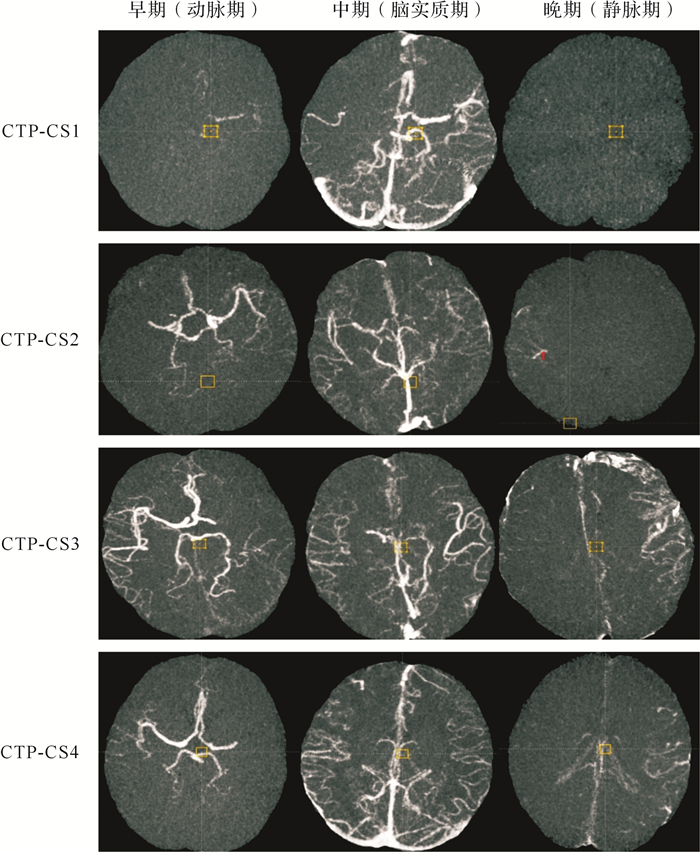
基于CT灌注成像的侧支评分(collateral score based on CT perfusion,CTP-CS)见 图 1。由MIStar软件(澳大利亚Apollo公司)重建10.0 mm序列获取动静脉曲线,并根据曲线确定动、静脉期的时间分布。健侧外侧裂出现造影剂高密度至动脉曲线高峰定为早期(动脉期)、动脉曲线高峰至静脉曲线高峰定为中期(脑实质期)、静脉曲线高峰之后定为晚期(静脉期);对1.5 mm序列相应各期采用最大密度投影(maximum intensity projection,MIP)重建动态CTA,选用轴位图像参考ASITN/SIR评分系统进行评分。CTP-CS 3~4分定义为侧支良好,CTP-CS 1~2分定义为侧支不良。

既往ESCAPE(endovascular treatment for small core and proximal occlusion ischemic stroke)研究使用的侧支评分为多相CTA侧支评分(collateral score based on multiphase CT angiography,MP-CS) [ 9] 。MP-CS 4~5分定义为侧支良好,MP-CS 0~3分定义为侧支不良。
静脉溶栓后责任血管再通评估主要通过对比患者溶栓前及溶栓后24 h血管影像学表现,根据动脉闭塞病变(arterial occlusive lesion,AOL)量表进行分级。其中AOL量表 0分提示责任血管闭塞部位完全无再通;AOL量表 1分提示责任血管闭塞部位部分再通,但无远端血流;AOL量表 2分提示责任血管闭塞部位不完全闭塞或部分再通,伴有远端血流;AOL量表 3分提示责任血管闭塞部位完全再通,伴有远端血流。定义AOL量表至少2分为血管再通,否则为血管未再通 [ 10] 。溶栓后3个月使用mRS评定神经功能,mRS不超过2分为预后优,否则为预后不优 [ 11] 。
运用SPSS 22.0软件进行统计分析。符合正态分布的计量资料用均数±标准差( x ± s)表示,两组间比较采用独立样本 t检验;不符合正态分布的计量资料用中位数(四分位数区间)[ M( Q 1~ Q 3)]表示,两组间比较采用独立样本Mann-Whitney U非参数检验;计数资料用例数和百分比表示,行 χ 2检验及Fisher检验。单因素分析中 P<0.1的变量进入二元logistic回归分析模型。利用二元logistic回归分析筛选预后优的独立预测因素,以优势比(odds ratio, OR)及其95% CI表示。CTP-CS与DSA-CS对比采用一致性分析,用 κ值表示。采用受试者工作特征(ROC)曲线评估CTP-CS对患者预后的预测能力。 P<0.05为差异有统计学意义。
单因素分析显示,预后优患者起病至再通时间较短,基线NIHSS评分较低,CTP-CS和DSA-CS侧支良好比例较高(均 P<0.05),见 表 1。提示CTP-CS和DSA-CS侧支良好与急性缺血性卒中取栓患者的预后优相关。
表1 影响急性前循环大血管闭塞取栓患者临床预后相关因素的单因素分析
Table 1 Univariate analysis of influencing factors for prognosis in patients with anterior circulation ischemic stroke after thrombectomy
|
[ n(%)或( x ± s)或 M( Q 1~ Q 3)] | ||||
|
相关因素 |
预后不优( n=24) |
预后优( n=16) |
检验值 |
P值 |
|
女性例数 |
10(41.7) |
8(50.0) |
0.269 |
>0.05 |
|
年龄(岁) |
63±11 |
64±15 |
-0.343 |
>0.05 |
|
起病至再通时间(min) |
401±96 |
291±68 |
3.638 |
<0.01 |
|
吸烟 |
7(29.2) |
5(31.3) |
0.020 |
>0.05 |
|
高血压 |
14(60.9) |
8(53.3) |
0.212 |
>0.05 |
|
糖尿病 |
3(12.5) |
3(18.8) |
0.294 |
>0.05 |
|
心房颤动 |
11(45.8) |
8(50.0) |
0.067 |
>0.05 |
|
高脂血症 |
13(54.2) |
7(43.8) |
0.417 |
>0.05 |
|
短暂性脑缺血发作或中风史 |
3(12.5) |
1(6.3) |
0.417 |
>0.05 |
|
抗血小板病史 |
4(16.7) |
3(18.8) |
0.029 |
>0.05 |
|
基线NIHSS评分 |
16(13~18) |
11(7~16) |
-2.782 |
<0.01 |
|
基线收缩压(mmHg) |
150±25 |
143±22 |
0.818 |
>0.05 |
|
基线血糖(mmol/L) |
7.11±1.27 |
7.46±2.15 |
-0.649 |
>0.05 |
|
血小板数(×10 9/L) |
179±49 |
199±53 |
-1.184 |
>0.05 |
|
国际标准化比值 |
1.17±0.48 |
0.98±0.59 |
1.573 |
>0.05 |
|
CTP-CS侧支良好 |
9(37.5) |
13(81.3) |
7.424 |
<0.01 |
|
CTP-CS评分 |
1(1~4) |
4(3~4) |
2.505 |
<0.05 |
|
DSA-CS侧支良好 |
9(37.5) |
13(81.3) |
7.424 |
<0.01 |
|
DSA-CS评分 |
3(1~4) |
4(4~4) |
-2.397 |
<0.05 |
|
MP-CS侧支良好 |
11(45.8) |
12(75.0) |
3.342 |
>0.05 |
|
MP-CS评分 |
3(3~4) |
4(3~4) |
-1.822 |
>0.05 |
|
再通例数 |
19(79.2) |
14(87.5) |
0.462 |
>0.05 |
共统计了32例患者的数据,预后不优组19例,预后优组14例.NIHSS:美国国立卫生研究院卒中量表;CTP-CS:基于CT灌注成像的侧支评分;DSA-CS:基于数字减影血管造影的侧支评分;MP-CS:ESCAPE研究中的侧支评分.1 mmHg=0.133 kPa.
二元logistic回归分析结果显示,在校正了年龄、起病至再通时间和基线NIHSS评分后,CTP-CS、DSA-CS均为预测患者临床预后优的独立因素,但MP-CS不是预测临床预后优的独立因素( 表 2)。提示CTP-CS、DSA-CS可作为急性缺血性卒中患者动脉取栓预后的独立预测因子。
表2 急性前循环大血管闭塞取栓患者临床预后相关因素的二元logistic回归分析
Table 2 Binary logistic regression analysis of influencing factors for prognosis in patients with anterior circulation ischemic stroke after thrombectomy
|
变 量 |
OR(95% CI)值 |
P值 |
|
|
模型1 |
年龄 |
0.952(0.869~1.044) |
>0.05 |
|
起病至再通时间 |
0.969(0.942~0.996) |
<0.05 |
|
|
基线NIHSS评分 |
0.765(0.536~1.092) |
>0.05 |
|
|
CTP-CS(1~2, 3~4) |
48.404(1.373~1706.585) |
<0.05 |
|
|
模型2 |
年龄 |
0.961(0.881~1.049) |
>0.05 |
|
起病至再通时间 |
0.968(0.942~0.996) |
<0.05 |
|
|
基线NIHSS评分 |
0.731(0.513~1.041) |
>0.05 |
|
|
DSA-CS(1~2, 3~4) |
34.651(1.147~1047.018) |
<0.05 |
|
|
模型3 |
年龄 |
0.923(0.835~1.021) |
>0.05 |
|
起病至再通时间 |
0.975(0.957~0.994) |
<0.01 |
|
|
基线NIHSS评分 |
0.689(0.492~0.966) |
<0.05 |
|
|
MP-CS(0~3, 4~5) |
8.441(0.580~122.826) |
>0.05 |
NIHSS:美国国立卫生研究院卒中量表;CTP-CS:基于CT灌注成像的侧支评分;DSA-CS:基于数字减影血管造影的侧支评分;MP-CS:ESCAPE研究中的侧支评分方法.
一致性分析结果显示,CTP-CS和DSA-CS均提示侧支良好19例,CTP-CS和DSA-CS均提示侧支不良15例,侧支评分一致共34例(85.0%);仅CTP-CS或DSA-CS提示侧支良好各3例。CTP-CS与DSA-CS两种评分间的一致性良好( κ=0.697, P<0.01)。
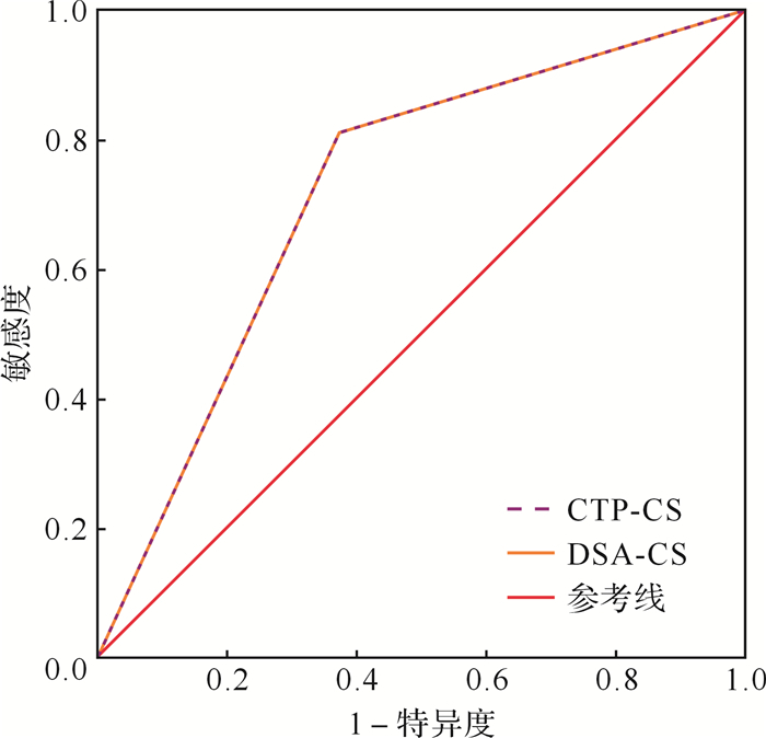
ROC曲线提示,CTP-CS预测患者临床预后的能力与DSA-CS相当(曲线下面积均为0.726,95% CI:0.559~0.893, P<0.05),见 图 2。

侧支循环是决定急性缺血性卒中患者最终梗死体积和缺血半暗带的主要因素,也是血管再灌注治疗中决定临床预后的主要指标。在脑血管病变导致血流量降低时,良好的侧支血流可以通过侧支或新形成的血管吻合到达缺血区域,从而使缺血脑组织得到不同程度的灌注代偿,也就是侧支良好的患者拥有相对较大的缺血半暗带和相对较小的核心梗死区,增加了获得良好预后的机会。另外,侧支良好的患者在溶栓过程中有更多的溶栓药物作用于血栓远端,从而提高了血管再通及获得良好预后的概率。Bang等 [ 12] 发现,侧支血流等级可帮助进行血管内治疗决策,治疗前良好的侧支循环与预后改善显著相关,且最终梗死体积小。
DSA是观察和评估颅内侧支血流的金标准,ASITN/SIR侧支血流评分系统是其经典评估方法,该评分越高,再灌注治疗的预后越好 [ 3, 13] 。在急性前循环大血管闭塞取栓患者中,为了尽早再通再灌,取栓前DSA多为单根动脉造影,曝光也多在静脉早期结束,而且患者多有躁动影响图像质量,不利于客观评价患者侧支循环。近年无创血管造影和灌注研究已被用来评价侧支循环。Kim等 [ 14] 研究证实,多相CT灌注可以预测急性大脑中动脉主干闭塞患者的预后,但该研究选取统一的固定时间段作为早、中、晚三期,并使用CT灌注原始图像分析,容易受到非动脉性的高密度的干扰,且只能观察到单层截面的脑组织。本研究相对客观地选择早、中、晚三期,更接近动脉期、实质期、静脉期,并采用重建动态CTA来改进侧支观测水平。
ESCAPE研究使用的侧支评分为MP-CS,该评分是对造影剂流经脑血管过程中的三个时相进行侧支水平的综合评分,然而该断点式的影像采集方式无法全面获取侧支血流信息。我们将该方法应用到本研究人群中进行验证发现,患者MP-CS得分良好与预后优之间有相关趋势,但未达到统计学意义,可能与本研究样本量较小以及该评分方法相对复杂有关。
本研究首次基于CTP重建动态CTA评价侧支,该评分系统模拟ASITN/SIR侧支评分系统,从侧支血流范围和速度两方面评价颅内侧支血流,属无创操作,简便易行。本文资料表明,CTP-CS与DSA-CS评分的匹配度较好,并能预测急性前循环卒中取栓患者的神经功能和临床结局,未来有望在急性前循环大血管闭塞动脉取栓患者中应用,在患者进入导管室前进行侧支血流分级,快速做出相对有利的临床决策。
但是,本研究尚有一定局限性,第一,本研究为单中心研究,样本量小,需要扩大样本进行多中心的前瞻性研究进一步证实;第二,本研究仅限于急性前循环大血管闭塞动脉取栓患者,因为后循环供血区域脑组织体量相对较小,且侧支较多,在后循环卒中动脉取栓患者中评价较困难,预测价值尚有待进一步研究。
综上所述,本研究基于CT灌注建立的模拟DSA侧支评分能独立预测急性前循环缺血性卒中动脉取栓患者的临床预后,且其预测能力与DSA相似,未来或可用于筛选血管内治疗适宜人群的术前评估。
Funding Statement
国家自然科学基金(81471170, 81622017)
References
- 1.SCHRAMM P, SCHELLINGER P D, FIEBACH J B, et al. Comparison of CT and CT angiography source images with diffusion-weighted imaging in patients with acute stroke within 6 hours after onset. Stroke. 2002;33(10):2426–2432. doi: 10.1161/01.STR.0000032244.03134.37. [SCHRAMM P, SCHELLINGER P D, FIEBACH J B, et al. Comparison of CT and CT angiography source images with diffusion-weighted imaging in patients with acute stroke within 6 hours after onset[J]. Stroke, 2002, 33(10):2426-2432.] [DOI] [PubMed] [Google Scholar]
- 2.SOUZA L C S, YOO A J, CHAUDHRY Z A, et al. Malignant CTA collateral profile is highly specific for large admission DWI infarct core and poor outcome in acute stroke. AJNR Am J Neuroradiol. 2012;33(7):1331–1336. doi: 10.3174/ajnr.A2985. [SOUZA L C S, YOO A J, CHAUDHRY Z A, et al. Malignant CTA collateral profile is highly specific for large admission DWI infarct core and poor outcome in acute stroke[J]. AJNR Am J Neuroradiol, 2012, 33(7):1331-1336.] [DOI] [PMC free article] [PubMed] [Google Scholar]
- 3.BANG O Y, SAVER J L, KIM S J, et al. Collateral flow predicts response to endovascular therapy for acute ischemic stroke. Stroke. 2011;42(3):693–699. doi: 10.1161/STROKEAHA.110.595256. [BANG O Y, SAVER J L, KIM S J, et al. Collateral flow predicts response to endovascular therapy for acute ischemic stroke[J]. Stroke, 2011, 42(3):693-699.] [DOI] [PMC free article] [PubMed] [Google Scholar]
- 4.CHRISTOFORIDIS G A, MOHAMMAD Y, KEHAGIAS D, et al. Angiographic assessment of pial collaterals as a prognostic indicator following intra-arterial thrombolysis for acute ischemic stroke. http://europepmc.org/abstract/MED/16091531. AJNR Am J Neuroradiol. 2005;26(7):1789–1797. [CHRISTOFORIDIS G A, MOHAMMAD Y, KEHAGIAS D, et al. Angiographic assessment of pial collaterals as a prognostic indicator following intra-arterial thrombolysis for acute ischemic stroke[J]. AJNR Am J Neuroradiol, 2005, 26(7):1789-1797.] [PMC free article] [PubMed] [Google Scholar]
- 5.HIGASHIDA R T, FURLAN A J, ROBERTS H, et al. Trial design and reporting standards for intra-arterial cerebral thrombolysis for acute ischemic stroke. Stroke. 2003;34(8):e109–e137. doi: 10.1161/01.STR.0000082721.62796.09. [HIGASHIDA R T, FURLAN A J, ROBERTS H, et al. Trial design and reporting standards for intra-arterial cerebral thrombolysis for acute ischemic stroke[J]. Stroke, 2003, 34(8):e109-e137.] [DOI] [PubMed] [Google Scholar]
- 6.MORTIMER A M, SIMPSON E, BRADLEY M D, et al. Computed tomography angiography in hyperacute ischemic stroke:prognostic implications and role in decision-making. Stroke. 2013;44(5):1480–1488. doi: 10.1161/STROKEAHA.111.679522. [MORTIMER A M, SIMPSON E, BRADLEY M D, et al. Computed tomography angiography in hyperacute ischemic stroke:prognostic implications and role in decision-making[J]. Stroke, 2013, 44(5):1480-1488.] [DOI] [PubMed] [Google Scholar]
- 7.中华医学会神经病学分会脑血管病学组急性缺血性脑卒中诊治指南撰写组 中国急性缺血性脑卒中诊治指南2010. http://group.medlive.cn/topic/3714. 中华神经科杂志. 2010;43(2):146–153. [中华医学会神经病学分会脑血管病学组急性缺血性脑卒中诊治指南撰写组.中国急性缺血性脑卒中诊治指南2010[J].中华神经科杂志, 2010, 43(2):146-153.] [Google Scholar]
- 8.CAMPBELL B C, MITCHELL P J, KLEINIG T J, et al. Endovascular therapy for ischemic stroke with perfusion-imaging selection. N Engl J Med. 2015;372(11):1009–1018. doi: 10.1056/NEJMoa1414792. [CAMPBELL B C, MITCHELL P J, KLEINIG T J, et al. Endovascular therapy for ischemic stroke with perfusion-imaging selection[J]. N Engl J Med, 2015, 372(11):1009-1018.] [DOI] [PubMed] [Google Scholar]
- 9.MENON B K, D'ESTERRE C D, QAZI E M, et al. Multiphase CT angiography:a new tool for the imaging triage of patients with acute ischemic stroke. Radiology. 2015;275(2):510–520. doi: 10.1148/radiol.15142256. [MENON B K, D'ESTERRE C D, QAZI E M, et al. Multiphase CT angiography:a new tool for the imaging triage of patients with acute ischemic stroke[J]. Radiology, 2015, 275(2):510-520.] [DOI] [PubMed] [Google Scholar]
- 10.ZAIDAT O O, YOO A J, KHATRI P, et al. Recommendations on angiographic revascularization grading standards for acute ischemic stroke:a consensus statement. Stroke. 2013;44(9):2650–2663. doi: 10.1161/STROKEAHA.113.001972. [ZAIDAT O O, YOO A J, KHATRI P, et al. Recommendations on angiographic revascularization grading standards for acute ischemic stroke:a consensus statement[J]. Stroke, 2013, 44(9):2650-2663.] [DOI] [PMC free article] [PubMed] [Google Scholar]
- 11.ERIKSSON M, NORRVING B, TERÉNT A, et al. Functional outcome 3 months after stroke predicts long-term survival. Cerebrovasc Dis. 2008;25(5):423–429. doi: 10.1159/000121343. [ERIKSSON M, NORRVING B, TERÉNT A, et al. Functional outcome 3 months after stroke predicts long-term survival[J]. Cerebrovasc Dis, 2008, 25(5):423-429.] [DOI] [PubMed] [Google Scholar]
- 12.BANG O Y, SAVER J L, KIM S J, et al. Collateral flow predicts response to endovascular therapy for acute ischemic stroke. Stroke. 2011;42(3):693–699. doi: 10.1161/STROKEAHA.110.595256. [BANG O Y, SAVER J L, KIM S J, et al. Collateral flow predicts response to endovascular therapy for acute ischemic stroke[J]. Stroke, 2011, 42(3):693-699.] [DOI] [PMC free article] [PubMed] [Google Scholar]
- 13.LAU A Y, WONG E H, WONG A, et al. Significance of good collateral compensation in symptomatic intracranial atherosclerosis. Cerebrovasc Dis. 2012;33(6):517–524. doi: 10.1159/000337332. [LAU A Y, WONG E H, WONG A, et al. Significance of good collateral compensation in symptomatic intracranial atherosclerosis[J]. Cerebrovasc Dis, 2012, 33(6):517-524.] [DOI] [PubMed] [Google Scholar]
- 14.KIM S J, NOH H J, YOON C W, et al. Multiphasic perfusion computed tomography as a predictor of collateral flow in acute ischemic stroke:comparison with digital subtraction angiography. Eur Neurol. 2012;67:252–255. doi: 10.1159/000334867. [KIM S J, NOH H J, YOON C W, et al. Multiphasic perfusion computed tomography as a predictor of collateral flow in acute ischemic stroke:comparison with digital subtraction angiography[J]. Eur Neurol, 2012, 67:252-255.] [DOI] [PubMed] [Google Scholar]


