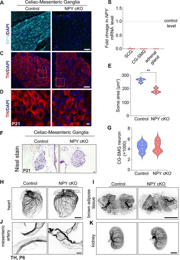
Figure 3. Loss of NPY in sympathetic neurons does not affect neuron survival and target innervation but results in reduced soma size.
(A) Immunostaining shows a marked reduction in NPY protein in CG-SMG from NPY cKO mice compared to control mice. Scale bar, 50 μm. (B) qPCR analyses show a loss of Npy transcript in SCG, CG-SMG, and adrenal glands from NPY cKO mice. Results are means ± s.e.m from n= 4 animals per genotype for SCG, CG-SMG and n=3 animals per genotype for adrenal glands. (C, D) TH expression appears unaffected by NPY loss as shown by immunofluorescence. Shown in D are higher magnification views of the insets in C. Scale bar, 50 μm for C and 5 μm for D. (E) Quantification of soma areas from CG-SMG tissue sections shows that neuronal soma size is reduced in NPY cKO sympathetic ganglia. Results are means ± s.e.m from n=3 mice per genotype. **p<0.01, unpaired t-test. (F) Nissl staining of CG-SMG in control and NPY cKO mice. Scale bar, 50 μm. (G) Quantification of Nissl-stained neurons shows that neuron numbers are comparable between control and mutant mice. Results are means ± s.e.m from n=4 per genotype. (H-K) Whole organ TH immunostaining imaged by light sheet microscopy shows that sympathetic axon innervation of target tissues including the heart, brown adipose tissue, mesenteric arteries, and kidneys is comparable between NPY cKO and control mice. Scale bars, mesenteric arteries-50 μm; heart, and kidney-800 μm, brown adipose tissue-1500 μm.

