Abstract
Background
Although schistosomiasis is a public health issue in Mali, little is known about the parasite genetic profile. The purpose of this study was to analyze the genetic profile of the schistosomes of Schistosoma haematobium group in school-aged children in various sites in Mali.
Methods
Urine samples were collected from 7 to 21 November 2021 and subjected to a filtration method for the presence S. haematobium eggs. The study took place in two schistosomiasis endemic villages (Fangouné Bamanan and Diakalèl), qualified as hotspots according to the World Health Organization (WHO) definition. Molecular genotyping on both Cox1 and ITS2/18S was used for eggs' taxonomic assignation.
Results
A total of 970 miracidia were individually collected from 63 school-aged children and stored on Whatman FTA cards for molecular analysis. After genotyping 42.0% (353/840) and 58.0% (487/840) of miracidia revealed Schistosoma bovis and S. haematobium Cox1 profiles, respectively; 95.7 (885/925) and 4.3% (40/925) revealed S. haematobium and S. haematobium/S. curassoni profiles for ITS/18S genes, respectively. There was a significant difference in the Cox1 and ITS2/18S profile distribution according to the village (P < 0.0001). Overall, 45.6% (360/789) were hybrids, of which 72.0% (322/447) were from Diakalèl. Three hybrids’ profiles (Sb/Sc_ShxSc with 2.3%; Sb/Sc_ShxSh with 40.5%; Sh_ShxSc with 2.8%) and one pure profile (Sh_ShxSh with 54.4%) were identified.
Conclusion
Our findings show, for the first time to our knowledge, high prevalence of hybrid schistosomes in Mali. More studies are needed on population genetics of schistosomes at the human and animal interface to evaluate the parasite’s gene flow and its consequences on epidemiology of the disease as well as the transmission to humans.
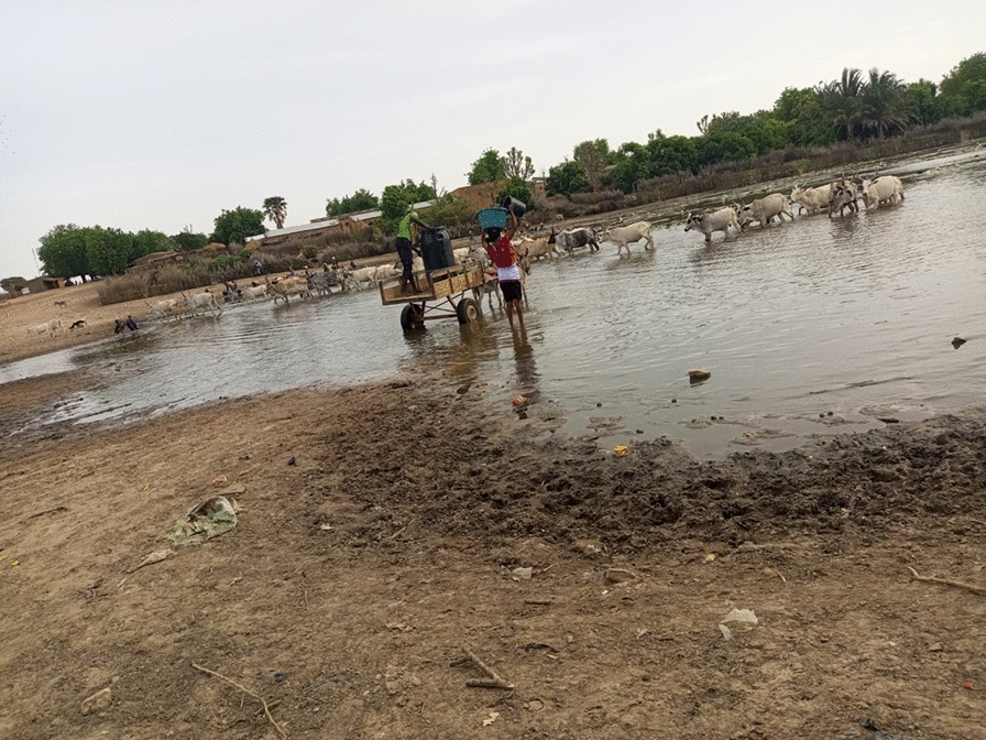
Graphical Abstract

Supplementary Information
The online version contains supplementary material available at 10.1186/s13071-023-05860-8.
Keywords: Schistosoma haematobium, Schistosoma bovis, Schistosoma curassoni, Hybridization, Cox1, ITS/18S, Mali
Introduction
Schistosomiasis is a parasitic disease of medical and veterinary importance that mainly affects tropical and subtropical areas. According to WHO [37], schistosomiasis affects almost 240 million people worldwide, with 85% of them living in Africa, and > 700 million people live in endemic areas. In Mali, the national prevalences of infection estimated in 2004–2006 were 38.3% and 6.7% for Schistosoma haematobium (Sh) and S. mansoni (Sm), respectively [10]. Whereas high Sh prevalence is widespread in all Malian regions, Sm infections are mainly restricted to small clusters in the center of the country (Macina and Niono districts in the Office du Niger irrigation area) and in Baguineda, 30 km from Bamako [8, 10, 33]. In addition to human vertebrate hosts, some Schistosoma species can also affect livestock. Overall, approximately 165 million parasitized animals worldwide are estimated to suffer from hemorrhagic enteritis, anemia and cachexia, and most often die [12]. In Africa, three Schistosoma species are involved in livestock infestation: S. bovis (Sb), S. mattheei (Sma) and S. curassoni (Sc) [23]. (S.b) is the most studied animal schistosome. Sb was first reported in Mali in 1988 in the Central Niger Delta where prevalence in animals is up to 80% [31]. Two years later, prevalences of 62.5% and 85.1% for Sb and Sc were reported in the slaughterhouses of Bamako and Mopti cities [26]. Beyond these cases reported in Mali, Sb occurs in the Mediterranean zone and throughout sub-Saharan Africa, especially in West Africa where it has been reported in almost all countries (Burkina Faso, Gambia, Ghana, Guinea, Bissau-Guinea, Mauritania, Niger, Nigeria, Senegal, Mali, Ivory Coast and Togo) [17, 21].
Hybridization is a biological phenomenon that corresponds to the meeting and crossing between two distinct genetic entities previously defined as different species [15, 20]. Hybridization can represent a real concern in terms of parasite transmission, epidemiology and morbidity. Hybridization in parasites can complicate the prevention, effective control and treatment of the disease [3] because hybrid forms sometimes exhibit higher virulence and/or resistance to treatments compared to their progenitor species [16, 30]. The study of hybridization has received renewed interest since the widespread use of molecular tools in parasite identification. Interestingly, eggs with typical (Sb) shape have been found in human feces [25], but to date no molecular tools are available to determine the hybrid status of these eggs. Today, natural hybridizations between schistosomes have already been identified, including between different species of animal-infecting schistosomes like SbxSc crosses in Senegal and Mali [26, 34], between human and animal-infecting schistosome species like ShxSb crosses in several West African countries [2, 14, 22, 27, 34] and between human-infecting schistosome ShxSm crosses in Senegal and Ivory Coast [13, 19].
In Mali, cases of hybridization have been reported in both animals and humans. In animals, cases of ScxSb hybrids were reported in cattle from slaughterhouses in Mopti and Bamako cities [26]. In humans, the only case of hybrid between Sh and Sb was reported in ten Belgian travelers who stayed on the Dogon Plateau, one of the most important endemic areas for Sh [29]. The hybrid status of the parasite was inferred by gene sequencing of two eggs recovered in the stool of patients. The nuclear ITS gene was assigned to Sh species while the Cox1 mitochondrial gene was assigned to Sb species [11].
The present study proposes the first epidemiological study to our knowledge on the possible presence of schistosome hybrids of the S. haematobium group in hyper-endemic areas of Mali.
Methodology
Site and period of study
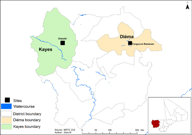
The study was conducted in November 2021 in two Sh-endemic villages (Fangouné Bamanan and Diakalèl) in the Kayes region of western Mali. These two villages are 300 km apart. They were chosen based on their proximity to water sources (ponds in the Diéma district, the Senegal River and its tributaries in the vicinity of the city of Kayes) (Fig. 1). The Kayes region is characterized by a northern Sudanese climate in the south and a Sahelian climate in the north with two main seasons: the rainy season (May–June to October), marked by average annual rainfall of up to 1000 mm in the south and 600 to 800 mm in the north, and the dry season, which extends from November to April–May [24]. Agriculture and livestock are the two main economic activities of the population [24]. The Sudano-Sahelian climate of the region is indeed favorable to the cultivation of dry cereals (millet, sorghum, maize) and groundnuts, and especially to extensive livestock farming where numerous herds of cattle, sheep and goats cohabit.
Fig. 1.
Kayes region (Mali, West Africa) and location of the two study sites
Parasitological methods
Urine samples were collected from 393 children (251 in Diakalèl and 142 in Fangouné Bamanan) aged 6–14 years. Each child was assigned an identification number based on the first two letters of the village name. Urine samples were collected in 60-ml sterile jars between 9 a.m. and 2 p.m. After homogenization, 10 ml urine was taken with a syringe and filtered through a numbered Whatman filter paper (catalog no. 1001–025, 25 mm) previously placed in a filter holder. The filter was then stained with 3% ninhydrin, dried and rewetted with tap water before being read under a microscope with a 4× or 10× objective for Sh eggs. The eggs were counted and classified according to WHO [36] recommendation as light (1–49 eggs/10 ml) or heavy (≥ 50 eggs/10 ml) infection [36]. Urine samples from 63 heavily infected children were used for miracidia capture on FTA cards (GE Healthcare Life Sciences; Amersham, UK). Urine samples were filtered through a 40-µm filter (Fisherbrand™ Sterile Cell Strainers, USA, lot 100082019) and then rewashed in a watch glass using spring water. After 10 to 20 min contact with spring water, the miracidia started to hatch. Miracidia were individually collected using a P10 micropipette set to a volume of 3 µl and then captured on a FTA card. FTA cards were stored at room temperature in a ziplock bag protected from moisture until used for molecular analysis. A total of 30–35 miracidia were captured per child.
Molecular analysis
Three-mm2 disks containing single miracidium were recovered from the FTA card using a Craft Punch and put in 1.5-ml Eppendorf tubes [2]. The DNA was then extracted using a previously published Chelex protocol [4]. A RD-PCR (rapid diagnostic multiplex PCR) was used to genotype the mitochondrial DNA Cox1 gene (mtDNA) [35], and ARMS (amplification-refractory mutation system-polymerase chain reaction) was used to simultaneously genotype the ITS2 and 18S nuclear genes [5]. RD-PCR produces two profiles (Sh and Sb/Sc) according to the band size (120 pb or 260 pb) in agarose gel. Note that the RD-PCR cannot differentiate between Sb and Sc. ARMS-PCR produces more complex profiles (4 to 6 bands) and allows discriminating Sc, Sb, Sh and all hybrid combinations. RD-PCR and ARMS-PCR genotyping methods were double checked by sequencing 136 parasite profiles. The resulting sequences were assembled and manually edited using Sequencher 4.5 (Gene Codes Corp.). Sequence polymorphisms were checked and confirmed by visualizing the chromatograms of the raw sequences and then aligned with the reference sequences using MEGA software. Then, the sequences were grouped by species and gene and aligned with previously published sequences with accession numbers. For the Cox1 gene (~ 1200 bp), the reference sequences for Sh were JQ397330.1 from Senegal and AY157209.1 from Mali, Sb (AJ519521.1 from Senegal and MT159594.1 from Benin) and Sc) (MT579424.1 and MT579422.1 from Senegal. We compared our sequences with those of Sh, Sb and Sc, already published in Genbank with the following respective accession numbers: GU257398.1, MT580950.1 and MT580946.1 for ITS (~ 630) and Z11976.1, AY157238.1 and AY157236.1 for 18S (~ 330). Species-specific SNPs allowed us to differentiate them [5]. Combining the species assignment of the mitochondrial and nuclear genes gives the following possible genotypes: Sb_SbxSb; Sh_ShxSh; Sc_ScxSc (considered pure) and Sb_SbxSh; Sb_SbxSc; Sh_ShxSb; Sh_ShxSc; Sc_ScxSh and Sc_ScxSb (considered hybrid).
Statistical analysis
Gene proportions were tested using the Chi-square or Fisher’s exact tests depending on the data. Probability values (P) < 0.05 were considered statistically significant.
Ethical approval
The project was reviewed and approved by the Institutional Review Board (IRB) of the Faculty of Medicine, Pharmacy and Dentistry of the University of Bamako (no. 2018/71/CE/FMPOS). Community consent was obtained before the study began. Only children whose parents or guardians and who themselves accepted to participate in the study were registered and requested to provide urine samples. Individuals who tested positive for schistosomiasis infection were treated with praziquantel (40 mg/kg of body weight) according to the Schistosomiasis National Control Program (PNLSH) guideline.
Results
A total of 393 urine samples were examined for Sh egg diagnostic (Table 1). The overall prevalence was 69.2% (272/393). The prevalence and intensity of infection were significantly higher in Diakalèl compared to Fangouné Bamanan (P < 0.0001). Urine from 63 heavily positive children (48 in Diakalèl and 15 in Fangouné Bamanan) was used to collect and store miracidia on FTA cards.
Table 1.
Prevalence and intensity of Schistosoma haematobium in the two study sites
| Villages | Number of children examined | Positive | Prevalence (%) | P-value | Negative (%) | Light infection (%) | Heavy infection (%) | P-value |
|---|---|---|---|---|---|---|---|---|
| Diakalèl | 251 | 196 | 78.1 | < 0.0001 | 55 (21.9) | 148 (59.0) | 48 (19.1) | < 0.0001 |
| Fangouné Bamanan | 142 | 76 | 53.5 | 66 (46.5) | 61 (43.0) | 15 (10.5) | ||
| Total | 393 | 272 | 69.2 | 121 (30.8) | 209 (53.2) | 63 (16.0) |
A total of 970 samples were genotyped, from which only 840 gave consistent results using the RD-PCR (Table 2 and Additional file 1: Table S1). From these 840 samples, 353 (42.0%) showed a Sb/Sc band size, and 487 (58.0%) showed a Sh band size. Fifty-five samples from 353 (about 15%) randomly chosen (47 from Diakalèl and 8 from Fangouné Bamanan) with Sb/Sc profile were sequenced, and all showed a Sb profile. Forty samples on 487 (8%) randomly chosen (17 from Diakalèl and 23 from Fangouné Bamanan) with Sh profile were sequenced, and all showed a Sh profile. The relative repartition of each haplotype was different between Diakalèl and Fangouné Bamanan with three times more Sh haplotype in Fangouné Bamanan compared to Diakalel. Of 970 genotyped samples, 925 gave consistent results using the ARMS-PCR, of which 885 (95.7%) showed a ShxSh profile and 40 (4.3%) a ShxSc profile (Table 2). Twenty-six samples of 885 (3%) randomly chosen (7 from Diakalèl and 19 from Fangouné Bamanan) with ShxSh profile were sequenced, and all showed a ShxSh profile. Fifteen samples of 40 (37.5%) randomly chosen (10 from Diakalèl and 5 from Fangouné Bamanan) with ShxSc profile were sequenced, and all showed a ShxSc profile.
Table 2.
Number and percentage (in parentheses) of genes identified by rapid diagnostic PCR (RD_PCR) for Cox 1, ARMS_PCR for IT2/18S and parasite genetic profiles in the two Schistosoma haematobium parasites collected in two villages in Mali
| Haplotype Cox1 | Genotype ITS/18S | ||||||||||||||
|---|---|---|---|---|---|---|---|---|---|---|---|---|---|---|---|
| District | Village | Total | Sb/Sc | Sh | < 0. 0001 | Total | ShxSh | ShxSc | = 0.14 | Total | Sb/Sc_ShxSc | Sb/Sc_ShxSh | Sh_ShxSc | Sh_ShxSh | Total hybrides |
| Kayes | Diakalèl | 475 (56.5) | 333 (70.0) | 142 (30.0) | 526 (56.9) | 508 (96.6) | 18 (3.4) | 447 | 16 (3.6) | 304 (68.0) | 2 (0.4) | 125 (28.0) | 322 (72.0) | ||
| Diéma | Fangouné Bamanan | 365 (43.5) | 20 (5.5) | 345 (94.5) | 399 (43.1) | 377 (94.5) | 22 (5.5) | 342 | 2 (0.6) | 16 (4.7) | 20 (5.8) | 304 (88.9) | 38 (11.1) | ||
| Total | 840 | 353 (42.0) | 487 (58.0) | 925 | 885 (95.7) | 40 (4.3) | 789 | 18 (2.3) | 320 (40.5) | 22 (2.8) | 429 (54.4) | 360 (45.6) | |||
There was a significant difference in the Cox1 profile distribution between the two villages (P < 0.0001). The frequency of Sh ITS2/18S alleles was > 90% in both villages. The distribution of ShxSh and ShxSc genotypes was comparable between the two villages (P = 0.14) (Table 2). Four genetic profiles were identified after genotyping miracidia using Cox1 and ITS/18S: Sb/Sc cox1 × Sh ITS 2/Sc_18S (Sb/Sc_ShxSc); Sb/Sc_cox1 × Sh_ITS 2/ Sh _18S (Sb/Sc_ShxSh); Sh_cox1 × Sh_ITS2/Sc _18S (Sh_ShxSc); Sh_cox1 × Sh_ITS 2/Sh _18S (Sh_ShxSh). Three hybrid profiles [Sb/Sc_ShxSc (2.3%); Sb/Sc_ShxSh (40.5%); Sh_ShxSc (2.8%)] and one pure genetic profile [Sh_ShxSh (54.4%)] have been described. Among the three hybrid profiles, the Sb/Sc_ShxSh profile was more widely represented in Diakalèl compared to Fangouné Bamanan (68.0% vs. 4.7%). In contrast, the non-hybrid Sh_ShxSh genetic profile was more common in Fangouné Bamanan (88.9%) (Table 2: Additional file 1: Table S1). Overall, 45.6% (360/789) of hybrids were recorded. The highest prevalence of hybrids was observed in Diakalèl with 72.0%.
Discussion
We carried out parasitological and molecular study focused on collecting urine from children from a schistosomiasis-endemic area subjected to several years of mass drug treatment (MDT) with PZQ. With an average S. haematobium prevalence of 69.2%, the rate is higher than that recently reported in six villages in the Kayes region (50.1%) [1]. The prevalence and intensity were significantly higher in Diakalèl compared to Fangouné Bamanan (P < 0.0001). This difference between the villages could be explained by the type of watercourse, the Senegal River and its tributaries in Diakalel being more frequented than ponds in Fangouné Bamanan.
The molecular analysis allowed determining the genetic profiles of both a mitochondrial (Cox1) and two nuclear (ITS2 and 18S) genes. Of the 840 miracidia genotyped, which gave consistent results using the RD-PCR in both villages, (42.0%) and (58.0%) had Sb/Sc and Sh Cox1 profiles, respectively. RD-PCR can discriminate Sh and Sm but cannot differentiate Sb from Sc. Although 15% of the randomly selected sequences were identified as Sb regardless of nuclear profile, we suppose that in the overall sampling, the vast majority of Cox1 came from Sb and not Sc. The percentage of Sh (58.0%) we found was higher than those observed in Nigeria (11%) [22] and Ivory Coast (49.3%) [2] but lower than those described in Senegal (79.0%) [7] and Cameroon (95.8%) [32]. For ITS2/18S genes, two profiles were identified with 95.7% for ShxSh and 4.3% for ShxSc. This percentage of ShxSh is higher than observed in Nigeria (40.7%) [22] and Ivory Coast (87.9%) [2] but lower than found in Senegal (97.9%). [7] and Cameroon (92.5%) [32]. All these studies showed that Sh nuclear alleles are dominant in parasites collected from humans.
Analysis of Cox1 and ITS2/18S genetic profiles was performed on 789 miracidia, of which 360 (45.6%) yielded three types of hybrids: Sb/Sc_ShxSc, Sb/Sc_ShxSh and Sh_ShxSc. This percentage of hybrids is lower than those observed in Ivory Coast (62.7%) [2] and Nigeria (89%) [22] but higher than that observed in Cameroon (11.3%) [32]. The hybrids with Sb/Sc_ShxSc profiles indicate gene flow among the three different schistosome species. The observation of the three-species hybrid (Sb_ShxSc) in our study suggests the possibility of interactions and pairing between these species as previously reported by [18] in Niger. Such results confirm the potential for repeated interactions and cross-pairing among these three species and support the hypothesis that they are not first-generation hybrids but rather relatively recent parental and/or hybrid backcrossing [18]. The hybrid genotype profile observed in our study could be explained by the use of the same water points by human populations and animals, especially in the Sahel where water scarcity is a recurring phenomenon. Beyond the hybridization between Sb and Sh previously shown by several authors [2, 14, 22, 27, 29, 32], we also observed Sh_ShxSc hybrids in children, suggesting clearly that Sh can hybridize naturally with Sc, as has also been shown in Senegal [34]. The absence of samples from animals is the main limitation of this study. Genotyping of miracidia from livestock or rodents could provide information on the zoonotic transmission as was the case in Benin with cows [28] and rodents [27] and only in rodents in Senegal [9]. Our research can be considered a pilot study that needs to be replicated across the country to confirm or not the frequency of the three hybrid Sb_ShxSc species.
Conclusion
Our results showed, for the first time to ouir knowledge, high prevalence of hybrid schistosomes in human infections in the Kayes region of Mali. The presence of hybrids underscores the importance of the livestock reservoir of schistosomes, hampering schistosomiasis control and elimination. More studies are needed on population genetics of schistosomes at the human and animal interface to evaluate the parasite’s gene flow and its impact on epidemiology of the disease, transmission to humans and disease control.
Supplementary Information
Additional file 1. Table S1: Genetic data of parasite per child and sentinel site.
Acknowledgements
We thank M. Mamadou Traore, chief of Fangouné Bamanan village, all the breeders, M. Mamadou Sidibe, Director of Diakalel School, and all the schoolchildren, school and health authorities and the population of Kayes for their appreciable contribution to the success of the study. The authors mainly thank Amadou Dabo, called Boua, for his tireless efforts, the transport of the team and equipment; the staff of the MRTC (Maria Research and Training Center); the staff of the Faculty of Medicine and Dentistry and the Faculty of Pharmacy. We thank the Malian government for the funding and the IHPE Laboratory, University Montpellier, CNRS, Ifremer, University Perpignan Via Domitia, Perpignan, France, for the welcome. This work was financially supported by ARES Trading S.A., an affiliate of Merck KGaA, Darmstadt, Germany. This study is set within the framework of the “Laboratoires d’Excellence (LABEX)” TULIP (ANR-10-LABX-41) and the HySWARM (ANR-18-CE35-0001) program of the French Research National Agency.
Abbreviations
- S
Schistosoma
- h
haematobium
- c
curassoni
- b
bovis
- WHO
World Health Organization
- MDA
Mass Drug Administration
- FTA
Find the agent
- ITS
Internal transcribed spacer
- ARMS
Amplification refractory mutation system
- CAT
Catalogue
- DNA
Deoxyribonucleic acid
- mt
Mitochondrial
- IR
Interne reverse
- IF
Interne forward
- OR
Out reverse
- OF
Out forward
- rDNA
Recombinant deoxyribonucleic
- SRB
Senegal River basin
Author contributions
Conceptualization: AD, LD, TS, JB, AKP, MI, DSN. Material preparation, data collection and analysis: PA, MB, SD, BS, AD, DSN, AA, HG, AD, JB. Funding acquisition: AD, LD. TS. Investigation: AD, AKP, BS, AD, AAB. Methodology: DSN, JB, AD, MI, PA, MB, SD. Project administration: AD. Supervision: AD, JB, DSN, MI. Validation: AD, LD. JB, DSN, MI. All authors read and approved the final manuscript.
Funding
This work was financially supported by ARES Trading S.A., an affiliate of Merck KGaA, Darmstadt, Germany.
Availability of data and materials
All data and materials are available in the article.
Declarations
Ethics approval and consent to participate
The study protocol was approved by the Institutional Ethics Committee of the Faculty of Medicine and Dentistry of Bamako under the reference number no. 2018/71/CE/FMPOS. School authorities, teachers, parents/guardians and children were informed about the objectives, procedures and potential risks and benefits of the study. Written informed consent was obtained from children’s parents or legal guardians, while children provided oral assent. After sampling, a praziquantel treatment following WHO guidelines (40 mg/kg) was provided to children found to have a Schistosoma infection. For data protection purposes, an identification number was assigned to each participant.
Informed consent
The Directors of Fangouné Bamanan and Diakalel School obtained informed consent from the parents of all the schoolchildren who participated to the study. Data set availability: Sequence data will be deposited in the NCBI GenBank database Cox 1 DNA.
Consent for publication
Not applicable.
Competing interests
The authors declare that they have no conflict of interest. Ethical approval Ethical permission was obtained from the Ethics Committee of the Faculté de Médecin et d’Odontostomatologie FMOS de Bamako, Mali. T.S. is an employee of Ares Trading S.A., an affiliate of Merck KGaA, Darmstadt, Germany.
Footnotes
Publisher's Note
Springer Nature remains neutral with regard to jurisdictional claims in published maps and institutional affiliations.
Change history
12/13/2023
This article has been corrected since original publication; please see the linked erratum for further details.
Change history
12/15/2023
A Correction to this paper has been published: 10.1186/s13071-023-06061-z
References
- 1.Agniwo P, Sidibé B, Diakité AT, Niaré SD, Guindo H, Akplogan A, et al. Ultrasound aspects and risk factors associated with urogenital schistosomiasis among primary school children in mali. Infect Dis Poverty. 2022;3:80. doi: 10.1186/s40249-023-01071-6. [DOI] [PMC free article] [PubMed] [Google Scholar]
- 2.Angora EK, Vangraefschepe A, Allienne J-F, Menan H, Coulibaly JT, Meïté A, et al. Population genetic structure of Schistosoma haematobium and Schistosoma haematobium × Schistosoma bovis hybrids among school-aged children in Côte d’Ivoire. Parasite. 2022;29:23. doi: 10.1051/parasite/2022023. [DOI] [PMC free article] [PubMed] [Google Scholar]
- 3.Arnold ML. Natural hybridization and the evolution of domesticated, pest and disease organisms. Mol Ecol. 2004;13:997–1007. doi: 10.1111/j.1365-294X.2004.02145.x. [DOI] [PubMed] [Google Scholar]
- 4.Beltran S, Galinier R, Allienne JF, Boissier J. Cheap, rapid and efficient DNA extraction method to perform multilocus microsatellite genotyping on all Schistosoma mansoni stages. Mem Inst Oswaldo Cruz. 2008;103:501–503. doi: 10.1590/S0074-02762008000500017. [DOI] [PubMed] [Google Scholar]
- 5.Blin M, Dametto S, Agniwo P, Webster BL, Angora E. A duplex tetra-primer ARMS-PCR assay to discriminate three species of the Schistosoma haematobium group: Schistosoma curassoni, S. bovis, S. haematobium and their hybrids. Parasites Vectors. 2023;16:1–11. doi: 10.1186/s13071-023-05754-9. [DOI] [PMC free article] [PubMed] [Google Scholar]
- 6.Boon A, Mbow M, Paredis L, Moris P, Sy I, Maes T, et al. No barrier breakdown between human and cattle Schistosome species in the senegal river basin in the face of hybridisation. Int J Parasitol. 2019;49:1039–1048. doi: 10.1016/j.ijpara.2019.08.004. [DOI] [PubMed] [Google Scholar]
- 7.Boon N, Van Den Broeck F, Faye D, Volckaert F, Mboup S, Polman K, Huyse T. Barcoding hybrids: heterogeneous distribution of Schistosoma haematobium × Schistosoma bovis hybrids across the senegal river basin. Parasitology. 2018;145:634–645. doi: 10.1017/S0031182018000525. [DOI] [PubMed] [Google Scholar]
- 8.Brinkmann U, Werler C, Traore M, Korte R. The national schistosomiasis control programme in mali, objectives, organization, results. Trop Med Parasitol. 1988;39:157–161. [PubMed] [Google Scholar]
- 9.Catalano S, Sène M, Diouf ND, Fall CB. Rodents as natural hosts of zoonotic schistosoma species and hybrids: an epidemiological and evolutionary perspective from West Africa. J Infect Dis. 2018;218:429–433. doi: 10.1093/infdis/jiy029. [DOI] [PubMed] [Google Scholar]
- 10.Clements ACA, Bosqué-Oliva E, Sacko M, Landouré A, Dembélé R. A comparative study of the spatial distribution of schistosomiasis in Mali in 1984–1989 and 2004–2006. PLoS Negl Trop Dis. 2009;3:1–11. doi: 10.1371/journal.pntd.0000431. [DOI] [PMC free article] [PubMed] [Google Scholar]
- 11.Cnops L, Soentjens P, Clerinx J, Van Esbroeck M. A Schistosoma haematobium-specific real-time PCR for diagnosis of urogenital schistosomiasis in serum samples of international travelers and migrants. PLoS Negl Trop Dis. 2013 doi: 10.1371/journal.pntd.0002413. [DOI] [PMC free article] [PubMed] [Google Scholar]
- 12.Jan DB, Vercruysse J. Schistosomiasis in cattle. Amsterdam: Elsevier; 1998. [Google Scholar]
- 13.Huyse T, Van den Broeck F, Hellemans B, Volckaert FAM, Polman K. Hybridisation between the two major African Schistosome species of humans. Int J Parasitol. 2013;43:687–689. doi: 10.1016/j.ijpara.2013.04.001. [DOI] [PubMed] [Google Scholar]
- 14.Huyse T, Webster BL, Sarah Geldof J, Stothard R, Diaw OT, Polman K, Rollinson D. Bidirectional introgressive hybridization between a cattle and human schistosome species. PLoS Pathog. 2009 doi: 10.1371/journal.ppat.1000571. [DOI] [PMC free article] [PubMed] [Google Scholar]
- 15.Kincaid-Smith J, Tracey A, Augusto R, Bulla I, Holroyd N, Rognon A, et al. Whole Genome sequencing and morphological analysis of the human-infecting schistosome emerging in europe reveals a complex admixture between Schistosoma haematobium and Schistosoma bovis Parasites. 2018. 10.1101/387969.
- 16.King CH, Bertsch D. Historical perspective: snail control to prevent schistosomiasis. PLoS Negl Trop Dis. 2015 doi: 10.1371/journal.pntd.0003657. [DOI] [PMC free article] [PubMed] [Google Scholar]
- 17.Kouadio JN, Giovanoli Evack J, Achi LY, et al. Prévalence et répartition de la schistosomiase et de la fascioliase du bétail en Côte d’Ivoire: résults from a cross-sectional survey. BMC Vet Res. 2020;16:446. doi: 10.1186/s12917-020-02667-y. [DOI] [PMC free article] [PubMed] [Google Scholar]
- 18.Leger E, Webster JP. Hybridizations within the genus schistosoma: implications for evolution, epidemiology and control. Parasitology. 2016;144:65–80. doi: 10.1017/S0031182016001190. [DOI] [PubMed] [Google Scholar]
- 19.Govic Le, Yohann J-S, Allienne JF, Rey O, de Gentile L, Boissier J. Schistosoma haematobium–Schistosoma mansoni hybrid parasite in migrant boy, France, 2017. Emerg Infect Dis. 2019;25:365–367. doi: 10.3201/eid2502.172028. [DOI] [PMC free article] [PubMed] [Google Scholar]
- 20.Mallet J. Hybridization as an invasion of the genome. Trends Ecol Evol. 2005 doi: 10.1016/j.tree.2005.02.010. [DOI] [PubMed] [Google Scholar]
- 21.Moné H, Mouahid G, Morand S. The distribution of schistosoma bovis sonsino, 1876 in relation to intermediate host mollusc-parasite relationships. Adv Parasitol. 1999 doi: 10.1016/S0065-308X(08)60231-6. [DOI] [PubMed] [Google Scholar]
- 22.Onyekwere A, Rey O, Allienne J, Nwanchor MC, Alo M, Uwa C, Boissier J. Population genetic structure and hybridization of schistosoma haematobium in Nigeria. Pathogens. 2022 doi: 10.3390/pathogens11040425. [DOI] [PMC free article] [PubMed] [Google Scholar]
- 23.Panzner U, Boissier J. Natural intra- and interclade human hybrid schistosomes in Africa with considerations on prevention through vaccination. Microorganisms. 2021;9:1465. doi: 10.3390/microorganisms9071465. [DOI] [PMC free article] [PubMed] [Google Scholar]
- 24.Philibert A, Tourigny C, Coulibaly A, Fournier P. Birth seasonality as a response to a changing rural environment (Kayes Region, Mali) J Biosoc Sci. 2013;45:547–565. doi: 10.1017/S0021932012000703. [DOI] [PubMed] [Google Scholar]
- 25.Pitchford RJ. A check list of definitive hosts exhibiting evidence of the genus Schistosoma weinland, 1858 acquired naturally in Africa and the Middle East. J Helminthol. 1977;51:229–251. doi: 10.1017/S0022149X00007574. [DOI] [PubMed] [Google Scholar]
- 26.Rollinson D, Southgate VR, Vercruysse J, Moore PJ. Observations on natural and experimental interactions between Schistosoma boris and S. curassoni from West Africa. Acta Trop. 1990;47:101–114. doi: 10.1016/0001-706X(90)90072-8. [DOI] [PubMed] [Google Scholar]
- 27.Savassi BAES, Dobigny G, Etougbétché JR, Avocegan TT, Quinsou FT, Gauthier P, et al. Mastomys natalensis (Smith, 1834) as a natural host for Schistosoma haematobium (Bilharz, 1852) Weinland, 1858 x Schistosoma bovis Sonsino, 1876 introgressive hybrids. Parasitol Res. 2021;120:1755–1770. doi: 10.1007/s00436-021-07099-7. [DOI] [PMC free article] [PubMed] [Google Scholar]
- 28.Savassi BAES, Mouahid G, Lasica C, Mahaman S-D, Garcia A, Courtin D, et al. Cattle as natural host for Schistosoma haematobium (Bilharz, 1852) Weinland, 1858 x Schistosoma bovis Sonsino, 1876 interactions, with new cercarial emergence and genetic patterns. Parasitol Res. 2020 doi: 10.1007/s00436-020-06709-0. [DOI] [PubMed] [Google Scholar]
- 29.Soentjens P, Cnops L, Huyse T, Yansouni C, De Vos D, Bottieau E, et al. Diagnosis and clinical management of Schistosoma haematobium – Schistosoma bovis hybrid infection in a cluster of travelers returning from Mali. Clin Infect Dis. 2016;63:15–18. doi: 10.1093/cid/ciw493. [DOI] [PubMed] [Google Scholar]
- 30.Taylor MG. Hybridisation experiments on five species of African Schistosomes. J Helminthol. 1970;44:253–314. doi: 10.1017/S0022149X00021969. [DOI] [PubMed] [Google Scholar]
- 31.Tembely S, Galvin TJ, Craig TM, Traoré S. Liver fluke infectious of cattle in Mali. an abattoir survey on prevalence and géagraphic distribution. Trop Anim Hlth Prod. 1988;20:117–212. doi: 10.1007/BF02242240. [DOI] [PubMed] [Google Scholar]
- 32.Teukeng FF, Djuikwo MB, Bech N, Gomez MR, Zein-Eddine R, Simo AMK, et al. Hybridization increases genetic diversity in Schistosoma haematobium populations infecting humans in cameroon. Infect Dis Poverty. 2022;11:1–11. doi: 10.1186/s40249-022-00958-0. [DOI] [PMC free article] [PubMed] [Google Scholar]
- 33.Traoré M, Maude G, Bradley D. Schistosomiasis haematobia in Mali: prevalence rate in school-age children as index of endemicity in the community. Tropical Med Int Health. 1998;3:214–221. doi: 10.1111/j.1365-3156.1998.tb00274.x. [DOI] [PubMed] [Google Scholar]
- 34.Webster BL, Diaw OT, Seye MM, Webster JP, Rollinson D. Introgressive hybridization of Schistosoma haematobium group species in Senegal: species barrier break down between ruminant and human Schistosomes. PLoS Negl Trop Dis. 2013;4:1–9. doi: 10.1371/journal.pntd.0002110. [DOI] [PMC free article] [PubMed] [Google Scholar]
- 35.Webster BL, Rollinson D, Stothard JR, Huyse T. Rapid diagnostic multiplex PCR (RD-PCR) to discriminate Schistosoma haematobium and S. bovis. J Helminthol. 2010;84:107–114. doi: 10.1017/S0022149X09990447. [DOI] [PubMed] [Google Scholar]
- 36.WHO . Control of schistosomiasis: report of a WHO expert committee techn. Geneva: Report Series; 1985. [Google Scholar]
- 37.WHO. 2022. “Schistosomiasis and Soil-Transmitted Helminthiases: Progress Report, 2021.” https://www.who.int/Publications/i/Item/Who-Wer9748-621-632 (48):621–32.
Associated Data
This section collects any data citations, data availability statements, or supplementary materials included in this article.
Supplementary Materials
Additional file 1. Table S1: Genetic data of parasite per child and sentinel site.
Data Availability Statement
All data and materials are available in the article.


