Abstract
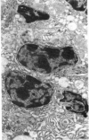
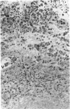
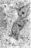
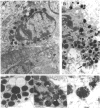
Light and electron microscopy were used to examine tissue excised during surgery from eight patients with advanced destructive scleral disease. These comprised two cases of scleromalacia perforans, three cases of anterior necrotising scleritis alone or in conjunction with other systemic diseases, and three cases in which scleritis developed following ocular surgery. It was not possible to distinguish between these three categories by histological or cytopathological criteria. All showed extensive granulomatous infiltration of the conjunctiva, episclera, and sclera by plasma cells and lymphocytes. Mast cells were abundant throughout these inflamed tissues. Examination of scleral stroma from sites in advance of the granuloma revealed active fibroblastic cells in the absence of other inflammatory cells. Fibroblastic transformation of scleral cells may be one of the earliest events in scleral degradation during necrotising disease.
Full text
PDF


Images in this article
Selected References
These references are in PubMed. This may not be the complete list of references from this article.
- ASHTON N., HOBBS H. E. Effect of cortisone on rheumatoid nodules of the sclera (scleromalacia perforans). Br J Ophthalmol. 1952 Jul;36(7):373–384. doi: 10.1136/bjo.36.7.373. [DOI] [PMC free article] [PubMed] [Google Scholar]
- Fell H. B., Jubb R. W. The effect of synovial tissue on the breakdown of articular cartilage in organ culture. Arthritis Rheum. 1977 Sep-Oct;20(7):1359–1371. doi: 10.1002/art.1780200710. [DOI] [PubMed] [Google Scholar]
- François J. Ocular manifestations in collagenoses (with colour plates I and II). Adv Ophthalmol. 1970;23:1–54. [PubMed] [Google Scholar]
- Henriquez A. S., Bloch K. J., Kenyon K. R., Baird R. S., Hanninen L. A., Allansmith M. R. Ultrastructure of mast cells in rat ocular tissue undergoing anaphylaxis. Arch Ophthalmol. 1983 Sep;101(9):1439–1446. doi: 10.1001/archopht.1983.01040020441023. [DOI] [PubMed] [Google Scholar]
- Lyne A. J., Lloyd-Jones D. Necrotizing scleritis after ocular surgery. Trans Ophthalmol Soc U K. 1979 Apr;99(1):146–149. [PubMed] [Google Scholar]
- Saklatvala J., Sarsfield S. J. Lymphocytes induce resorption of cartilage by producing catabolin. Biochem J. 1982 Jan 15;202(1):275–278. doi: 10.1042/bj2020275. [DOI] [PMC free article] [PubMed] [Google Scholar]
- Sevel D. Necrogranulomatous scleritis. Clinical and histologic features. Am J Ophthalmol. 1967 Dec;64(6):1125–1134. [PubMed] [Google Scholar]
- Taylor D. J., Yoffe J. R., Woolley D. E. Histamine H2 receptors on foetal-bovine articular chondrocytes. Biochem J. 1983 May 15;212(2):517–520. doi: 10.1042/bj2120517. [DOI] [PMC free article] [PubMed] [Google Scholar]
- Watson P. G. Doyne Memorial Lecture, 1982. The nature and the treatment of scleral inflammation. Trans Ophthalmol Soc U K. 1982 Jul;102(Pt 2):257–281. [PubMed] [Google Scholar]
- Young R. D., Watson P. G. Microscopical studies of necrotising scleritis. II. Collagen degradation in the scleral stroma. Br J Ophthalmol. 1984 Nov;68(11):781–789. doi: 10.1136/bjo.68.11.781. [DOI] [PMC free article] [PubMed] [Google Scholar]


