

Abstract
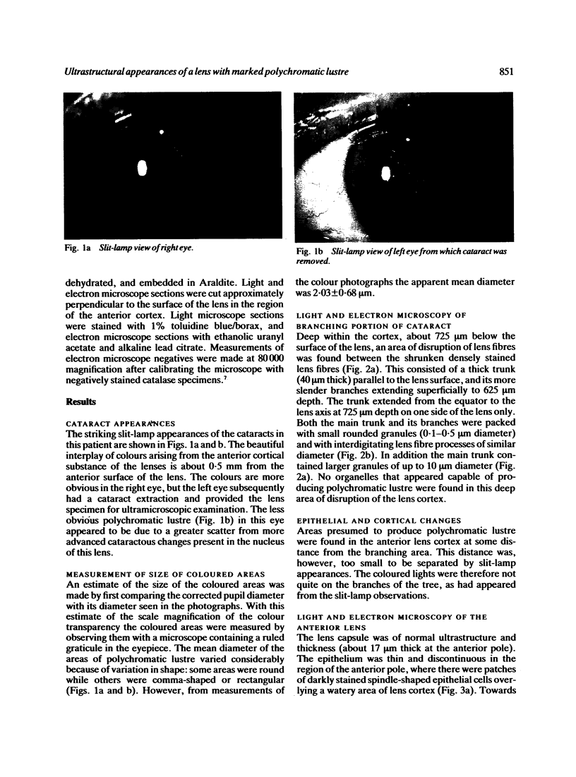
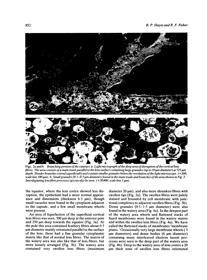
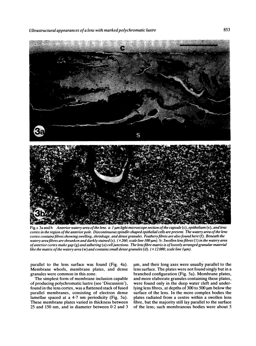
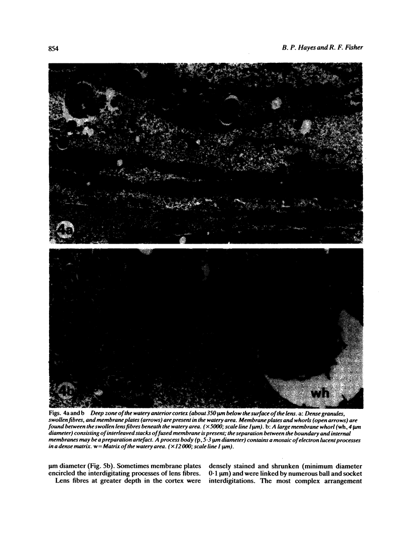
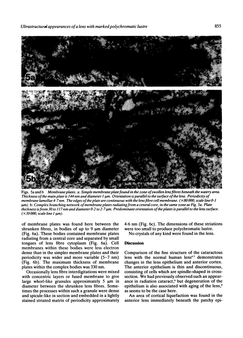
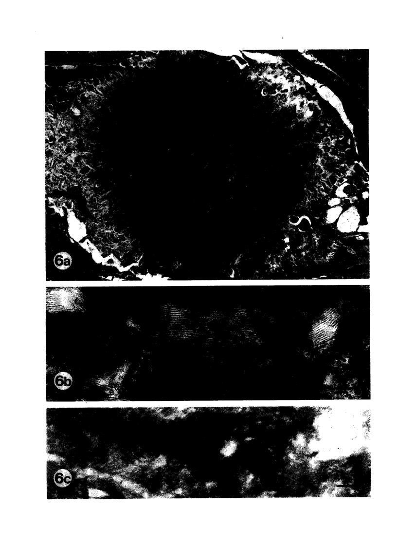
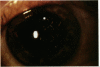
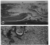
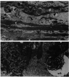
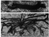
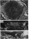
A human lens showing polychromatic lustre associated with cortical opacities (Christmas tree cataract) has been examined by slit-lamp photography, thin-section light microscopy, and electron microscopy. Anterior epithelial cells were fibroblast-like, and an area of breakdown of cortical lens fibres was observed in the anterior lens, containing feathery fibres, whorls, and process bodies. Parallel sided stacks of fused cell membranes were found beneath the watery area at the same depth as the polychromatic lustre seen in the slit-lamp. The dimensions of these membrane plates are consistent with the diffraction of light by a parallel thin film to give coloured points of light. Deeper areas of small granules between the lens fibres are associated with the main trunk-like opacity and its branches seen with the slit-lamp.
Full text
PDF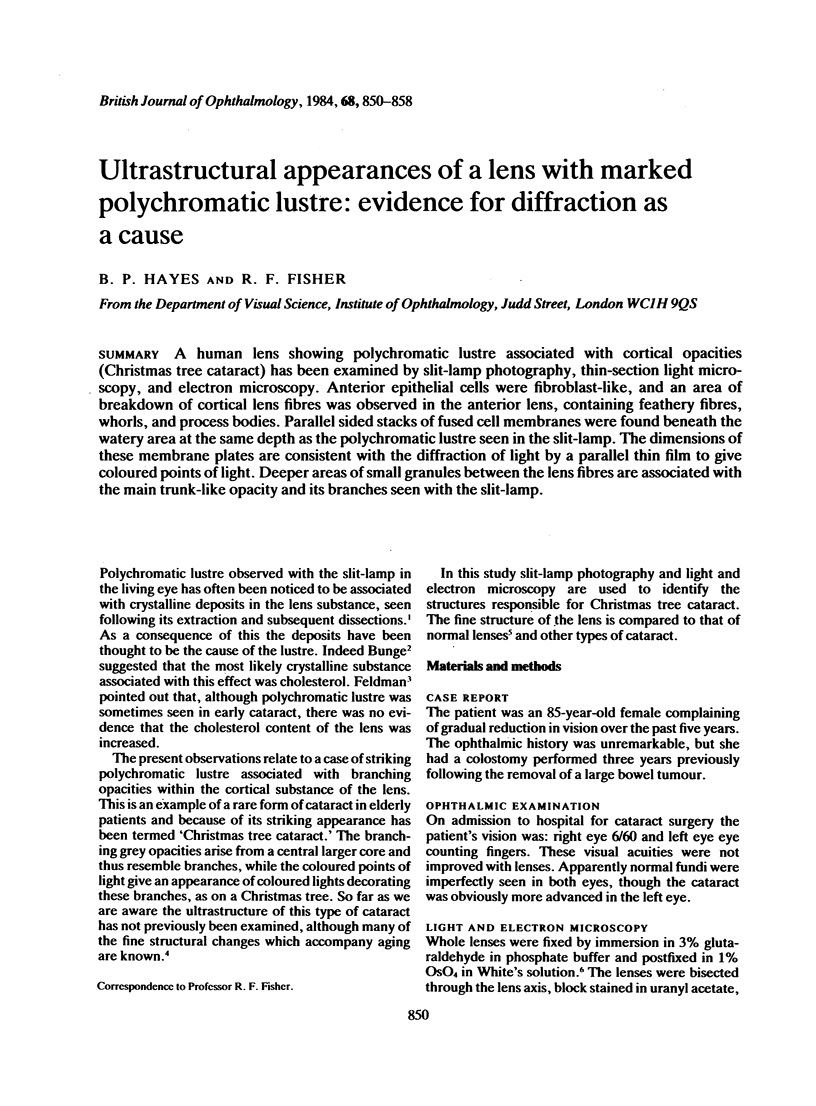
Images in this article
Selected References
These references are in PubMed. This may not be the complete list of references from this article.
- BRINI A., PORTE A., STOECKEL M. E. MODIFICATIONS ULTRASTRUCTURALES DU CRISTALLIN DANS CERTAINES CATARACTES EXP'ERIMENTALES ET HUMAINES. Bull Mem Soc Fr Ophtalmol. 1963;76:193–208. [PubMed] [Google Scholar]
- Fisher R. F., Hayes B. P. Ultrastructure and elastic changes of regenerated basement membrane in normal and diabetic rats. Br J Exp Pathol. 1982 Jun;63(3):341–350. [PMC free article] [PubMed] [Google Scholar]
- Fisher R. F. The water permeability of basement membrane under increasing pressure: evidence for a new theory of permeability. Proc R Soc Lond B Biol Sci. 1982 Nov 22;216(1205):475–496. doi: 10.1098/rspb.1982.0087. [DOI] [PubMed] [Google Scholar]
- Hayes B. P., Fisher R. F. Influence of a prolonged period of low-dosage x-rays on the optic and ultrastructural appearances of cataract of the human lens. Br J Ophthalmol. 1979 Jul;63(7):457–464. doi: 10.1136/bjo.63.7.457. [DOI] [PMC free article] [PubMed] [Google Scholar]