

Abstract
The gonioscopic findings of 76 patients with the exfoliation syndrome were reviewed. A high frequency of narrowness of the anterior chamber (AC) angle was found (32%). 18% had angles considered occludable, and 14% had obvious angle-closure glaucoma as shown by the presence of peripheral anterior synechias (PAS). Increased pigmentation of the posterior trabecular meshwork (PTM) was noted in all cases. When this pigmentation was markedly asymmetrical, unilateral exfoliation with glaucoma was common in the more pigmented eye. In addition heavy angle pigmentation in the absence of exfoliation was noted in the fellow eye of patients with characteristic exfoliated material in the other eye. Increased pigmentation of the PTM may be the earliest detectable sign of the exfoliation syndrome (ES). The clinical significance of our estimating PTM pigmentation at the 12 o'clock position is discussed. In view of the accelerated optic nerve damage associated with the development of glaucoma secondary to ES, routine estimation of the pigmentation of the PTM at 12 o'clock is recommended in the hope of early detection of cases of otherwise inapparent ES.
Full text
PDF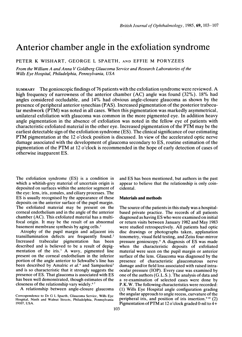
Images in this article
Selected References
These references are in PubMed. This may not be the complete list of references from this article.
- AMALRIC P., SAMPAOLESI R., BESSOU P. [On the early diagnosis and heredity of capsular pseudo-exfoliation]. Bull Soc Ophtalmol Fr. 1960 May-Jun;6:341–350. [PubMed] [Google Scholar]
- Aasved H. Incidence of defects in the pigmented pupillary ruff in eyes with and without fibrillopathia epitheliocapsularis (so-called senile exfoliation or pseudoexfoliation of the anterior lens capsule). Acta Ophthalmol (Copenh) 1973;51(5):710–715. doi: 10.1111/j.1755-3768.1973.tb08264.x. [DOI] [PubMed] [Google Scholar]
- Aasved H. Intraocular pressure in eyes with and without fibrillopathia epitheliocapsularis (so-called senile exfoliation or pseudoexfoliation). Acta Ophthalmol (Copenh) 1971;49(4):601–610. doi: 10.1111/j.1755-3768.1971.tb02967.x. [DOI] [PubMed] [Google Scholar]
- Bartholomew R. S. Anterior chamber depth in eyes with pseudoexfoliation. Br J Ophthalmol. 1980 May;64(5):322–323. doi: 10.1136/bjo.64.5.322. [DOI] [PMC free article] [PubMed] [Google Scholar]
- Eagle R. C., Jr, Font R. L., Fine B. S. The basement membrane exfoliation syndrome. Arch Ophthalmol. 1979 Mar;97(3):510–515. doi: 10.1001/archopht.1979.01020010254014. [DOI] [PubMed] [Google Scholar]
- Forbes M. Gonioscopy with corneal indentation. A method for distinguishing between appositional closure and synechial closure. Arch Ophthalmol. 1966 Oct;76(4):488–492. doi: 10.1001/archopht.1966.03850010490005. [DOI] [PubMed] [Google Scholar]
- Haddad R., Strasser G., Heilig P., Jurecka W. Decompensation of chronic open-angle glaucoma following mydriasis-induced pigmentary dispersion into the aqueous humour: a light and electron microscopic study. Br J Ophthalmol. 1981 Apr;65(4):252–257. doi: 10.1136/bjo.65.4.252. [DOI] [PMC free article] [PubMed] [Google Scholar]
- Kozart D. M., Yanoff M. Intraocular pressure status in 100 consecutive patients with exfoliation syndrome. Ophthalmology. 1982 Mar;89(3):214–218. doi: 10.1016/s0161-6420(82)34804-6. [DOI] [PubMed] [Google Scholar]
- LOWE R. F. PRIMARY ANGLE-CLOSURE GLAUCOMA WITH CAPSULAR EXFOLIATION OF THE LENS. Br J Ophthalmol. 1964 Sep;48:492–494. doi: 10.1136/bjo.48.9.492. [DOI] [PMC free article] [PubMed] [Google Scholar]
- Layden W. E., Shaffer R. N. Exfoliation syndrome. Am J Ophthalmol. 1974 Nov;78(5):835–841. [PubMed] [Google Scholar]
- Madden J. G., Crowley M. J. Factors in the exfoliation syndrome. Br J Ophthalmol. 1982 Jul;66(7):432–437. doi: 10.1136/bjo.66.7.432. [DOI] [PMC free article] [PubMed] [Google Scholar]
- Petersen H. P. Can pigmentary deposits on the trabecular meshwork increase the resistance of the aqueous outflow? Acta Ophthalmol (Copenh) 1969;47(3):743–749. doi: 10.1111/j.1755-3768.1969.tb08163.x. [DOI] [PubMed] [Google Scholar]
- Richardson T. M., Epstein D. L. Exfoliation glaucoma: a quantitative perfusion and ultrastructural study. Ophthalmology. 1981 Sep;88(9):968–980. [PubMed] [Google Scholar]
- Richardson T. M., Hutchinson B. T., Grant W. M. The outflow tract in pigmentary glaucoma: a light and electron microscopic study. Arch Ophthalmol. 1977 Jun;95(6):1015–1025. doi: 10.1001/archopht.1977.04450060101010. [DOI] [PubMed] [Google Scholar]
- Rodrigues M. M., Spaeth G. L., Weinreb S., Sivalingam E. Spectrum of trabecular pigmentation in open-angle glaucoma: a clinicopathologic study. Trans Sect Ophthalmol Am Acad Ophthalmol Otolaryngol. 1976 Mar-Apr;81(2):258–276. [PubMed] [Google Scholar]
- Roth M., Epstein D. L. Exfoliation syndrome. Am J Ophthalmol. 1980 Apr;89(4):477–481. doi: 10.1016/0002-9394(80)90054-9. [DOI] [PubMed] [Google Scholar]
- Spaeth G. L. Gonioscopy: uses old and new. The inheritance of occludable angles. Ophthalmology. 1978 Mar;85(3):222–232. doi: 10.1016/s0161-6420(78)35675-x. [DOI] [PubMed] [Google Scholar]
- Spaeth G. L. The normal development of the human anterior chamber angle: a new system of descriptive grading. Trans Ophthalmol Soc U K. 1971;91:709–739. [PubMed] [Google Scholar]
- Sugar H. S., Harding C., Barsky D. The exfoliation syndrome. Ann Ophthalmol. 1976 Oct;8(10):1165–1181. [PubMed] [Google Scholar]