

Introduction and importance:
Macrodactyly is an uncommon, not inherited congenital malformation of the digit with unknown prevalence and path of pathogenesis. The condition was described in 1940 and since then 107 cases were reported. Manifestations may mislead the diagnosis of hemangiomas and lymphangiomatosis. There are different options for treatment without a clear consensus. The authors are presenting a macrodactyly case that improved the quality of his life after he underwent surgical amputation of the toes.
Case presentation:
The authors had a case of a 2-year and 4-month-old male child presented with progressive growth of the left foot toes; which started since birth in the 4th toe and then involved 3rd and 5th toe later; resulting in deformity and difficulty in wearing shoes. Physical examination; showed left foot enlargements of the 3rd–5th toes. X-ray of the left foot was done he was diagnosed to have macrodactyly. Under general anesthesia metatarsophalangeal joint of the 4th–5th toe and distal interphalangeal joint of the 3rd toe, disarticulation was done. The patient is doing okay on follow-up for the last year.
Clinical discussion:
Consistently with other case reports from Korea, Tanzania, and Congo our patient presented with a primary type of left foot macrodactyly in his early life, and he was successfully managed with amputation of the affected digits.
Conclusion:
This is one of the rare cases which needs a high index of suspicion to diagnose and treat early to improve quality of life. Amputation is the most important management in resources limited areas.
Keywords: amputation, case report, foot, macrodactyly
Introduction
Highlights
Macrodactyly is one of the rare and not inherited congenital anomalies of the extremities.
Macrodactyly results in deformity and functional limitation of the affected limb.
It needs a high index of suspicion to diagnose and treat early to improve quality of life.
Amputation is the most important management in resources limited areas.
Macrodactyly is an uncommon congenital malformation of the digit with no clearly identified cause but is not inherited1. The condition was described in 1940 and since then 107 cases were reported2. Due to the rarity of the condition, the exact prevalence and clear path of pathogenesis are unknown. It is associated with males more than females and is more common in feet than hands1,3.
There are two types of macrodactyly categorized by different criteria: Static versus progressive: static macrodactyly is when the affected digit grows proportionally to the other body; whereas progressive macrodactyly is manifested by disproportionate growth of the affected limb when compared to the child’s growth2,3. Primary or true versus secondary: True/primary macrodactyly is when all tissues of the affected digit increase in size but secondary macrodactyly is when some components like soft tissue and skin or bone only are affected which shows the presence of other underlying causes. All kinds of manifestations may mislead the diagnosis as hemangioma, fibro lipomatous hamartoma, and lymphangiomatosis3.
Patients complain of deformity and local dysfunctionality especially when the toes are affected which hampers the wearing of shoes. The target of treatment is a pain-free and functional foot that accommodates shoes. There are different options for treatment without clear consensus and treatment is individualized but mainly for those countries that have limited resources amputation is the option4,5. In the early era, the only option of treatment was partial or complete amputation of the affected digit till 1956 when defatting procedure and epiphyseal destruction were introduced. Later, in 1965 stage wise procedures were also introduced as other better alternative management options2.
There were reviewed cases in Korea and out of 31 macrodactyly patients, 20 cases were idiopathic (primary) and 11 cases were secondary and the majority of them (93%) were under 18 years. Both feet and hands were involved with no difference on the right and left sides. Most of the patients had unilateral involvement but with multiple digit involvements2. Another study from Congo reported four cases of feet Macrodactyly, half of them were female, all were under 18 years old, and all were managed by ray amputation of the affected toe4. There was also a 5-year progressive type of feet macrodactyly case report from Tanzania that involved two toes since birth, which impairs his shoe wearing and amputation was done5. The objective of this paper is to present a macrodactyly case that improved the quality of his life after he underwent surgical amputation of the toes. This case report has been reported in line with the Surgical CAse REport (SCARE) 2020 Checklist6. Agha RA, Franchi T, Sohrabi C, Mathew G, for the SCARE Group. The SCARE 2020 Guideline: Updating Consensus Surgical Case Report (SCARE) Guidelines, International Journal of Surgery 2020;84:226-230.
Case report
MH is a 2-year and 4-month-old male patient who presents to our hospital with progressive right 3–5th toe enlargement and change in shape. The enlargement was present in the 4th toe since birth and then later involved the other toe with a progressive increment in size, which makes it difficult to wear shoes. He was able to walk independently without waking aids. Neither the left foot nor the upper extremity was involved. No family history of a similar illness or no other known familial illness. He is right-handed. He is from a low socio-economic family and lives in the countryside. No previous known medical illness and no surgical intervention. No skin allergy, no self or family history of asthma or smoking exposure. No other pertinent history. The family visited health facilities at the age of 1 year but they were told it will improve by itself. Currently referred to our hospital for better management arrived at regular OPD walking on foot.
Physical exam
Conscious with stable vital signs and pertinent fining was on the lower extremity, which showed significant enlargements of the 3–5th toes prominently in the middle and distal phalanx with the biggest toe being the 4th toe (Fig. 1). A left foot radiograph was done and showed thickening and hypertrophying of the three distal toes (Fig. 2). He was diagnosed to have macrodactyly left 3–5th toes. There were no difficulties in reaching the diagnosis and no other alternative diagnosis was considered. Preoperatively: The patient was assessed for any underlying medical condition preoperatively by a pediatrician and anesthesiologist and there were no findings. He was categorized as fit for surgery. He was prepared for surgery and informed that written consent for the operation was taken from his mother. The patient was managed per the schedule. Intraoperative: After positioning supine., under general anesthesia; the right foot was cleaned and draped. The metatarsophalangeal joint of the 4–5th toe and the distal interphalangeal joint of the 3rd toe disarticulation and amputation was done (Fig. 3). The wound was closed layer by layer. There was no accident intraoperatively and the patient’s vital sign was stable. The operation was done in a tertiary hospital setting by an orthopedic surgeon. Postoperative: The patient was transferred to the pediatric ward after the surgery, kept NPO, and put on maintenance fluid for 12 h. He continued paracetamol 125 mg suppository and Ceftriaxone 250 mg IV for 48 h and the hospital course was smooth. During discharge, the patient was advised to wear shoes when the wound completely heals. The parents were adherent to the advice provided during discharge. He was discharged after 5 days of hospital stay and he had subsequent follow-ups in the orthopedic clinic every three months for the last year and patient was comfortable enough to wear shoes. Parents were happy about the management as their child is able to wear shoes and they were curious about the genetic transmissibility but after they were advised they were happy and reassured.
Figure 1.
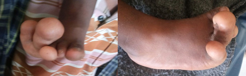
Front and Lateral view photo of a 2-year and 4-month-old patient’s left foot showing enlargement of 3–5th toes (preoperative).
Figure 2.
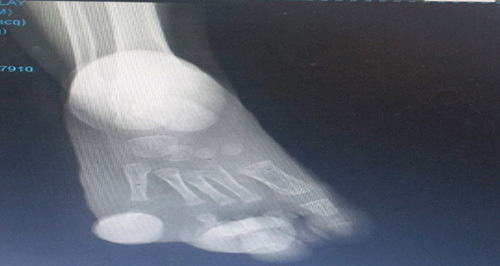
Left foot radiograph of 2 years and 4 months old showing thickened and hypertrophic distal 3–5th toe (preoperative).
Figure 3.
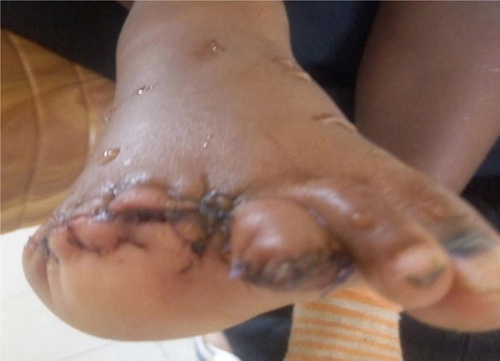
Postoperation picture of the left feet after 3–5th toes were amputated.
Discussion and conclusion
Macrodactyly is one of the rare not inherited anomalies that may present since birth and may increase after that proportionally with the child’s physical growth or out of proportion7,8. Though the etiology is unknown, the possible explanation is due to the nerve stimulated with abnormal neuronal control in the sensory distribution of the peripheral nerve as the enlarged site coincides with nerve enlargement7,8. Like our patients did not have a family history of similar illnesses. In the majority of reports found in different literature, patients visit at an early age since it is congenital and cause deformity of the extremities leading to difficulty in wearing shoes and limited limb functions9,10. This was consistent with our patient that was congenital and presented at 2 years and 4 months with difficulty wearing shoes. There were different types of macrodactyly; which may be primary or secondary. A review of Korean hospitals showed 64.5% were primary and the majority of them were also progressive types2. Our patient had a progressive type and had no underlying secondary cause. This was a similar finding to a case report from Tanzania5. There are no clear and concise management guidelines; though the majority especially in the limited resources areas use amputation of the affected limb as the better option of treatment4,5. Our patient had been managed by an orthopedic surgeon, and disarticulation and amputation at Metatarsophalangeal joint for 4 and 5th and at distal interphalangeal joint for the 3rd toe was done. Similar management options were used in Tanzania’s case and in some of the Korean patients2,5. There were four cases seriously reported from Congo with macrodactyly and all were managed with amputation and achieved well in terms of outcome measured by the ability to walk and wear their shoe4. In our case, the wound healed well and started to wear shoes and ambulate well. Early detection and management of the overgrowth of the affected digit will improve the quality of life; avoid the stigma due to disfigurement of the limb. Amputation of the affected digit may be an effective treatment with limited resources like in our case.
Strength and limitation
The strength of this case report, the diagnosis of macrodactyly was made on his first visit as such kind of cases are rare and may be missed the diagnosis. His outcome was good as functionally and disfigurement improved after amputation. The limitation of this case report is a complete loss of his toes as this may be avoidable with serial debulking of the overgrowth of the affected digits in a well-equipped setting.
Ethics approval and consent for publication
Written informed consent was obtained from the patient’s father for publication of this case with the inclusion of symptoms, signs, and images of the patients in the manuscript paper. A copy of the written consent is available for review by the Editor-in-Chief of this journal. There is no need to have Ethical approval of case reports in our institution.
Consent
Written informed consent was obtained from the patient's father for publication of this case report and accompanying images. A copy of the written consent is available for review by the Editor-in-Chief of this journal on request.
Sources of funding
No specific funding was received.
Author contribution
All authors made a significant contribution to the work reported, whether that is in the conception, study design, execution, acquisition of data, analysis, and interpretation, or in all these areas; took part in drafting, revising, or critically reviewing the article; gave final approval of the version to be published; have agreed on the journal to which the article has been submitted; and agree to be accountable for all aspects of the study.
Conflicts of interest disclosure
The authors declare that they have no competing interests.
Research registration unique identifying number (UIN)
The case report is registered with a unique identifying number of researchregistry8833.
Guarantor
Hansa Haftu (MD, Pediatrician), Mekelle University, College of Health Science, Pediatric, and Child Health, Tigray, Mekelle, Ethiopia. Tel: +251 948487877. E-mail: hansahaftu21@gmail.com.
Availability of data and material
Please contact the author for data requests. Data never presented before in any conferences or regional meetings.
Provenance and peer review
Not commissioned, externally peer-reviewed.
Acknowledgements
The authors thank pediatric residents, seniors, ortho-pediatricians, and other staff in Ayder hospitals who contributed to the hospital management of our patients. The authors are also thankful to the patient and his parents for their willingness to the publication.
Footnotes
Sponsorships or competing interests that may be relevant to content are disclosed at the end of this article.
Published online 20 June 2023
Contributor Information
Hansa Haftu, Email: hansahaftu21@gmail.com.
Atsede Gebrekidan, Email: atse40@gmail.com.
Teklu Gebrehiwot, Email: tekluu2020@gmail.com.
Niguse Tsegay Gebre, Email: nigusetsg@gmail.com.
Gebreegziabher Mahtsun, Email: gereamd2008@gmail.com.
References
- 1.Natarajan M, Dhua S, Garg C. Macrodactyly of lower limbs-an update. J Evolution Med Dent Sci 2016;5:3806–381.0. [Google Scholar]
- 2.Chung M, Park Y, Chung P. Clinical study of Macrodactyly. Jof Korean Orthop 1985;20:1169–1175. [Google Scholar]
- 3.Monteleone G, Maccaferro M, Roselli M. Isolated macrodactyly of the foot, a review. J Pathol Locomot Appar - Riv Patol Appar Locomot 2014;13:7–10. [Google Scholar]
- 4.Kibadi K. Ray amputation for macrodractyly of the foot: an effective treatment in an environment with limited resources. Surgery Curr Res 2014;6:1–4. [Google Scholar]
- 5.Mlay E, Jusabani A, Minja F, et al. Foot macrodactyly with simple syndactyly: a case report from Northern Tanzania. J Orthop Sports Med 2021;3:047–051. [Google Scholar]
- 6.Agha RA, Franchi T, Sohrabi C, Mathew G. for the SCARE Group. The SCARE 2020 guideline: updating consensus Surgical CAse REport (SCARE) guidelines. Int J of Surg 2020;84:226–230. [DOI] [PubMed] [Google Scholar]
- 7.Hardwicke J, Khan M, Richards H, et al. Macrodactyly — options and outcomes. J Hand Surg (Eur) 2012;0:1–7. [DOI] [PubMed] [Google Scholar]
- 8.McNamara C, Lanni J, Daane J, et al. Characterization of coordinated growth in macrodactyly caused by somatic mosaic activating mutations in PIK3CA. medRxiv 2022:06.07.22275709 [Google Scholar]
- 9.Franzblau LE, Chung KC, Carlozzi N, et al. Coping with congenital hand differences. Plast Reconstr Surg 2015;135:1067–1075. [DOI] [PMC free article] [PubMed] [Google Scholar]
- 10.Oishi S Stutz C, and Hovius S et al. , IFSSH Scientific Committee on Congenital Conditions, Macrodactyly Update, 2015.
Associated Data
This section collects any data citations, data availability statements, or supplementary materials included in this article.
Data Availability Statement
Please contact the author for data requests. Data never presented before in any conferences or regional meetings.