

Abstract
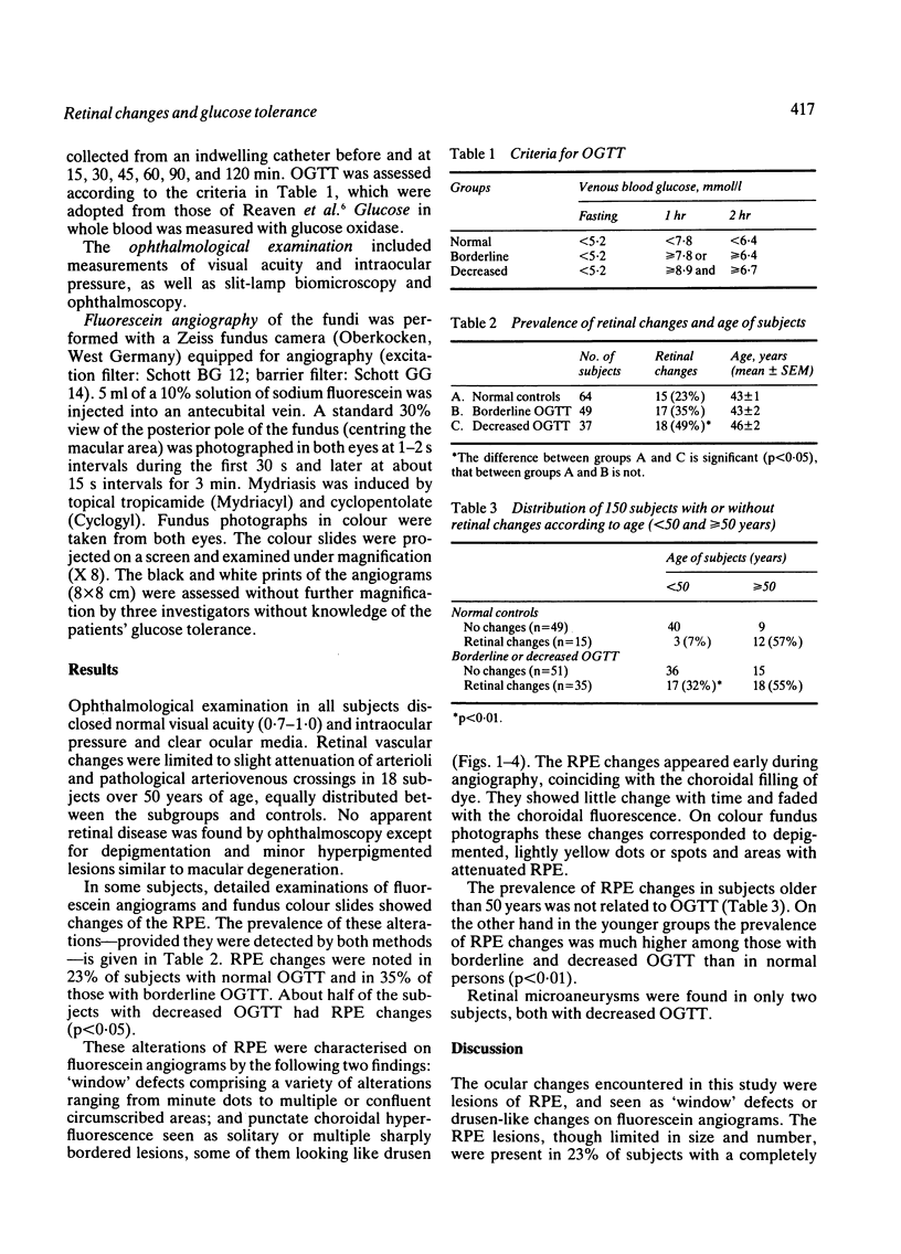
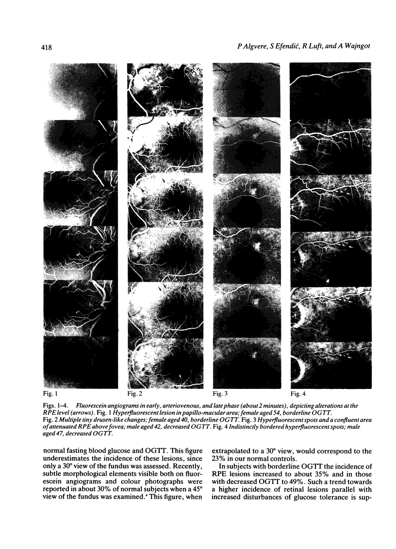
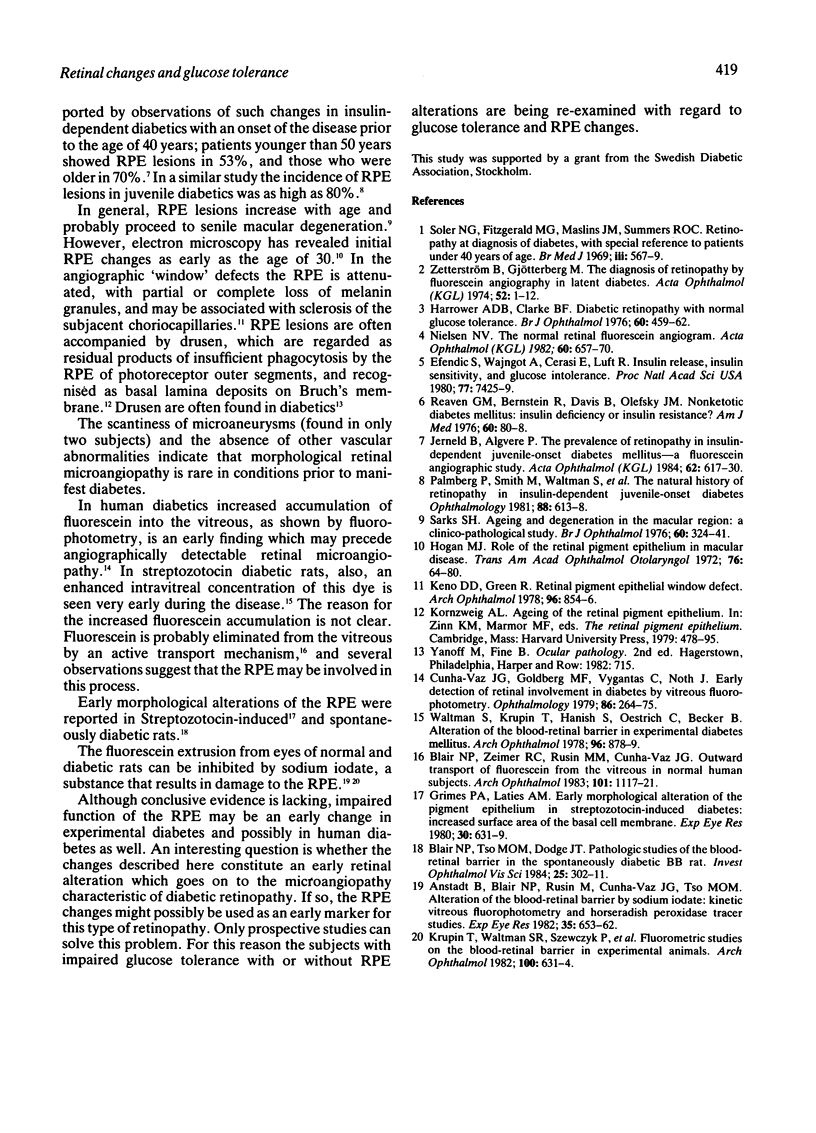
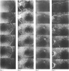
Retinal fluorescein angiography was performed in 150 subjects: 64 with normal fasting blood glucose and normal oral glucose tolerance test (OGTT), 49 with borderline, and 37 with decreased OGTT. Microaneurysms were noted in only two subjects, both with decreased OGTT. Minute changes in the retinal pigment epithelium (RPE) were seen in 23% of the 64 normal persons, in 35% of those with borderline, and 49% of those with decreased OGTT (p less than 0.05). The impact of glucose intolerance was more pronounced in subjects under the age of 50 years, RPE changes being rare (7%) in those with normal OGTT but occurring in 32% of those with borderline or decreased OGTT (p less than 0.01). The corresponding figures among subjects aged 50 or more were 55% and 57%, respectively. We conclude that at least half of the subjects above 50 years show RPE alterations, and that minimal changes in glucose metabolism may precipitate the development of such changes at an earlier age.
Full text
PDF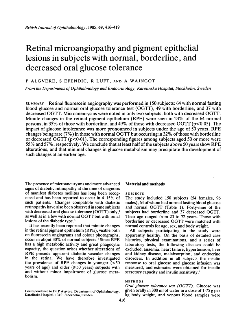
Images in this article
Selected References
These references are in PubMed. This may not be the complete list of references from this article.
- Anstadt B., Blair N. P., Rusin M., Cunha-Vaz J. G., Tso M. O. Alteration of the blood-retinal barrier by sodium iodate: kinetic vitreous fluorophotometry and horseradish peroxidase tracer studies. Exp Eye Res. 1982 Dec;35(6):653–662. doi: 10.1016/s0014-4835(82)80077-8. [DOI] [PubMed] [Google Scholar]
- Blair N. P., Tso M. O., Dodge J. T. Pathologic studies of the blood--retinal barrier in the spontaneously diabetic BB rat. Invest Ophthalmol Vis Sci. 1984 Mar;25(3):302–311. [PubMed] [Google Scholar]
- Blair N. P., Zeimer R. C., Rusin M. M., Cunha-Vaz J. G. Outward transport of fluorescein from the vitreous in normal human subjects. Arch Ophthalmol. 1983 Jul;101(7):1117–1121. doi: 10.1001/archopht.1983.01040020119021. [DOI] [PubMed] [Google Scholar]
- Cunha-Vaz J. G., Goldberg M. F., Vygantas C., Noth J. Early detection of retinal involvement in diabetes by vitreous fluorophotometry. Ophthalmology. 1979 Feb;86(2):264–275. doi: 10.1016/s0161-6420(79)35516-6. [DOI] [PubMed] [Google Scholar]
- Efendić S., Wajngot A., Cerasi E., Luft R. Insulin release, insulin sensitivity, and glucose intolerance. Proc Natl Acad Sci U S A. 1980 Dec;77(12):7425–7429. doi: 10.1073/pnas.77.12.7425. [DOI] [PMC free article] [PubMed] [Google Scholar]
- Grimes P. A., Laties A. M. Early morphological alteration of the pigment epithelium in streptozotocin-induced diabetes: increased surface area of the basal cell membrane. Exp Eye Res. 1980 Jun;30(6):631–639. doi: 10.1016/0014-4835(80)90062-7. [DOI] [PubMed] [Google Scholar]
- Harrower A. D., Clarke B. F. Diabetic retinopathy with normal glucose tolerance. Br J Ophthalmol. 1976 Jun;60(6):459–463. doi: 10.1136/bjo.60.6.459. [DOI] [PMC free article] [PubMed] [Google Scholar]
- Hogan M. J. Role of the retinal pigment epithelium in macular disease. Trans Am Acad Ophthalmol Otolaryngol. 1972 Jan-Feb;76(1):64–80. [PubMed] [Google Scholar]
- Jerneld B., Algvere P. The prevalence of retinopathy in insulin-dependent juvenile-onset diabetes mellitus--a fluorescein-angiographic study. Acta Ophthalmol (Copenh) 1984 Aug;62(4):617–630. doi: 10.1111/j.1755-3768.1984.tb03974.x. [DOI] [PubMed] [Google Scholar]
- Keno D. D., Green W. R. Retinal pigment epithelial window defect. Arch Ophthalmol. 1978 May;96(5):854–856. doi: 10.1001/archopht.1978.03910050456012. [DOI] [PubMed] [Google Scholar]
- Krupin T., Waltman S. R., Szewczyk P., Koloms B., Farber M., Silverstein B., Becker B. Fluorometric studies on the blood-retinal barrier in experimental animals. Arch Ophthalmol. 1982 Apr;100(4):631–634. doi: 10.1001/archopht.1982.01030030633021. [DOI] [PubMed] [Google Scholar]
- Nielsen N. V. The normal retinal fluorescein angiogram I. A study of the fluoresceinangiographic appearance of the retina in normal subjects without ophthalmoscopically obvious pathological changes. Acta Ophthalmol (Copenh) 1982 Oct;60(5):657–670. doi: 10.1111/j.1755-3768.1982.tb06726.x. [DOI] [PubMed] [Google Scholar]
- Palmberg P., Smith M., Waltman S., Krupin T., Singer P., Burgess D., Wendtlant T., Achtenberg J., Cryer P., Santiago J. The natural history of retinopathy in insulin-dependent juvenile-onset diabetes. Ophthalmology. 1981 Jul;88(7):613–618. doi: 10.1016/s0161-6420(81)34975-6. [DOI] [PubMed] [Google Scholar]
- Reaven G. M., Bernstein R., Davis B., Olefsky J. M. Nonketotic diabetes mellitus: insulin deficiency or insulin resistance? Am J Med. 1976 Jan;60(1):80–88. doi: 10.1016/0002-9343(76)90536-2. [DOI] [PubMed] [Google Scholar]
- Sarks S. H. Ageing and degeneration in the macular region: a clinico-pathological study. Br J Ophthalmol. 1976 May;60(5):324–341. doi: 10.1136/bjo.60.5.324. [DOI] [PMC free article] [PubMed] [Google Scholar]
- Soler N. G., Fitzgerald M. G., Malins J. M., Summers R. O. Retinopathy at diagnosis of diabetes, with special reference to patients under 40 years of age. Br Med J. 1969 Sep 6;3(5670):567–569. doi: 10.1136/bmj.3.5670.567. [DOI] [PMC free article] [PubMed] [Google Scholar]
- Waltman S., Krupin T., Hanish S., Oestrich C., Becker B. Alteration of the blood-retinal barrier in experimental diabetes mellitus. Arch Ophthalmol. 1978 May;96(5):878–879. doi: 10.1001/archopht.1978.03910050480018. [DOI] [PubMed] [Google Scholar]
- Zetterström B., Gjötterberg M. The diagnosis of retinopathy by fluorescein angiography in latent diabetes. Acta Ophthalmol (Copenh) 1974;52(1):1–12. doi: 10.1111/j.1755-3768.1974.tb00350.x. [DOI] [PubMed] [Google Scholar]