Abstract
A system has been developed for generating chimeric yellow fever/Japanese encephalitis (YF/JE) viruses from cDNA templates encoding the structural proteins prM and E of JE virus within the backbone of a molecular clone of the YF17D strain. Chimeric viruses incorporating the proteins of two JE strains, SA14-14-2 (human vaccine strain) and JE Nakayama (JE-N [virulent mouse brain-passaged strain]), were studied in cell culture and laboratory mice. The JE envelope protein (E) retained antigenic and biological properties when expressed with its prM protein together with the YF capsid; however, viable chimeric viruses incorporating the entire JE structural region (C-prM-E) could not be obtained. YF/JE(prM-E) chimeric viruses grew efficiently in cells of vertebrate or mosquito origin compared to the parental viruses. The YF/JE SA14-14-2 virus was unable to kill young adult mice by intracerebral challenge, even at doses of 106 PFU. In contrast, the YF/JE-N virus was neurovirulent, but the phenotype resembled parental YF virus rather than JE-N. Ten predicted amino acid differences distinguish the JE E proteins of the two chimeric viruses, therefore implicating one or more residues as virus-specific determinants of mouse neurovirulence in this chimeric system. This study indicates the feasibility of expressing protective antigens of JE virus in the context of a live, attenuated flavivirus vaccine strain (YF17D) and also establishes a genetic system for investigating the molecular basis for neurovirulence determinants encoded within the JE E protein.
Within the genus Flavivirus of the family Flaviviridae, yellow fever (YF) and Japanese encephalitis (JE) viruses are distinguishable by a number of properties. The viruses are antigenically distinct (2), lack common mosquito vectors and vertebrate reservoirs (22, 24), and cause dissimilar disease syndromes in humans (15). Such differences presumably reflect evolutionary divergence of these viruses from common ancestors (16). To begin investigation of the molecular basis for the differences in biological properties of these two flaviviruses, we have engineered chimeric YF/JE viruses in which the structural proteins prM and E of JE virus were exchanged for the homologous proteins of YF virus within a molecular clone of the YF17D strain (21). This strategy was based on the previous observation that chimeric viruses between distantly related members of this family, such as TBE and DEN, can be recovered from engineered cDNA templates (19). Because of the conserved features of flavivirus genome organization and replication (4), it may be possible to genetically engineer a range of such chimeric viruses. This approach is relevant for the potential use of recombinant flaviviruses as live-attenuated vaccine candidates (1, 14) and also for studying protein-protein and RNA-protein interactions important for flavivirus replication and pathogenesis.
MATERIALS AND METHODS
Cells and viruses.
SW-13 (derived from human adenocarcinoma originating from adrenal cortex), Vero, LLC-MK2, C6/36, and NB41A3 (mouse neuroblastoma) cells were originally obtained from the American Type Culture Collection (ATCC). YF5.2iv (a molecular clone of the YF17D strain), has been previously described (21). The JE Nakayama (JE-N) strain was obtained from ATCC and was amplified in LLC-MK2 cells. JE SA14-14-2 was obtained at passage level PHK-5 (courtesy of Kenneth H. Eckels) and amplified in LLC-MK2 cells.
Plasmid constructions.
JE SA14-14-2 cDNA was derived by reverse transcription (RT)-PCR from infected LLC-MK2 cells based on the published nucleotide sequence of this strain (18). The structural region was cloned into pBluescript-KS(+) (pBS) (Stratagene) by using two primer sets. The 5′ terminus through nucleotide 1132 was cloned into pBS with primers containing EcoRI sites to yield pBS/JE(1–1131). The 3′ primer contained a nested NheI restriction site for subsequent insertion of an engineered chimeric YF/JE fragment containing the YF nucleotide sequence from 1 to 481 joined to the JE nucleotide sequence from 477 to 1131 (see below). The region from nucleotide 1108 to nucleotide 2472 was cloned by using primers containing XbaI restriction sites to produce pBS/JE(1108–2472). The 5′ primer contained a nested NsiI restriction site, and the 3′ primer contained a nested KasI site to facilitate insertion of an NsiI-KasI digestion fragment of pBS/JE(1108–2472) into YFM5.2[KasI] (see below). The cDNA for the JE-N has been previously described (13).
A two-plasmid system used for generation of infectious YF17D virus (21) was modified for construction of the chimeric YF/JE viruses. Heterologous YF-5′ untranslated region/JE-capsid or YF-capsid/JE-prM junctions were engineered by PCR. The 3′ chimeric primers corresponding to the desired junctions and a 5′ primer representing nucleotide positions 6625 to 6639 in pYF5′3′IV (21) were used to generate 426- and 785-bp PCR products, respectively. These products were gel purified and used as 5′ primers, together with a 3′ primer corresponding to the T3 promoter [flanking the JE insert in pBS/JE(1–1132)], to amplify a chimeric YF/JE PCR product from pBS/JE(1–1132) incorporating the JE capsid or prM region and the E region through nucleotide 1132. The resulting PCR products were inserted into pYF5′3′IV by using NotI and EcoRI restriction sites to create YF5′3′IV/JE-S plasmids encoding the YF/JE chimeric 5′ untranslated region/capsid and capsid/prM junctions. These plasmids were used for in vitro ligation.
To modify pYFM5.2 (21) to encode a chimeric JE E/YF-NS1 region, pYFM5.2 was first engineered by site-directed mutagenesis to contain a unique KasI restriction site at the YF E/NS1 junction while preserving a signalase cleavage site at this position. JE cDNA was inserted into pYFM5.2(KasI) by using an NsiI-KasI restriction fragment from pBS/JE(1108–2472) to create pYFM5.2/JE-S. Unique BspEI restriction sites were created in both pYF5′3′IV/JE-S and pYFM5.2/JE-S at position 8579 (YF numbering) by site-directed mutagenesis. An additional NheI site in YFM5.2/JE at nucleotide position 5459 (YF numbering) was eliminated by site-directed mutagenesis. These modifications allowed use of the NheI and BspEI sites in pYF5′3′IV/JE-S and pYFM5.2/JE-S for assembly of in vitro-ligated cDNA templates for synthesis of full-length RNA transcripts.
Plasmids for generation of chimeric YF/JE-N templates were constructed by exchanging appropriate restriction fragments in pYF5′3′IV/JE-S and pYFM5.2/JE-S with the corresponding fragments of JE-N cDNA (13). These fragments included from HindIII to PvuII (nucleotides 501 to 1061 [JE numbering]) and BpmI to MfeI (nucleotides 1220 to 2413). A serine residue at position 52 of the JE SA14-14-2 E protein was retained in pYF/5′3′IV/JE-N and YFM5.2/JE-N to allow use of the NheI site at this position for in vitro ligation.
Recovery of infectious virus.
Full-length cDNA templates were assembled by in vitro ligation of restriction fragments after digestion of the YF/JE plasmids with NheI and BspEI and gel purification of the appropriate DNA products. The in vitro-ligated templates encoding chimeric YF/JE viruses were used for synthesis of SP6 RNA transcripts based on methods described previously (21). RNA transfection of Vero cells was done in the presence of Lipofectin (Gibco/BRL), using between 100 and 250 ng of RNA transcript. Virus was harvested after onset of cytopathic effect and titrated by plaque assay on LLC-MK2 cells.
Protein labelling.
Vero cells or LLC-MK2 cells were infected with virus at a multiplicity of 1 PFU/cell and labelled at approximately 48 h postinfection with [35S]methionine for 10 to 12 h. The media from the culture or lysates made from the cell monolayers were used for immunoprecipitation of the viral proteins under either denaturing conditions for YF polyclonal anti-E antisera (5) or nondenaturing conditions for JE monoclonal antibody (12) and with hyperimmune antisera against YF and JE. Immunoprecipitates were processed and analyzed by sodium dodecyl sulfate-polyacrylamide gel electrophoresis (SDS-PAGE) and fluorography as previously described (5).
Neutralization assays.
Mouse hyperimmune antisera to YF, JE, and nonimmune ascitic fluid were obtained from the ATCC. Anti-YF immunoglobulin G (IgG) to the YF E protein and a nonimmune isotype-matched IgG were purified from ascites fluid containing monoclonal antibody 2E10 or 2C5 (23 [courtesy of Jack Schlesinger]) by using a monotrap protein A column (Pharmacia). IgG concentrations were estimated by using stained protein markers (Sigma) as standards for quantitation by SDS-PAGE. For neutralization assays, antisera or IgGs were diluted in minimal essential medium plus 3% fetal bovine serum. Plaque reduction titers were calculated as the highest dilution of serum or IgG which neutralized 50% of the input virus (100 PFU). Plaque assays were performed on LLC-MK2 cells.
Growth curves.
Growth curves were done by infecting confluent LLC-MK2, C6/36, SW-13, or NB41A3 cells at a multiplicity of 0.5 PFU/cell and harvesting the media at successive 12 or 24-h intervals postinfection. Yields of virus in each sample were then quantitated by plaque titration on LLC-MK2 cells.
Mouse experiments.
Three- to four-week-old outbred male and female mice (ICR and C57BL/6 strains; Harlan-Sprague Dawley) were used. Neurovirulence was assessed by intracerebral inoculation of virus diluted in sterile phosphate-buffered saline plus 5% fetal bovine serum into the left cerebral hemisphere of anesthetized mice. Virus doses were confirmed by back-titration of the inocula on LLC-MK2 cells. Neuroinvasiveness was assessed by inoculation of virus by the intraperitoneal route. Endpoints were scored as either the day of onset of a moribund condition or, alternatively, survival to 3 weeks after inoculation. Mice found in a moribund state were euthanized.
Nucleotide sequence analyses.
DNA sequences of the plasmids encoding the YF/JE chimeras were determined by dideoxy sequencing by the Sequenase (U.S. Biochemical Corp.) protocol. Duplicate clones were sequenced for each chimeric virus clone.
RESULTS
Recovery of chimeric YF/JE viruses.
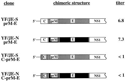
We chose to engineer chimeric viruses encoding the prM and E proteins of the JE SA14-14-2 human vaccine strain and the virulent JE-N strain. The SA14-14-2 strain is an attenuated derivative of the JE SA-14 strain, obtained after serial passage in cell culture (6, 17). JE SA-14 is a virulent strain originally isolated from mosquitoes in China (17). The JE-N strain was selected rather than JE SA-14 because of availability of a well-characterized cDNA clone encoding the structural proteins of this virus (12, 13). Figure 1 shows the structural region of the plasmids encoding the chimeric viruses and the recovery of virus from cells transfected with synthetic RNA derived from the plasmid templates. High titers of infectious virus were recovered from transcripts derived from templates encoding the YF capsid protein together with the JE prM and E proteins of the SA14-14-2 strain (YF/JE-S) or the Nakayama strain (YF/JE-N): 6.8 and 7.3 log10 PFU/ml, respectively. The yield of YF5.2iv virus after transfection of Vero cells under similar conditions was 6.1 log10 PFU/ml (data not shown). Templates encoding the entire JE structural region (C-prM-E) did not yield detectable virus by this assay. Virus recovered from the transfections was passaged onto new cell monolayers, and total intracellular RNA was used for RT-PCR to verify the chimeric structure. Primer pairs which amplify the YF genome from nucleotide 1 to nucleotide 2980 were used to generate PCR products which were analyzed by restriction enzyme digestion to verify the presence of JE sequences in the recovered virus (data not shown). Moreover, nucleotide sequence analysis of the PCR products across the chimeric C/prM junction revealed the predicted structure of the chimeric virus (data not shown).
FIG. 1.
Structure of cDNA templates for chimeric YF/JE viruses (truncated within the NS1 protein for clarity). YF/JE-S and YF/JE-N refer to the two chimeric viruses as described in the text. The 5′ nontranslated region is derived from YF5.2iv (6). Hatched regions are from JE SA14-14-2, and solid regions are from JE-N. The remainder of the nonstructural region is derived from YF5.2iv. The amount of virus recovered is indicated as the titer of virus (log10 PFU per milliliter) in the media at the time of harvest at 96 h after RNA transfection of Vero cells.
Properties of viral proteins.
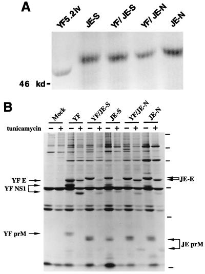
Figure 2A illustrates the immunoprecipitation of viral E proteins present in the media of cells infected with either the parental or chimeric viruses. The YF E protein migrates with a molecular mass of approximately 50 kDa, consistent with previous observations (5). The E proteins of the chimeric viruses and parental JE viruses migrated with molecular masses of approximately 52 kDa, consistent with the presence of an N-linked glycan on the E protein (12). To determine if this difference in the apparent molecular masses of the JE E proteins relative to the YF E protein resulted from N-linked glycosylation, the profiles of intracellular viral glycoproteins produced in the presence and absence of tunicamycin were analyzed. Figure 2B illustrates that the apparent molecular mass of the YF E protein is not affected by tunicamycin, whereas the molecular masses of the JE E proteins are reduced from 52 kDa to 50 kDa when labelled in the presence of this drug. This is consistent with utilization of a single N-linked glycosylation site on these proteins. The YF prM protein migrated with an apparent molecular mass of 24 kDa, consistent with addition of two N-linked glycans (5), whereas the JE prM protein migrated with a molecular mass of 19 kDa, consistent with a single N-linked glycan (12). In the presence of tunicamycin, the molecular mass of the JE prM protein was reduced by 2 kDa.
FIG. 2.
Immunoprecipitation of viral proteins from chimeric YF/JE viruses. (A) Parental or chimeric viruses were grown in LLC-MK2 cells, labelled with [35S]methionine, and harvested from the media at 48 to 60 h postinfection. Viral E proteins were immunoprecipitated from the media as described in Materials and Methods, and proteins were analyzed on an SDS–9% polyacrylamide gel. The E protein of YF5.2iv virus was immunoprecipitated with rabbit polyclonal antiserum against YF. The E proteins of the JE-S (JE SA14-14-2), YF/JE-S, YF/JE-N, and JE-N viruses were immunoprecipitated with mouse hyperimmune ascitic fluid against JE virus. (B) Viral proteins produced in Vero cells. Proteins were labelled with [35S]methionine for 6 h, and lysates were prepared at 48 h postinfection as described in Materials and Methods. Virus in the media was harvested and immunoprecipitated with hyperimmune ascitic fluid to either YF or JE, as described for panel A. Mock-infected lysates were immunoprecipitated with a mixture of YF and JE hyperimmune ascitic fluids. Proteins were analyzed on 13% SDS–polyacrylamide gels. Tunicamycin was added (+) or not added (−) at the time of labelling at a concentration of 7.5 μg/μl. Molecular mass markers, indicated by small lines in the right margin, represent 220, 97.4, 66, 46, 30, and 14.3 kDa, respectively.
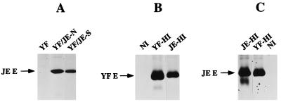
A smaller form of the YF prM protein was not clearly visible after tunicamycin treatment, even with longer exposures. Presumably the protein was unstable under these conditions. The YF NS1 glycoprotein was also detectable and was immunoprecipitated by antisera to both YF and JE viruses. The molecular mass of the NS1 protein was reduced by approximately 4 kDa by treatment with tunicamycin, consistent with the presence of two N-linked glycans on this protein (5). The JE E protein was detectable by immunoprecipitation with a monoclonal antibody to JE, as shown by reactivity with the E protein produced by the YF/JE-N chimera (Fig. 3A). No reactivity of this antibody was observed against the YF E protein. Polyclonal antiserum against YF was able to immunoprecipitate the JE E protein produced by YF/JE-N (Fig. 3C), and polyclonal antisera against the JE E protein also immunoprecipitated the YF E protein (Fig. 3B), indicating the presence of many cross-reactive, nonneutralizing epitopes (see below) on the E proteins of the YF and JE viruses.
FIG. 3.
Immunoprecipitation of YF and JE proteins with monoclonal and polyclonal antisera. LLC-MK2 cells were infected and labelled as described for Fig. 2A. Viral proteins were immunoprecipitated from the media with monoclonal antibody to JE (A) or polyclonal antisera to the YF and JE viruses (B and C), as described in Materials and Methods. In panel A, YF, YF/JE-N, and YF/JE-S refer to media from cells infected with these respective viruses. In panel B, YF-infected cells were used. In panel C, YF/JE-N-infected cells were used. NI refers to nonimmune ascites fluid. YF-HI and JE-HI refer to hyperimmune ascites fluid to the YF and JE viruses, respectively. Proteins were analyzed on 10% SDS gels.
The capacity of antisera to YF and JE viruses to neutralize the chimeric viruses was tested in a plaque reduction neutralization assay as shown in Table 1. Polyclonal sera against JE efficiently neutralized JE viruses and the YF/JE chimeras, but no difference in neutralization against YF was observed compared to the effect with nonimmune sera. Polyclonal antisera to YF exhibited neutralization activity against YF5.2iv at a dilution of 1:2,560, but against JE and the YF/JE chimeras, no difference was observed relative to nonimmune sera. A purified anti-YF IgG exhibited high neutralization activity against YF, but not JE or the chimeric viruses, whereas nonimmune IgG had no neutralizing activity against any virus at the lowest dilution tested.
TABLE 1.
Plaque reduction-neutralization titers for YF/JE chimeras and parental viruses
| Virus | Result fora:
|
||||
|---|---|---|---|---|---|
| NI-AS | YF-AS | JE-AS | NI-IgG | YF-IgG | |
| YF5.2iv | <1.3 | 3.7 | <1.3 | <2.2 | >4.3 |
| JE SA14-14-2 | <1.3 | <1.3 | 3.4 | <2.2 | <2.2 |
| YF/JE-S | <1.3 | <1.3 | 3.1 | <2.2 | <1.9 |
| YF/JE-N | <1.3 | <1.3 | 3.4 | <2.2 | <2.2 |
| JE-N | <1.3 | <1.3 | 3.4 | <1.5 | <1.5 |
Values are the log reciprocal of the dilution yielding 50% neutralization, based on 100 PFU on LLC-MK2 cells. NI-AS, YF-AS, and JE-AS are nonimmune, YF hyperimmune, and JE hyperimmune ascitic fluids, respectively. NI-IgG and YF-IgG are purified Ig as described in Materials and Methods.
Growth efficiency in cell culture.
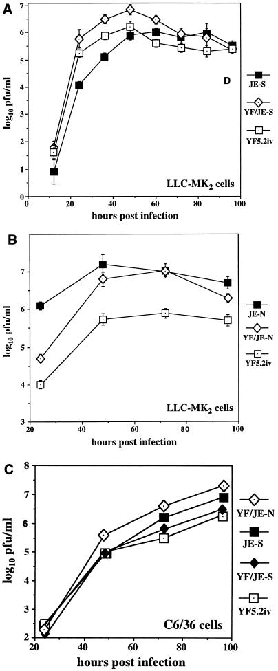
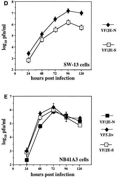
Growth kinetics of the chimeras were initially examined on LLC-MK2 and C6/36 cells. As shown in Fig. 4A and B, differences were observed between the chimeras and the parental viruses in terms of the maximal yield of virus production on LLC-MK2 cells, based on single-step growth analysis. All viruses reached a peak of production at approximately 48 h postinfection. The maximal virus production from the chimeric viruses was higher than that of the parental YF5.2iv virus. YF/JE-S replicated more efficiently than JE SA14-14-2, whereas YF/JE-N replicated less efficiently than JE-N. Both chimeric viruses replicated better than YF5.2iv. On mosquito cells, the YF/JE chimeric viruses also exhibited efficient replication compared to the parental YF5.2iv and JE SA14-14-2 viruses (Fig. 4C). The growth properties of the chimeras were examined on two other cell lines. Comparison of the growth rates of the chimeras on SW-13 cells (Fig. 4D) revealed a difference in peak titer, with the YF/JE-N virus exhibiting a higher replication efficiency than YF/JE-S in this cell line. This virus also exhibited higher plaque efficiency than YF/JE-S on SW-13 cells (data not shown). On NB41A3 cells (Fig. 4E), the YF/JE-S and parental YF5.2iv viruses exhibited roughly similar growth kinetics, whereas YF/JE-N appeared to replicate slightly less efficiently.
FIG. 4.
Growth curves of chimeric viruses in cell culture. Cells were infected at a multiplicity of 0.5 PFU/cell, and media were collected at 12- or 24-h intervals, followed by plaque titration on LLC-MK2 cells. (A) YF/JE-S compared to its parental viruses on LLC-MK2 cells. (B) YF/JE-N compared to its parental viruses on LLC-MK2 cells. The titers represent averages of triplicate samples for both experiments. (C) Chimeric virus growth on C6/36 cells, with values representing an average of two samples for each virus. (D) Growth of YF/JE-S and YF/JE-N viruses on SW-13 cells. (E) Growth of YF5.2iv, YF/JE-S, and YF/JE-N on NB41A3 cells. The titers represent averages of triplicate samples, except for the last time point, which was determined in duplicate.
Mouse virulence testing.
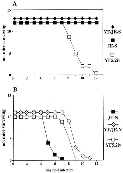
Mice 3 to 4 weeks old were used to assess virulence phenotypes. Figure 5A compares the results obtained with the YF/JE-S chimera with those obtained with the YF5.2iv and JE SA14-14-2 viruses. At a dose of 104 PFU delivered intracerebrally, parental YF caused 100% mortality within 7 to 12 days, whereas neither the JE SA14-14-2 strain nor the YF/JE-S chimera caused any mortality. No signs of illness were observed over the period of 3 weeks after inoculation for the two latter viruses. The neurovirulence of the YF/JE-N virus in this assay is compared to the YF5.2iv and JE-N virus in Fig. 5B. The YF/JE-N chimera exhibited a level of neurovirulence similar to the YF5.2iv virus in this model (average time of survival, 7 days), whereas the JE-N virus required less time to cause fatal disease (average time of survival, 4 days). The attenuation phenotype of the YF/JE-S virus was also demonstrated at a higher-dose intracerebral challenge (Table 2). Compared to the 100% mortality of mice receiving 104 PFU of YF5.2iv virus, no mortality or signs of illness were observed in mice receiving between 104 and 106 PFU of the YF/JE-S chimera. In contrast, the YF/JE-N virus was lethal for 60% of mice at a dose of only 10 PFU (Table 2).
FIG. 5.
Mouse neurovirulence assay. A fixed-dose intracerebral challenge with 104 PFU was carried out in 4-week-old ICR mice. (A) YF/JE-S compared with its parental viruses. (B) YF/JE-N compared with its parental viruses. Differences in mortality between YF/JE-S and YF5.2iv were significant (P < .005), based on χ2 analysis of the proportion of survivors.
TABLE 2.
Neurovirulence analysis of YF/JE chimerasa
| Virus | Dose (log10 PFU) | % Mortality (no. dead/no. tested) |
|---|---|---|
| YF5.2iv | 4 | 100 (7/7) |
| YF/JE-S | 6 | 0 (0/8) |
| 5 | 0 (0/7) | |
| 4 | 0 (0/7) | |
| YF/JE-N | 4 | 100 (5/5) |
| 3 | 100 (5/5) | |
| 2 | 100 (5/5) | |
| 1 | 60 (3/5) | |
| 0 | 0 (0/5) |
ICR mice (4 weeks old) received YF5.2iv, YF/JE-S, or YF/JE-N at the indicated doses by intracerebral inoculation.
Histologic analysis of the cerebral cortex and subcortical regions (hippocampus) of mice inoculated with 104 PFU of the YF/JE-S virus revealed only focal areas of inflammation, and virus could be recovered at 10 days postinoculation at only very low titers (data not shown). In contrast, brains infected with the YF/JE-N virus exhibited a histologic picture similar to that of those infected with the YF5.2iv virus, with extensive inflammation, necrosis, and virus titers in excess of 107 PFU/g of brain (data not shown).
Both chimeric viruses were also tested for neuroinvasiveness in 3-week-old mice (Table 3). At doses of 106 PFU delivered by intraperitoneal inoculation, YF/JE-S and YF/JE-N caused no mortality in ICR mice. In C57BL/6 mice, YF/JE-N was partially invasive, with 3 of 15 mice succumbing to infection. In contrast, the JE-N virus was lethal for 100% of mice of both strains at the same dose.
TABLE 3.
Neuroinvasiveness of YF/JE virusesa
| Virus | % Mortality (no. dead/no. tested)b | Strain |
|---|---|---|
| YF/JE-S | 0 (0/4) | ICR |
| YF/JE-N | 0 (0/13) | ICR |
| 20 (3/15) | C57BL/6 | |
| JE-N | 100 (12/12) | ICR |
| 100 (11/11) | C57BL/6 |
YF/JE-S, YF/JE-N, or JE-N was inoculated at 6 log10 PFU into 3-week-old mice by the intraperitoneal route.
For both ICR and C57BL/6 mice, the difference in mortality of YF/JE-N versus JE-N was significant (P < 0.005, by χ2 analysis of the proportion of survivors).
Nucleotide sequence analysis of chimeric viruses.
Plasmids encoding the chimeric YF/JE-N and YF/JE-S viruses were sequenced through the JE prM-E portion of the respective clones (Table 4). The E protein sequence of YF/JE-N agreed with that of the JE-N strain (13). The sequence of YF/JE-S agreed with that of JE SA14-14-2 (18), except at two positions (residues 177 and 264, as discussed below). Comparison of the predicted amino acid sequences reveals a total of 10 differences between the E proteins of the chimeric viruses. Four of these residues (227, 244, 315, and 439) are common to YF/JE-S and its virulent JE SA-14 parent. The remaining six residues (107, 138, 176, 177, 264, and 279) are common between the sequences of the YF/JE-N virus and the virulent JE-SA14 virus. In the prM region, three residues of the YF/JE-S chimera (I14, I129, and V140) differed from those in YF/JE-N (V14, V129, and I140) (data not shown).
TABLE 4.
Predicted amino acid differences (single-letter code) in the E proteins of the YF/JE chimeras compared with parental viruses
| Virusa | Predicted difference from:
|
|||||||||
|---|---|---|---|---|---|---|---|---|---|---|
| E-107 | E-138 | E-176 | E-177 | E-227 | E-244 | E-264 | E-279 | E-315 | E-439 | |
| JE SA14-14-2 | F | K | V | T | S | G | Q | M | V | R |
| YF/JE-S | F | K | V | A | S | G | H | M | V | R |
| YF/JE-N | L | E | I | T | P | E | Q | K | A | K |
| JE-N | L | E | I | T | P | E | Q | K | A | K |
| JE SA-14 | L | E | I | T | S | G | Q | K | V | R |
DISCUSSION
This study demonstrates that a molecular clone of YF17D virus can be engineered to express the structural proteins prM and E of the heterologous flavivirus, JE virus. These findings are similar to those originally reported for a chimeric DEN/TBE virus showing that the prM-E region of TBE could be expressed in the context of the dengue-4 virus genome (19). Although chimeric YF/JE(prM-E) viruses could be readily recovered, failure to generate YF/JE(C-prM-E) chimeric virus could be due to several possible causes. These include incompatibility of predicted cis-acting RNA structures, such as conserved sequences found within the capsid region (7), inefficient processing of the JE C/prM junction by the YF protease, or other steps, such as defective packaging or deleterious effects of the JE capsid on RNA synthesis driven by YF nonstructural proteins. It has been shown, for instance, that Kunjin virus RNA replication requires between 2 and 20 amino acid residues of the homologous capsid protein, although it is not known whether nonhomologous sequences can substitute in the capsid region (11). Similar requirements may exist for other flaviviruses.
Immunoprecipitation and plaque-reduction neutralization assays were used to determine whether the JE E protein is expressed in an antigenically intact form in the context of chimeric YF/JE viruses. The JE prM and E proteins appeared to be properly glycosylated, as indicated by detection of radiolabelled proteins in infected cells, and the E protein exhibited both JE-specific as well as cross-reactive flavivirus epitopes. The neutralization data suggest that protective antibody epitopes of the JE E protein are likely to be preserved in the chimeric viruses, although we cannot rule out subtle structural differences between their E proteins relative to those of the parental JE viruses. Thus, it may be possible to elicit JE-specific neutralizing antibodies by using these chimeras as experimental vaccines.
Differences in growth efficiency of the chimeric viruses were observed in cell culture in LLC-MK2 cells. Both chimeric viruses exhibited higher growth efficiency than the YF5.2iv parent. This may reflect the higher efficiency of the JE prM-E proteins for steps involved in replication and spread of virus in these cells, particularly since infection was done at relatively low multiplicity. Efficient replication of the chimeric viruses was also observed in mosquito cells (C6/36). Growth of the chimeric viruses in NB41A3 cells was examined to determine if the difference in neurovirulence of the viruses would be reflected by different replication efficiencies in this cell line, but no correlation could be established. However, a difference was observed in SW-13 cells, suggesting that these cells may serve as a surrogate for investigation of molecular mechanisms governing the difference in mouse neurovirulence which exists between the two chimeric viruses. Taken together, these preliminary analyses have not revealed any unexpected changes in host range due to the combination of YF nonstructural proteins and JE structural glycoproteins.
A striking difference between the neurovirulence levels of the YF/JE-S and YF/JE-N viruses was observed in young adult mice. Previous studies have demonstrated that the JE SA14-14-2 virus is noneurovirulent compared to its JE SA-14 parent (6, 8), but sequence differences have been identified throughout their genomes (18), making it unclear which protein (or proteins) is a principal virulence factor. The data reported here with the chimeric viruses suggest that the prM-E proteins may be major determinants of neurovirulence, since YF/JE-N was more virulent than YF/JE-S. The YF/JE-N virus exhibited partial neuroinvasiveness in these experiments, which is consistent with other studies indicating that the JE E protein contains determinants of mouse neuroinvasiveness (3, 9). Failure to observe neuroinvasiveness in ICR mice presumably indicates that there are strain-specific differences in the susceptibility of young mice to the chimeric YF/JE-N virus. Further studies are needed to fully characterize the neuropathogenic properties of this chimeric virus.
Nucleotide sequence analysis of the clones encoding the YF/JE chimeric viruses suggests that 10 amino acid residues within the E protein and possibly 3 residues within the prM protein account for the difference in neurovirulence. We cannot exclude the potential contribution of mutations outside of the structural region as determinants of this difference; however, some of the predicted differences in the E proteins map to positions which have been implicated as virulence determinants among neuropathogenic flaviviruses (20). In particular, these include position 138, which has been reported to be a critical determinant of JE virulence in the context of the JaOArS982 strain (25). Since the 10 residues which differ in the YF/JE chimeras are distributed through all three structural domains of the E protein as predicted from the TBE model (20), it is possible that the difference in neurovirulence is dependent on more than a single functional property of the E protein. Such steps may include those involved in virus attachment, the acid pH-catalyzed conformational change, and/or membrane fusion events associated with virus entry. In this regard, attenuation of the JE SA-14 strain is believed to involve sequential accumulation of several mutations in the E protein (17). Substitutions at positions 107 (L→F), 138 (E→K), 176 (I→V), and 279 (K→M) occurred early and remained stable during subsequent passage of attenuated derivatives of the JE SA-14 strain. Substitutions at positions 177 (T→A) and 264 (Q→H) occurred during passage in PHK cells and were unstable upon subsequent passage in PDK cells, reverting to the original residues (18). Since the YF/JE-S virus was derived from JE virus at the PHK passage level, its E protein contains alanine at position 177 and histidine at position 264, rather than those of the JE SA14-14-2 virus which was produced from the PDK passage (6). This suggests that the four positions 107, 138, 176, and 279 may harbor residues which are critical for determining the neurovirulence phenotype. As a first step toward investigating these possibilities, the genetic basis for the attenuation can now be defined by constructing a series of intertypic YF/JE chimeric viruses containing one or more reversions of the YF/JE-S virus to the sequence of the JE-N strain and testing these engineered viruses for their mouse neurovirulence phenotypes.
The high degree of attenuation of the YF/JE-S virus, demonstrated by lack of virulence even at very high doses given by either the intraperitoneal or intracerebral route, is of interest because it mimics the properties of the attenuated JE SA14-14-2 virus (6). This virus has been used extensively for vaccine production in the People’s Republic of China; however, there is evidence that complete protection against JE may require multiple-dose immunization (10, 26). Incorporation of the protective antigens against JE into the YF virus may circumvent problems with replication efficiency that could explain the less than optimal vaccine properties of JE SA14-14-2. This raises the possibility that the YF/JE-S virus could be used as a second-generation live, attenuated vaccine for JE. Because of incomplete knowledge of the mechanisms of protection associated with live, attenuated JE vaccine, this question warrants further investigation by careful assessment of the properties of YF/JE-S as an experimental vaccine in murine and primate models. If this system proves successful, use of engineered YF17D chimeric viruses may have promise in vaccine development for other important flavivirus diseases, including dengue fever and tick-borne encephalitis.
ACKNOWLEDGMENTS
This study was supported by the WHO Global Program for Vaccines and Immunization and the Edward Mallinckrodt, Jr., Foundation.
The advice and assistance of many colleagues, including Tom Monath, Dennis Trent, Jack Schlesinger, Alan Barrett, John Roehrig, Ted Tsai, and Beth Levy, are gratefully acknowledged.
REFERENCES
- 1.Bray M, Men R, Lai C-J. Monkeys immunized with intertypic chimeric dengue viruses are protected against wild-type virus challenge. J Virol. 1996;70:4162–4166. doi: 10.1128/jvi.70.6.4162-4166.1996. [DOI] [PMC free article] [PubMed] [Google Scholar]
- 2.Calisher C H, Karabatsos N, Dalrymple J M, Shope R E, Porterfield J S, Westaway E G, Brandt W E. Antigenic relationships between flaviviruses as determined by cross-neutralization tests with polyclonal antisera. J Gen Virol. 1989;70:37–43. doi: 10.1099/0022-1317-70-1-37. [DOI] [PubMed] [Google Scholar]
- 3.Cecilia D, Gould E A. Nucleotide changes responsible for loss of neuroinvasiveness in Japanese encephalitis virus neutralization-resistant mutants. Virology. 1991;181:70–77. doi: 10.1016/0042-6822(91)90471-m. [DOI] [PubMed] [Google Scholar]
- 4.Chambers T J, Hahn C S, Galler R, Rice C M. Flavivirus genome organization, expression and replication. Annu Rev Microbiol. 1990;44:649–688. doi: 10.1146/annurev.mi.44.100190.003245. [DOI] [PubMed] [Google Scholar]
- 5.Chambers T J, McCourt D W, Rice C M. Production of yellow fever virus proteins in infected cells: identification of discrete polyprotein species and analysis of cleavage kinetics using region-specific polyclonal antisera. Virology. 1990;177:159–174. doi: 10.1016/0042-6822(90)90470-c. [DOI] [PubMed] [Google Scholar]
- 6.Eckels K H, Yu Y X, Dubois D R, Marchette N J, Trent D W, Johnson A J. Japanese encephalitis virus live attenuated vaccine, Chinese strain SA14-14-2: adaptation to primary canine kidney cell cultures and preparation of a vaccine for human use. Vaccine. 1988;6:513–518. doi: 10.1016/0264-410x(88)90103-x. [DOI] [PubMed] [Google Scholar]
- 7.Hahn C S, Hahn Y S, Rice C M, Lee E, Dalgarno L, Strauss E G, Strass J H. Conserved elements in the 3′ untranslated region of flavivirus RNAs and potential cyclization sequences. J Mol Biol. 1987;198:33–41. doi: 10.1016/0022-2836(87)90455-4. [DOI] [PubMed] [Google Scholar]
- 8.Hase T, Dubois D R, Summers P L, Downs M B, Ussery M A. Comparison of replication rates and pathogenicities between the SA14 parent and SA14-14-2 vaccine strains of Japanese encephalitis virus in mouse brain neurons. Arch Virol. 1993;130:131–143. doi: 10.1007/BF01319002. [DOI] [PubMed] [Google Scholar]
- 9.Hasegawa H, Yoshida M, Shiosaka T, Fujita S, Kobayashi Y. Mutations in the envelope protein of Japanese encephalitis virus affect entry into cultured cells and virulence in mice. Virology. 1992;191:158–165. doi: 10.1016/0042-6822(92)90177-q. [DOI] [PubMed] [Google Scholar]
- 10.Hennessy S, Zhengle L, Tsai T F, Strom B L, Chao-Min W, Hui-Lian L, Tai-Xiang W, Hong-Ji Y, Qi-Mau L, Karabatsos N, Bilker W B, Halstead S B. Effectiveness of live-attenuated Japanese encephalitis vaccine (SA14-14-2): a case control study. Lancet. 1996;347:1583–1586. doi: 10.1016/s0140-6736(96)91075-2. [DOI] [PubMed] [Google Scholar]
- 11.Khromykh A A, Westaway E G. Subgenomic replicons of the flavivirus Kunjin: construction and applications. J Virol. 1997;71:1497–1505. doi: 10.1128/jvi.71.2.1497-1505.1997. [DOI] [PMC free article] [PubMed] [Google Scholar]
- 12.Mason P W. Maturation of Japanese encephalitis virus glycoproteins produced by infected mammalian and mosquito cells. Virology. 1989;169:354–364. doi: 10.1016/0042-6822(89)90161-X. [DOI] [PMC free article] [PubMed] [Google Scholar]
- 13.McAda P C, Mason P W, Schmaljohn C S, Dalrymple J M, Mason T L, Fournier M J. Partial nucleotide sequence of the Japanese encephalitis virus genome. Virology. 1987;158:348–360. doi: 10.1016/0042-6822(87)90207-8. [DOI] [PubMed] [Google Scholar]
- 14.Men R, Bray M, Clark D, Chanock R M, Lai C-J. Dengue type 4 virus mutants containing deletions in the 3′ noncoding region of the RNA genome: analysis of growth restriction in cell culture and altered viremia pattern and immunogenicity in rhesus monkeys. J Virol. 1996;70:3930–3937. doi: 10.1128/jvi.70.6.3930-3937.1996. [DOI] [PMC free article] [PubMed] [Google Scholar]
- 15.Monath T P. Pathobiology of the flaviviruses. In: Schlesinger S, Schlesinger M J, editors. The Togaviridae and the Flaviviridae. New York, N.Y: Plenum Press; 1986. pp. 375–440. [Google Scholar]
- 16.Monath T P. Dengue: the risk to developed and developing countries. Proc Natl Acad Sci USA. 1994;91:2395–2400. doi: 10.1073/pnas.91.7.2395. [DOI] [PMC free article] [PubMed] [Google Scholar]
- 17.Ni H, Burns N J, Chang G-J J, Zhang M-J, Wills M R, Trent D W, Sanders P G, Barrett A D T. Comparison of nucleotide and deduced amino acid sequence of the 5′ non-coding region and structural protein genes of the wild-type Japanese encephalitis virus strain SA14 and its attenuated vaccine derivatives. J Gen Virol. 1994;75:1505–1510. doi: 10.1099/0022-1317-75-6-1505. [DOI] [PubMed] [Google Scholar]
- 18.Nitayaphan S, Grant J A, Chang G-J J, Trent D W. Nucleotide sequence of the virulent SA-14 strain of Japanese encephalitis virus and its attenuated vaccine derivative, SA-14-14-2. Virology. 1990;177:541–552. doi: 10.1016/0042-6822(90)90519-w. [DOI] [PubMed] [Google Scholar]
- 19.Pletnev A G, Bray M, Huggins J, Lai C-J. Construction and characterization of chimeric tick-borne encephalitis/dengue type 4 viruses. Proc Natl Acad Sci USA. 1992;89:10532–10536. doi: 10.1073/pnas.89.21.10532. [DOI] [PMC free article] [PubMed] [Google Scholar]
- 20.Rey F A, Heinz F X, Mandl C, Kunz C, Harrison S C. The envelope glycoprotein from tick-borne encephalitis virus at 2 angstrom resolution. Nature. 1995;375:291–298. doi: 10.1038/375291a0. [DOI] [PubMed] [Google Scholar]
- 21.Rice C M, Grakoui A, Galler R, Chambers T J. Transcription of infectious yellow fever virus RNA from full-length cDNA templates produced by in vitro ligation. New Biol. 1989;1:285–296. [PubMed] [Google Scholar]
- 22.Rosen L. The natural history of Japanese encephalitis virus. Annu Rev Microbiol. 1986;40:395–414. doi: 10.1146/annurev.mi.40.100186.002143. [DOI] [PubMed] [Google Scholar]
- 23.Schlesinger J J, Brandriss M W, Monath T P. Monoclonal antibodies distinguish between wild and vaccine strains of yellow fever virus by neutralization, hemagglutination-inhibition and immune precipitation of the virus envelope protein. Virology. 1983;125:8–17. doi: 10.1016/0042-6822(83)90059-4. [DOI] [PubMed] [Google Scholar]
- 24.Strode G K, editor. Yellow fever. New York, N.Y: McGraw-Hill; 1951. pp. 233–289. [Google Scholar]
- 25.Sumiyoshi H, Tignor G H, Shope R E. Characterization of a highly attenuated Japanese encephalitis virus generated from molecularly cloned cDNA. J Infect Dis. 1995;171:1144–1151. doi: 10.1093/infdis/171.5.1144. [DOI] [PubMed] [Google Scholar]
- 26.Tsai T F. Japanese encephalitis vaccines. In: Plotkin S A, Mortimer E A, editors. Vaccines. 2nd ed. Philadelphia, Pa: W. B. Saunders; 1994. pp. 671–713. [Google Scholar]


