

Abstract
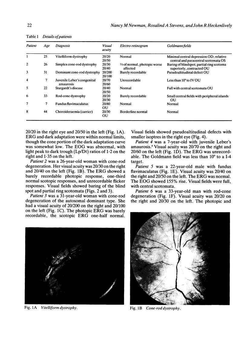
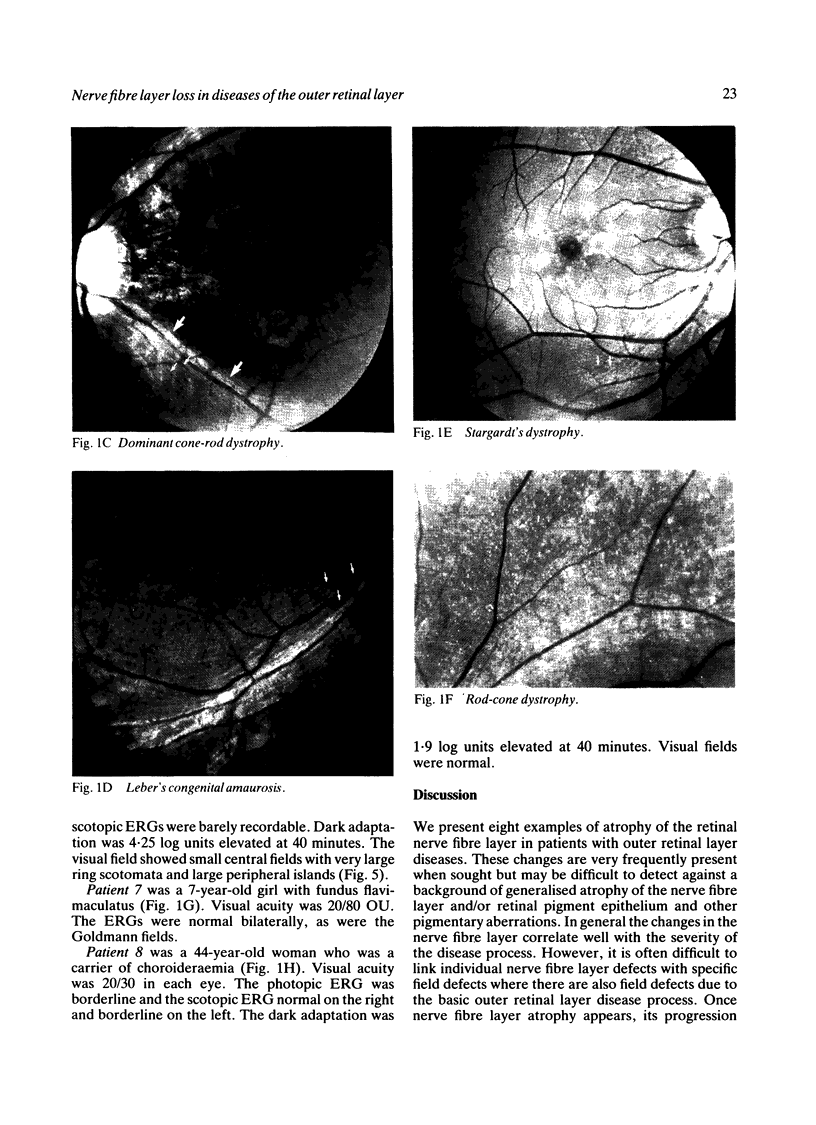
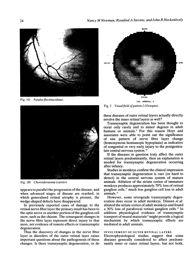
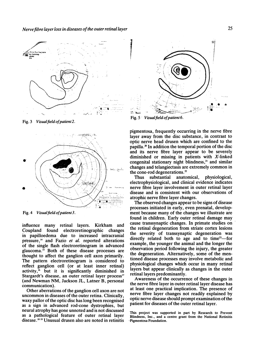
We present examples of nerve fibre layer changes in diseases thought to affect primarily the outer retinal layers. These disease processes include cone-rod dystrophies, rod-cone dystrophies, juvenile macular degeneration (Stargardt's disease) and fundus flavimaculatis, vitelliform macular dystrophy, and Leber's congenital amaurosis. All were associated with alterations in the retinal nerve fibre layer, either diffuse or focal. The presence of these nerve fibre layer changes raises the question of transsynaptic degeneration and of possible primary or associated disease of other retinal cells than the receptors-namely, bipolar, amacrine, Mueller, or ganglion cells--in these disease of the outer retinal layer. Involvement of the nerve fibre layer also indicates the need to examine patients with defects in the nerve fibre layer by electroretinograms and other tests for outer retinal layer disease when no obvious optic nerve disease is present.
Full text
PDF
Images in this article


Selected References
These references are in PubMed. This may not be the complete list of references from this article.
- Dineen J. T., Hendrickson A. E. Age correlated differences in the amount of retinal degeneration after striate cortex lesions in monkeys. Invest Ophthalmol Vis Sci. 1981 Nov;21(5):749–752. [PubMed] [Google Scholar]
- Dineen J., Hendrickson A., Keating E. G. Alterations of retinal inputs following striate cortex removal in adult monkey. Exp Brain Res. 1982;47(3):446–456. doi: 10.1007/BF00239362. [DOI] [PubMed] [Google Scholar]
- Fiorentini A., Maffei L., Pirchio M., Spinelli D., Porciatti V. The ERG in response to alternating gratings in patients with diseases of the peripheral visual pathway. Invest Ophthalmol Vis Sci. 1981 Sep;21(3):490–493. [PubMed] [Google Scholar]
- Foxman S. G., Heckenlively J. R., Bateman J. B., Wirtschafter J. D. Classification of congenital and early onset retinitis pigmentosa. Arch Ophthalmol. 1985 Oct;103(10):1502–1506. doi: 10.1001/archopht.1985.01050100078023. [DOI] [PubMed] [Google Scholar]
- Grafstein B., Laureno R. Transport of radioactivity from eye to visual cortex in the mouse. Exp Neurol. 1973 Apr;39(1):44–57. doi: 10.1016/0014-4886(73)90040-x. [DOI] [PubMed] [Google Scholar]
- Heckenlively J. R., Martin D. A., Rosales T. O. Telangiectasia and optic atrophy in cone-rod degenerations. Arch Ophthalmol. 1981 Nov;99(11):1983–1991. doi: 10.1001/archopht.1981.03930020859009. [DOI] [PubMed] [Google Scholar]
- Heckenlively J. R., Martin D. A., Rosenbaum A. L. Loss of electroretinographic oscillatory potentials, optic atrophy, and dysplasia in congenital stationary night blindness. Am J Ophthalmol. 1983 Oct;96(4):526–534. doi: 10.1016/s0002-9394(14)77917-6. [DOI] [PubMed] [Google Scholar]
- Hoyt W. F., Rios-Montenegro E. N., Behrens M. M., Eckelhoff R. J. Homonymous hemioptic hypoplasia. Fundoscopic features in standard and red-free illumination in three patients with congenital hemiplegia. Br J Ophthalmol. 1972 Jul;56(7):537–545. doi: 10.1136/bjo.56.7.537. [DOI] [PMC free article] [PubMed] [Google Scholar]
- Kirkham T. H., Coupland S. G. Abnormal electroretinograms and visual evoked potentials in chronic papilledema using time-difference analysis. Can J Neurol Sci. 1981 Aug;8(3):243–248. doi: 10.1017/s0317167100043274. [DOI] [PubMed] [Google Scholar]
- Miller N. R., Newman S. A. Transsynaptic degeneration. Arch Ophthalmol. 1981 Sep;99(9):1654–1654. doi: 10.1001/archopht.1981.03930020528032. [DOI] [PubMed] [Google Scholar]
- Newman N. M. Ophthalmoscopic observation of the retinal nerve fiber layer. Trans Sect Ophthalmol Am Acad Ophthalmol Otolaryngol. 1977 Sep-Oct;83(5):786–796. [PubMed] [Google Scholar]
- Newman N. M., Tornambe P. E., Corbett J. J. Ophthalmoscopy of the retinal nerve fiber layer. Use in detection of neurologic disease. Arch Neurol. 1982 Apr;39(4):226–233. doi: 10.1001/archneur.1982.00510160032006. [DOI] [PubMed] [Google Scholar]
- Quigley H. A., Miller N. R., George T. Clinical evaluation of nerve fiber layer atrophy as an indicator of glaucomatous optic nerve damage. Arch Ophthalmol. 1980 Sep;98(9):1564–1571. doi: 10.1001/archopht.1980.01020040416003. [DOI] [PubMed] [Google Scholar]
- Sommer A., Miller N. R., Pollack I., Maumenee A. E., George T. The nerve fiber layer in the diagnosis of glaucoma. Arch Ophthalmol. 1977 Dec;95(12):2149–2156. doi: 10.1001/archopht.1977.04450120055003. [DOI] [PubMed] [Google Scholar]
- Spencer W. H. Drusen of the optic disk and aberrant axoplasmic transport. The XXXIV Edward Jackson memorial lecture. Am J Ophthalmol. 1978 Jan;85(1):1–12. doi: 10.1016/s0002-9394(14)76658-9. [DOI] [PubMed] [Google Scholar]
- Stevens R. A., Newman N. M. Abnormal visual-evoked potentials from eyes with optic nerve head drusen. Am J Ophthalmol. 1981 Dec;92(6):857–862. doi: 10.1016/s0002-9394(14)75644-2. [DOI] [PubMed] [Google Scholar]
- VANBUREN J. M. TRANS-SYNAPTIC RETROGRADE DEGENERATION IN THE VISUAL SYSTEM OF PRIMATES. J Neurol Neurosurg Psychiatry. 1963 Oct;26:402–409. doi: 10.1136/jnnp.26.5.402. [DOI] [PMC free article] [PubMed] [Google Scholar]