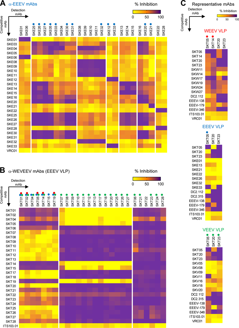
Figure 2. Competition ELISAs identify distinct binding groups of single-specific and triple-specific α-EEV mAbs.
Competitive binding ELISAs were performed to identify overlap in EEEV VLP surface binding areas within (A) ⍺-EEEV mAbs and (B) ⍺-WEVEEV mAbs. Data were hierarchically clustered to determine competition groups for each VLP and are representative of at least two independent experiments (C) Representative single-specific mAbs, as well as previously published broadly reactive ⍺-EEV mAbs (DC2.112, DC2.315, EEEV-138, EEEV-179, and EEEV-346),24,26 were competed against representative triple-specific mAbs. Heat maps display percent inhibition ranging from yellow (minimal competition) to orange (moderate competition) to purple (maximal competition). Negative control mAbs include either the human ⍺-HIV mAb VRC0160 or the NHP ⍺-SIV mAb ITS103.01.62 Colored circles above mAbs along x-axis represent the EEV pseudovirus that was neutralized (red: WEEV; blue: EEEV; green: VEEV). Data are representative of two independent experiments. See also Figures S2–S3, and Table S2.

