ABSTRACT
Aims and Objectives:
The study was carried out to evaluate the efficacy of four conservative therapeutic modalities on the mandibular range of motion (MRM) in subjects with anterior disc displacement with reduction (ADDwR) of the temporomandibular joint (TMJ).
Materials and Methods:
One hundred patients (64 women and 36 men) were selected, and randomly distributed into four groups. Group I: Subjects receiving behavioral therapy (BT). Group II: Subjects receiving low-level laser therapy (LLLT). Group III: Subjects receiving maxillary anterior repositioning splint (MARS). Group IV: Subjects receiving stabilization splint (SS). The MRM was evaluated for each patient before treatment and after 6 months. Paired t test and one-way analysis of variance (ANOVA) tests were used for statistical analysis followed by a post hoc Tukey test (P ≤ 0.05).
Results:
All groups showed significant improvement in MRM after 6 months of treatment (P ≤ 0.05) except for BT. There was a significant improvement for SS and MARS on the different movements of MRM, more than for LLLT and BT (P ≤ 0.05).
Conclusion:
The MARS and the SS are effective in increasing the MRM for patients with ADDwR.
Keywords: Anterior disc displacement with reduction, anterior repositioning splint, internal derangement, low-level laser therapy, occlusal splint, Stabilization splint, TMD
INTRODUCTION
The term “temporomandibular disorders” (TMD) is referred to a large group of disorders that affect the temporomandibular joint (TMJ) and its related musculoskeletal structures.[1] TMJ disc displacement is a disorder characterized by misalignment of the articular disc as related to the mandibular condyle and the articular fossa.[2,3] Disc displacement affects around 41% of patients with TMD.[4] TMJ Internal derangement can be classified into three categories according to the Research Diagnostic Criteria for TMD (RDC/TMD);[5] disc displacement with reduction and disc displacement without reduction with or without limited mouth opening.
Anterior disc displacement with reduction (ADDwR) is the most prevalent type, which is characterized by a shift occurring during mouth closing and reduction of the disc to its normal relationship with the mandibular condyle and the glenoid fossa that is evident during mouth opening.[6] Disc displacement disrupts the normal function of the TMJ and can cause pain, reduced mandibular movements, and deficient mastication. A clicking or popping sound during the opening and closure of the mouth (reciprocal click) can occur as a result of a displaced disc.[7,8] It has been stated that “One of the clinical characteristics of ADDwR is reduced mouth opening, which is generally accompanied by a mandibular deviation to the affected side until a pop or click (reduction) took place.”[9]
The primary treatment objectives for individuals with ADDwR are to reduce pain, restore chewing function, improve mandibular range of motion (MRM), and enhance patients’ quality of life.[10] The therapeutic options range from non-invasive reversible procedures to minimally invasive and invasive irreversible procedures. Although surgical procedures may be helpful in some cases, conservative therapy should be the first therapeutic option to avoid the risk of postoperative side effects. Conservative therapies include behavioral therapy (BT), thermal, and coolant therapy, repositioning splints, stabilization splints (SS), and low-level laser therapy (LLLT).[11,12]
BT is a psychologically based treatment that has been proposed for patients suffering from chronic TMD pain.[13] Such therapy is non-invasive, reversible, and safe therapeutic option that comprises a variety of techniques including; cognitive BT, biofeedback, re-education, and other techniques for relaxation which aim to reduce disabilities associated with pain and enhance pain coping skills by improving cognitive and adaptive behaviors.[14]
LLLT has been used recently as a conservative treatment approach for individuals with TMD and myofascial pain.[15] LLLT is a form of phototherapy that produces biostimulation and analgesia without causing temperature changes.[16] It is considered a successful, simple, and short-term therapeutic approach that has gained popularity as an alternative treatment for TMD due to its analgesic, anti-inflammatory, and regenerative properties.[17,18]
Occlusal splints can be divided into two categories according to the intended use: stabilizing and repositioning splints.[19] Treatment with occlusal splint aims to realign the articular disc between the mandibular condyle and the articular fossa, reduce TMJ pain and noise, enhance masticatory function, remove disc interference, and recapture the displaced disc. Even though many types of occlusal splints have been used to treat TMD, there is still much disagreement over the form of occlusal splints, how they should be worn, and their mechanism of action. Various forms of splints have been evaluated for the treatment of disc displacement, however, the most frequently employed types are anterior repositioning (maxillary anterior repositioning splint [MARS]) and SS.[20] Occlusal splints were originally constructed of acrylic resin and designed to cover most or all of the teeth in the maxillary or the mandibular arch. Nowadays, there have been significant advancements in materials, designs, and the use of occlusal splints as therapeutic appliances.[21]
There are various conservative treatment options available for cases of TMJ disc displacement with reduction (ADDwR) in the literature. Yet, there is limited data available regarding which treatment is more effective in improving MRM in ADDwR cases. Consequently, this study aimed to compare the efficacy of four non-invasive therapeutic interventions (BT, LLLT, MARS, and SS) on the different movements of MRM of ADDwR cases. The study hypothesized that none of the above different treatment modalities would have a significant effect on MRM for ADDwR cases.
MATERIALS AND METHODS
This randomized clinical trial included 100 patients, consisting of 64 women and 36 men, the age of patients ranged from 21 to 44 years old. These patients were recruited from the outpatient clinic of the Department of Removable Prosthodontics, Faculty of Dental Medicine, Al-Azhar University between March 2016 and April 2020. The Research Ethical Committee at the Faculty of Dental Medicine, Al-Azhar University granted ethical approval for the study under the number 502-04-16. Before their enrollment in the study, all participants provided their informed consent by signing appropriate forms.
All participants were diagnosed with TMJ ADDwR based on the research diagnostic criteria for temporomandibular disorders (RDC/TMD)[5]; Additionally, they underwent a bilateral magnetic resonance imaging (MRI) examination of their TMJ before treatment using the Ingenia S, MRI system from Philips, Amsterdam, Netherlands. Two examiners, blinded to the study grouping and treatment interventions and experienced in TMD made the diagnosis and treatment evaluations.
INCLUSION CRITERIA
To be included in the study, patients had to be diagnosed with unilateral TMJ disc displacement and have TMJ pain, noise, and restricted mouth opening. Additionally, they had to have persistent reciprocal joint clicking sounds during early or late opening and closing, a history of restricted mouth opening for more than 2 weeks, and pain in the TMJ area that worsened during jaw function or movement.
EXCLUSION CRITERIA
The study excluded individuals who had undergone previous TMJ surgery, had a history of head and neck radiotherapy or chemotherapy, had a systemic inflammatory joint disease, had a pathological lesion in the joint, had experienced direct trauma to the facial bones, had congenital disturbances such as hyperplasia or hypoplasia of the joint, had osteoarthritic changes in the TMJ bony components, or had lost the normal TMJ disc architecture due to thinning or perforations of the disc.
STUDY GROUPS
A total of 100 participants were recruited and randomly selected into four groups of 25 patients. All participants satisfying the inclusion and exclusion criteria over the period from March 2016 to April 2020 were included in the study. The randomization process was done using a block random allocation method through IBM SPSS version 20.0 software. All therapeutic interventions were provided by two skilled professionals who were also responsible for the randomization process.
Group I: Patients were treated using BT. Counseling was used to reduce the severity of symptoms and increase pain coping skills. Information, explanation, and reassurance during the first visit and also in later phases of management were given to increase the compliance of the patient. The study participants were provided with information about the pain pathology, dysfunction related to TMJ disc displacement, and the co-factors, which included psychological and behavioral aspects, as well as general medical conditions, that played a role in their TMD symptoms. They were also informed about the possible fluctuations in TMD symptoms. Patients were instructed to relax their TMJs as much as possible, to avoid tough foods, to use heat packs, and avoid incorrect working/sleeping and forehead positions and informed of their responsibility in the therapeutic process (compliance, motivation, coping). The patients were advised to adhere to the recommendations for a period of 6 months and provided with a pamphlet containing all the necessary instructions.
Group II: Patients were treated with LLLT according to the following protocol: laser was applied over the joint area twice a week for the 4 weeks of the treatment duration (a total of eight sessions). The equipment used was TOWER LIGHT LASER (ELETTRONICA PAGANI, Paderno Dugnano, Milan, Italy). The emitted radiation is obtained from a solid-state laser source with a wavelength of 808nm, in continuous emission. The output power was adjusted to 70 mW and doses of 105 J/cm2. During each session, laser therapy was administered at five specific points on the TMJ, which were predetermined to be the anterior, superior, posterior, and posteroinferior regions of the mandibular condylar, as well as the external acoustic canal. A self-adhesive plastic template was made for each participant to ensure replication of points of laser application each session. A central part was marked in the template with a 10 mm diameter to be positioned over the lateral edge of the condyle and the application points (anterior, posterior, superior, and posteroinferior) were marked 10 mm anterior, posterior, superior, and posteroinferior to the central part.
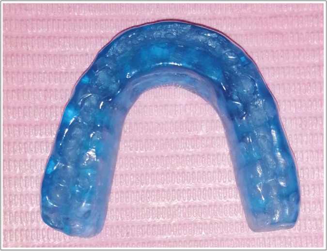
Group III: Patients were treated with MARS appliance, which was fabricated from a 2 mm Polyethylene terephthalate sheet (Folidur N/clear, Thermoforming Sheet, al dente Dental produkte GmbH, Horgenzell, Germany) and cold-curing acrylic resin (Vertex™ Orthoplast, Zeist, Netherlands) [Figure 1]. The splint was made with a guiding ramp in the anterior region that compels the mandible to adopt a more forward position in relation to the intercuspal position. Identifying the most appropriate position (therapeutic position) to alleviate the patient’s symptoms is crucial to the fabrication of an effective MARS appliance. The patient was directed to move the mandible slightly forward and perform opening and closing movements in this position. The TMJ was re-assessed for any symptoms, and the anterior position that prevented clicking was detected and identified using red articulating paper. An anterior ramp was constructed at this therapeutic position, and the patient was advised to wear the appliance for at least 12 h overnight every day for six months.
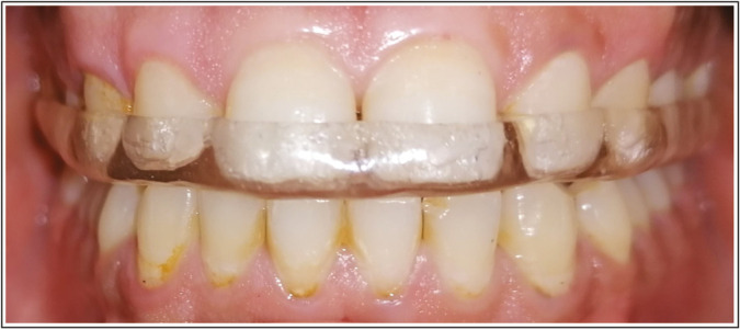
Group IV: Each patient received treatment with an SS appliance that was custom-made using a Polyethylene terephthalate sheet and self-curing acrylic resin [Figure 2]. The splint was modified orally so that mandibular buccal cusp tips and incisal edges have simultaneous occlusal contacts with the splint in a centric relation position, while during protrusive and lateral movements, canine-protected occlusion was established. During the fabrication of the splint, the excess self-curing acrylic resin surrounding the centric contacts was removed, except labial to the mandibular canines, to create eccentric guidance so that the canine was able to pass over smoothly and continuously during protrusive and latero-trusive excursions. The splint was used by patients during sleeping hours (at least 12 h per day) for the entire treatment period of 6 months. All patients were followed-up by telephone calls to ensure compliance with the treatment prescribed.
Figure 1.

Maxillary anterior repositioning splint
Figure 2.

Stabilization splint
MEASUREMENTS OF MRM
The measurements of MRM were registered at baseline and after six months of treatment, using a digital caliper according to the following criteria:
Measurements of mouth opening: Subjects were instructed to open their mouth comfortably to measure pain-free mouth opening and to open widely to measure the unassisted maximal mouth opening. In addition, the assisted mouth opening was recorded after a gentle pressure was applied to achieve the maximum mouth opening. The vertical inter-incisal distance, which refers to the space between the upper and lower incisors, was calculated and used for the registration of all mouth-opening measurements.[22]
Measurements of lateral movements: Subjects were instructed to open slightly and move the mandible as extensively as they could to the right or left. Measurements were registered using a digital caliper from the labio-incisal embrasure between the mandibular central incisors to the labio-incisal embrasure of the maxillary incisors; lateral movements were measured during contralateral and ipsilateral jaw movements.[22]
Measurement of protrusive movement: Subjects were instructed to open slightly from the rest position and to maximally protrude the mandible and the horizontal distance between the upper and lower central incisors’ incisal edges was observed and recorded. Additionally, the amount of horizontal overlap was assessed and included in the measurement of the distance between the incisal edges of the maxillary and mandibular incisor teeth.[22]
All measurements were recorded separately by two independent examiners for all participants, with the first examiner performing all measurements on each participant twice. The second examiner carried out the measurement procedure in the same manner as the first examiner. There was a 24-h period between the two measurements and the second examiner was blinded to the recordings of the first examiner.
The number of participants in the study was determined by using the findings of the previous research conducted by Haketa et al.[23] and the expected sample size was 21 individuals per group, with an α level of 0.05 (80% power) and an effect size of 36.93.
Statistical analyses
The means of different measurements of MRM of each group were calculated. Data were collected, organized into tables, and statistically analyzed by IBM SPSS version 20.0 software for Windows. The comparison between groups was performed by conducting a one-way analysis of variance (ANOVA) test, followed by a post hoc Tukey test at a 95% confidence level (P ≤ 0.05), and paired t test was used to compare baseline and post-treatment data within each group.
Pearson’s correlation coefficients were employed with a confidence interval of 95% to evaluate the test-retest reliability between the first and second measurements taken by the second examiner. Furthermore, to ensure the inter-observer reliability of the calibration process between the two examiners, the mean of two readings taken by each examiner for the initial ten participants was computed and then evaluated using the intra-class correlation coefficient (ICC) test.
RESULTS
The PC coefficient which indicates test-retest reliability had a value of 0.98 (P < 0.01), and the average measure ICC index between the two examiners was found to be 0.97 (P < 0.01).
Table 1 shows the effect of BT, LLLT, SS, and MARS on different movements of the MRM of the ADDwR cases. The paired t test revealed statistically significant differences between baseline and post-treatment values for various movements of the MRM of the ADDwR cases in all treatment groups (P < 0.05) except that of BT(P > 0.05).
Table 1.
Different movements of mandibular range of motion for different groups at baseline and after 6 months of treatment
| Group I (BT) | Group II (LLLT) | Group III (MARS) | Group IV (SS) | ||
|---|---|---|---|---|---|
| Mouth opening without pain | Baseline | 24.66 ± 6.56 | 23.67 ± 7.79 | 23.87 ± 7.35 | 25.44 ± 6.32 |
| 6 months | 25.45 ± 5.78 | 30.02 ± 8.22 | 34.72 ± 7.52 | 37.93 ± 7.88 | |
| P Value | 0.52ns | 0.00005* | 0.002* | 0.004* | |
| Unassisted maximum opening | Baseline | 28.22 ± 7.89 | 26.27 ± 9.55 | 27.84 ± 8.97 | 27.12 ± 7.88 |
| 6 months | 30.91 ± 8.98 | 34.92 ± 10.03 | 39.41 ± 9.56 | 42.89 ± 8.56 | |
| P Value | 0.522ns | 0.0255* | 0.0038* | 0.0001* | |
| Assisted maximum opening | Baseline | 32.67 ± 9.11 | 31.88 ± 7.62 | 33.66 ± 8.79 | 32.99 ± 9.94 |
| 6 months | 35.77 ± 8.96 | 40.64 ± 8.39 | 44.93 ± 7.47 | 49.94 ± 7.12 | |
| P Value | 0.39ns | 0.012* | 0.003* | 0.0001* | |
| Ipsilateral movement | Baseline | 7.77 ± 3.25 | 7.65 ± 2.74 | 7.42 ± 1.18 | 7.11 ± 2.27 |
| 6 months | 8.1 ± 2.94 | 9.5 ± 2.45 | 10.3 ± 2.12 | 11.9 ± 2.16 | |
| P Value | 0.511ns | 0.0004* | 0.00001* | 0.00001* | |
| Contralateral movement | Baseline | 5.91 ± 2.89 | 6.34 ± 3.37 | 5.79 ± 2.12 | 6.01 ± 1.83 |
| 6 months | 6.07 ± 1.74 | 7.13 ± 2.43 | 7.72 ± 2.16 | 8.39 ± 2.12 | |
| P Value | 0.75ns | 0.00003* | 0.002* | 0.006* | |
| Protrusive movement | Baseline | 4.5 ± 1.68 | 4.2 ± 1.24 | 3.8 ± 1.87 | 4.1 ± 1.74 |
| 6 months | 4.3 ± 1.97 | 5.5 ± 1.35 | 6.6 ± 1.64 | 7.3 ± 1.63 | |
| P Value | 0.698ns | 0.033* | 0.0000049* | 0.00001* | |
| Number of patients | 23 | 25 | 24 | 23 |
Ns = nonsignificant
*Denotes significant difference (P ≤ 0.05)
The mean changes between baseline and 6 months values of different measurements of MRM are shown in Table 2.
Table 2.
Mean changes and standard deviations (SD ±) of the different movements of the mandibular range of motion (mm) of the four treatment groups
| Group I (behavioral Therapy) | Group II (Low-level laser Therapy) | Group III (maxillary anterior repositioning splint) | Group IV (stabilization Splint) | |
|---|---|---|---|---|
| Mouth opening without pain | 0.79 ± 1.9 | 6.36 ± 4.1 | 10.85 ± 3.5 | 12.49 ± 3.8 |
| Unassisted maximum opening | 2.69 ± 4.5 | 8.65 ± 2.5 | 11.57 ± 3.6 | 15.77 ± 3.2 |
| Assisted maximum opening | 3.1 ± 1.1 | 8.76 ± 3.6 | 11.27 ± 3.8 | 16.95 ± 4.3 |
| Ipsilateral movement | 0.33 ± 0.65 | 1.85 ± 0.38 | 2.88 ± 0.63 | 4.79 ± 1.7 |
| Contralateral movement | 0.16 ± 0.46 | 0.79 ± 0.38 | 1.93 ± 0.68 | 2.38 ± 0.18 |
| Protrusive movement | –0.2 ± 1.1 | 1.3 ± 0.72 | 2.8 ± 0.76 | 3.2 ± 1.6 |
Table 3 shows the pair-wise comparisons of mean changes of different movements of MRM between the different treatment groups. The pair-wise comparisons using the post hoc Tukey test showed statistically significant differences for the mean changes of various movements of MRM (mouth opening without pain, unassisted maximum opening, maximum assisted opening, ipsilateral movement, contralateral movement) between all groups at 95% confidence level (P ≤ 0.05) except for protrusive movement between group MARS and group SS where P > 0.05. Serious complications such as alteration of occlusion or teeth mobility have not been observed in any participant.
Table 3.
Pair-wise comparisons of mean changes of different movements of mandibular range of motion between the different treatment groups
| Mouth opening without pain | Unassisted maximum opening | Assisted maximum opening | Ipsilateral movement | Contralateral movement | Protrusive movement | |
|---|---|---|---|---|---|---|
| Behavioral therapy vs. low-level laser therapy | 0.046* | 0.039* | 0.035* | 0.012* | 0.040* | 0.028* |
| Behavioral therapy vs. maxillary anterior repositioning splint | <0.001* | <0.001* | <0.001* | <0.001* | <0.001* | <0.001* |
| Behavioral therapy vs. stabilization splint | <0.001* | <0.001* | <0.001* | <0.001* | <0.001* | <0.001* |
| Low-level laser Therapy vs. Maxillary anterior repositioning splint | 0.047* | 0.045* | 0.029* | 0.039* | 0.006* | 0.006* |
| Low-level laser therapy vs. stabilization splint | <0.001* | <0.001* | <0.001* | <0.001* | <0.001* | <0.001* |
| Maxillary anterior repositioning splint vs. stabilization splint | 0.047* | 0.046* | 0.031* | 0.035* | 0.047* | 0.323ns |
Ns = nonsignificant
*Denotes significant difference (P ≤ 0.05)
DISCUSSION
Patients with TMD often experience restricted mandibular movements and pain in the TMJ area, masticatory muscles, and other related musculoskeletal structures in the head and neck area.[24,25]
Patients presently treated by LLLT, MARS, and SS showed a significant improvement of the different movements of the MRM (P ≤ 0.05), thus rejecting the null hypothesis that none of the different treatment modalities would have a significant effect on MRM for ADDwR cases. On the contrary, there was no corresponding improvement for the group treated with BT (P ˃ 0.05).
The pair-wise comparisons of mean changes of different movements of MRM between the different treatment groups revealed that there were significant differences in mean improvement of MRM of each movement of MRM between all the other treatment modalities (P ≤ 0.05) except that between protrusive movement of MARS and SS (P ˃ 0.05).
With the exception of protrusive movement, the SS improved MRM significantly more than MARS and the other treatment modalities used in this study (P ≤ 0.05).
These results are at variance with some,[26] but others[27,28] found a marked improvement on MARS in vertical jaw opening after treatment with an SS. These findings are also consistent with a prior study by Hosgor et al.,[29] who compared the efficacy of four non-invasive treatment approaches for ADDwR and reported significant improvement in maximum mouth opening after treatment with occlusal splint and LLLT.
LLLT is a conservative therapeutic approach for individuals suffering from TMD with analgesic, anti-inflammatory, muscle relaxant properties and bio-stimulation effects. Despite various theories that have been proposed to explain LLLT effects, the actual mechanism of action remains unknown.[30,31,32] The current findings revealed that LLLT caused a significant improvement in MRM. These were in accordance with previous findings,[33,34,35,36] which might be related to pain relief following LLLT, since restricted mandibular movement may be a pain-protective response. However, other studies [37,38] have reported no significant outcome difference between LLLT and placebo in patients with TMD. It has been suggested that pain score or mandibular movements may be attributed to variation in laser type, dosages, wavelength, treatment time, and sessions.
In the present study, the mean baseline values for the maximum unassisted opening were ranging from 26.27 ± 9.55 mm to 28.22 ± 7.89 mm for the different groups. These values improved after treatment with MARS and SS to 39.41 ± 9.56 mm and 42.89 ± 8.56 mm, respectively. This is in agreement with a previous study,[39] in which the corresponding baseline values were ranging from 27.6 ± 3.6 mm to 29.2 ± 2.9 mm and improved after treatment to 38.1 ± 4.1 mm to 43.3 ± 4.1 mm, respectively.
The baseline values of the protrusive movement in the present study were ranging from 3.8 ± 1.87 mm to 4.5 ± 1.68 mm and improved after treatment with MARS and SS to 6.6 ± 1.64 mm and7.3 ± 1.63 mm, respectively. This is in harmony with a previous study,[40,41] where the baseline values for the protrusive movement were 4.09 ± 1.41mm and improved after treatment to 7.3 ± 1.43 mm.
BT did not show any improvement in the various movements of the MRM among ADDwR cases (P > 0.05), which is consistent with a previous investigation by Okeson et al.,[28] where they compared relaxation therapy with occlusal splint and found that relaxation therapy is an ineffective treatment for limited MRM associated with TMD. On the contrary, others[26] have recommended BT as an effective method for treatment of limited jaw function in TMD if performed by a specialized therapist.
The current study showed better MRM improvement for subjects treated with SS than the other treatment modalities, this may be attributed to reducing the pain level.
The study’s limitation is a short follow-up period, which may not be sufficient to finally determine what conservative treatment modality of TMD is best. Another limitation was that due to high cost, there was no follow-up using MRI that could confirm repositioning of the TMJ disc in relation to the mandibular condyle. Therefore, additional investigations are required to assess the long-term effectiveness of these therapies.
CONCLUSION
Within the limitations of the study, it can be concluded that MARS and SS are effective treatment methods for improving the MRM of ADDwR cases. BT appears to have little effect on MRM of ADDwR cases. LLLT can be used as a complementary therapy to occlusal splints.
FINANCIAL SUPPORT AND SPONSORSHIP
The authors received no financial support for the research, authorship, and/or publication of this article.
CONFLICT OF INTEREST
The authors declare no conflict of interest.
AUTHORS CONTRIBUTIONS
Study conception and design: ANE, EAA, AAB, MAH. Data collection and acquisition: ANE,EAA,EA,MYF. Data analysis: ANE, MYF. Data interpretation: ANE,MAH, EA, AAB. Manuscript writing: ANE, EAA, EA, MYF. Review and editing: EAA, EA, AAB, MAH. All the authors approved the final version of the manuscript for publication.
ETHICAL POLICY AND INSTITUTIONAL REVIEW BOARD STATEMENT
The Research Ethical Committee atthe Faculty of Dental Medicine, Al-Azhar University granted ethical approval for the study under the number 502-04-16.
PATIENT DECLARATION OF CONSENT
Informed consent was obtained from all subjectsinvolved in the study.
DATA AVAILABILITY STATEMENT
The data that support the findings of this study are available on request from the corresponding author.
ACKNOWLEDGEMENT
None.
REFERENCES
- 1.Carrara S, Conti P, Stuginski-Barbosa J. Statement of the 1st consensus on temporomandibular disorders and orofacial pain. Dental Press J Orthod. 2010;15:114–20. [Google Scholar]
- 2.Warburton G. Internal Derangements of the Temporomandibular Joint. In: Bonanthaya K, Panneerselvam E, Manuel S, Kumar VV, Rai A, editors. Oral and Maxillofacial Surgery for the Clinician. Singapore: Springer Nature Singapore; 2021. pp. 1361–80. [Google Scholar]
- 3.Minervini G, D’Amico C, Cicciù M, Fiorillo L. Temporomandibular joint disk displacement: Etiology, diagnosis, imaging, and therapeutic approaches. J Craniofac Surg. 2022;10:1097. doi: 10.1097/SCS.0000000000009103. [DOI] [PubMed] [Google Scholar]
- 4.Manfredini D, Guarda-Nardini L, Winocur E, Piccotti F, Ahlberg J, Lobbezoo F. Research diagnostic criteria for temporomandibular disorders: A systematic review of axis I epidemiologic findings. Oral Surg Oral Med Oral Pathol Oral Radiol Endod. 2011;112:453–62. doi: 10.1016/j.tripleo.2011.04.021. [DOI] [PubMed] [Google Scholar]
- 5.Schiffman E, Ohrbach R, Truelove E, Look J, Anderson G, Goulet JP, et al. International RDC/TMD Consortium Network, International association for Dental Research. Diagnostic criteria for temporomandibular disorders (DC/TMD) for clinical and research applications: Recommendations of the international RDC/TMD consortium network* and orofacial pain special interest group†. J Oral Facial Pain Headache. 2014;28:6–27. doi: 10.11607/jop.1151. [DOI] [PMC free article] [PubMed] [Google Scholar]
- 6.Kellenberger CJ, Bucheli J, Schroeder-Kohler S, Saurenmann RK, Colombo V, Ettlin DA. Temporomandibular joint magnetic resonance imaging findings in adolescents with anterior disk displacement compared to those with juvenile idiopathic arthritis. J Oral Rehabil. 2019;46:14–22. doi: 10.1111/joor.12720. [DOI] [PubMed] [Google Scholar]
- 7.Okeson JP. Occlusal Appliance Therapy: Management of Temporomandibular Disorders and Occlusion. 7th ed. St. Louis, MO: Mosby; 2014. pp. 375–98. [Google Scholar]
- 8.Dworkin SF, LeResche L. Research diagnostic criteria for temporomandibular disorders: Review, criteria, examinations and specifications, critique. J Craniomandib Disord. 1992;6:301–55. [PubMed] [Google Scholar]
- 9.Poluha RL, Canales GT, Costa YM, Grossmann E, Bonjardim LR, Conti PCR. Temporomandibular joint disc displacement with reduction: A review of mechanisms and clinical presentation. J Appl Oral Sci. 2019;27:e20180433. doi: 10.1590/1678-7757-2018-0433. [DOI] [PMC free article] [PubMed] [Google Scholar]
- 10.de Leeuw R, Klasser GD. Diagnosis and Management of TMDs. In: de Leeuw R, Klasser GD, editors. Orofacial Pain: Guidelines for Assessment, Diagnosis, and Management. Chicago, USA: Quintessence Publishing Co, Inc; 2013. pp. 127–86. [Google Scholar]
- 11.Herpich CM, Leal-Junior ECP, Politti F, de Paula Gomes CAF, Dos Santos Glória IP, de Souza Amaral MFR, et al. Intraoral photobiomodulation diminishes pain and improves functioning in women with temporomandibular disorder: A randomized, sham-controlled, double-blind clinical trial: Intraoral photobiomodulation diminishes pain in women with temporomandibular disorder. Lasers Med Sci. 2020;35:439–45. doi: 10.1007/s10103-019-02841-1. [DOI] [PubMed] [Google Scholar]
- 12.Tournavitis A, Sandris E, Theocharidou A, Slini T, Kokoti M, Koidis P, et al. Effectiveness of conservative therapeutic modalities for temporomandibular disorders-related pain: A systematic review. Acta Odontol Scand. 2023;81:286–97. doi: 10.1080/00016357.2022.2138967. [DOI] [PubMed] [Google Scholar]
- 13.Huttunen J, Qvintus V, Suominen AL, Sipilä K. Role of psychosocial factors on treatment outcome of temporomandibular disorders. Acta Odontol Scand. 2019;77:119–25. doi: 10.1080/00016357.2018.1511057. [DOI] [PubMed] [Google Scholar]
- 14.Iwasaki S, Shigeishi H, Akita T, Tanaka J, Sugiyama M. Efficacy of cognitive-behavioral therapy for patients with temporomandibular disorder pain-systematic review of previous reports. Int J Clin Exp Med. 2018;11:500–9. [Google Scholar]
- 15.Desai AP, Roy SK, Semi RS, Balasundaram T. Efficacy of low-level laser therapy in management of temporomandibular joint pain: A double blind and placebo controlled trial. J Oral Maxillofac Surg. 2022;21:948–56. doi: 10.1007/s12663-021-01591-4. [DOI] [PMC free article] [PubMed] [Google Scholar]
- 16.Madani A, Ahrari F, Fallahrastegar A, Daghestani N. A randomized clinical trial comparing the efficacy of low-level laser therapy (LLLT) and laser acupuncture therapy (LAT) in patients with temporomandibular disorders. Lasers Med Sci. 2020;35:181–92. doi: 10.1007/s10103-019-02837-x. [DOI] [PubMed] [Google Scholar]
- 17.Jing G, Zhao Y, Dong F, Zhang P, Ren H, Liu J, et al. Effects of different energy density low-level laser therapies for temporomandibular joint disorders patients: A systematic review and network meta-analysis of parallel randomized controlled trials. Lasers Med Sci. 2021;36:1101–8. doi: 10.1007/s10103-020-03197-7. [DOI] [PubMed] [Google Scholar]
- 18.Abdel-Gawwad E, Abdullah A, Farhat M, Helal M. Effect of using Photobiomodulation, stabilization, and anterior repositioning splints on the pain level of subjects with temporomandibular joint disc displacement with reduction. Brazil Dent Sci. 2021;24:1–8. [Google Scholar]
- 19.Zhang S-H, He K-X, Lin C-J, Liu X-D, Wu L, Chen J, et al. Efficacy of occlusal splints in the treatment of temporomandibular disorders: A systematic review of randomized controlled trials. Acta Odontol Scand. 2020;78:580–9. doi: 10.1080/00016357.2020.1759818. [DOI] [PubMed] [Google Scholar]
- 20.Riley P, Glenny A-M, Worthington HV, Jacobsen E, Robertson C, Durham J, et al. Oral splints for temporomandibular disorder or bruxism: A systematic review. Br Dent J. 2020;228:191–7. doi: 10.1038/s41415-020-1250-2. [DOI] [PMC free article] [PubMed] [Google Scholar]
- 21.Greene CS, Menchel HFJO, Clinics MS. The use of oral appliances in the management of temporomandibular disorders. Oral Maxillofac Surg Clin North Am. 2018;30:265–77. doi: 10.1016/j.coms.2018.04.003. [DOI] [PubMed] [Google Scholar]
- 22.Celić R, Jerolimov V, Knezović Zlatarić D, Klaić B. Measurement of mandibular movements in patients with temporomandibular disorders and in asymptomatic subjects. Coll Antropol. 2003;27:43–9. [PubMed] [Google Scholar]
- 23.Haketa T, Kino K, Sugisaki M, Takaoka M, Ohta T. Randomized clinical trial of treatment for TMJ disc displacement. J Dent Res. 2010;89:1259–63. doi: 10.1177/0022034510378424. [DOI] [PubMed] [Google Scholar]
- 24.Li CX, Liu X, Gong ZC, Jumatai S, Ling B. Morphologic analysis of condyle among different disc status in the temporomandibular joints by three-dimensional reconstructive imaging: A preliminary study. BMC Oral Health. 2022;22:395. doi: 10.1186/s12903-022-02438-1. [DOI] [PMC free article] [PubMed] [Google Scholar]
- 25.Valesan LF, Da-Cas CD, Réus JC, Denardin ACS, Garanhani RR, Bonotto D, et al. Prevalence of temporomandibular joint disorders: A systematic review and meta-analysis. Clin Oral Investig. 2021;25:441–53. doi: 10.1007/s00784-020-03710-w. [DOI] [PubMed] [Google Scholar]
- 26.Shedden Mora MC, Weber D, Neff A, Rief W. Biofeedback-based cognitive-behavioral treatment compared with occlusal splint for temporomandibular disorder: A randomized controlled trial. Clin J Pain. 2013;29:1057–65. doi: 10.1097/AJP.0b013e3182850559. [DOI] [PubMed] [Google Scholar]
- 27.Kurt H, Mumcu E, Sülün T, Diraçoǧu D, Ünalan F, Aksoy C, et al. Comparison of effectiveness of stabilization splint, anterior repositioning splint and behavioral therapy in treatment of disc displacement with reduction. Turk J Phys Med Rehab. 2011;57:25–30. [Google Scholar]
- 28.Okeson JP, Moody PM, Kemper JT, Haley JV. Evaluation of occlusal splint therapy and relaxation procedures in patients with temporomandibular disorders. J Am Dent Assoc. 1983;107:420–4. doi: 10.14219/jada.archive.1983.0275. [DOI] [PubMed] [Google Scholar]
- 29.Hosgor H, Bas B, Celenk C. A comparison of the outcomes of four minimally invasive treatment methods for anterior disc displacement of the temporomandibular joint. Int J Oral Maxillofac Surg. 2017;46:1403–10. doi: 10.1016/j.ijom.2017.05.010. [DOI] [PubMed] [Google Scholar]
- 30.Sakurai Y, Yamaguchi M, Abiko Y. Inhibitory effect of low-level laser irradiation on LPS-stimulated prostaglandin E2 production and cyclooxygenase-2 in human gingival fibroblasts. Eur J Oral Sci. 2000;108:29–34. doi: 10.1034/j.1600-0722.2000.00783.x. [DOI] [PubMed] [Google Scholar]
- 31.Amaroli A, Ravera S, Parker S, Panfoli I, Benedicenti A, Benedicenti S. An 808-nm diode laser with a flat-top handpiece positively photobiomodulates mitochondria activities. Photomed Laser Surg. 2016;34:564–71. doi: 10.1089/pho.2015.4035. [DOI] [PubMed] [Google Scholar]
- 32.Carvalho CM, Lacerda JA, dos Santos Neto FP, de Castro IC, Ramos TA, de Lima FO, et al. Evaluation of laser phototherapy in the inflammatory process of the rat’s TMJ induced by carrageenan. Photomed Laser Surg. 2011;29:245–54. doi: 10.1089/pho.2009.2685. [DOI] [PubMed] [Google Scholar]
- 33.Mazzetto M, Hirono Hotta T, Pizzo R. Measurements of jaw movements and TMJ pain intensity in patients treated with GaAlAs laser. Braz Dent J. 2010;21:356–60. doi: 10.1590/s0103-64402010000400012. [DOI] [PubMed] [Google Scholar]
- 34.Rodrigues JH, Marques MM, Biasotto-Gonzalez DA, Moreira MS, Bussadori SK, Mesquita-Ferrari RA, et al. Evaluation of pain, jaw movements, and psychosocial factors in elderly individuals with temporomandibular disorder under laser phototherapy. Lasers Med Sci. 2015;30:953–9. doi: 10.1007/s10103-013-1514-z. [DOI] [PubMed] [Google Scholar]
- 35.Marini I, Gatto MR, Bonetti GA. Effects of superpulsed low-level laser therapy on temporomandibular joint pain. Clin J Pain. 2010;26:611–6. doi: 10.1097/AJP.0b013e3181e0190d. [DOI] [PubMed] [Google Scholar]
- 36.Kulekcioglu S, Sivrioglu K, Ozcan O, Parlak M. Effectiveness of low-level laser therapy in temporomandibular disorder. Scand J Rheumatol. 2003;32:114–8. doi: 10.1080/03009740310000139. [DOI] [PubMed] [Google Scholar]
- 37.Venancio Rde A, Camparis CM, Lizarelli Rde F. Low intensity laser therapy in the treatment of temporomandibular disorders: A double-blind study. J Oral Rehabil. 2005;32:800–7. doi: 10.1111/j.1365-2842.2005.01516.x. [DOI] [PubMed] [Google Scholar]
- 38.Hansen HJ, Thorøe U. Low power laser biostimulation of chronic oro-facial pain: A double-blind placebo controlled cross-over study in 40 patients. Pain. 1990;43:169–79. doi: 10.1016/0304-3959(90)91070-Y. [DOI] [PubMed] [Google Scholar]
- 39.Tatli U, Benlidayi M, Ekren O, Salimov F. Comparison of the effectiveness of three different treatment methods for temporomandibular joint disc displacement without reduction. Int J Oral Maxillofac Surg. 2017;46:603–9. doi: 10.1016/j.ijom.2017.01.018. [DOI] [PubMed] [Google Scholar]
- 40.Muhtarogullari M, Avci M, Yuzugullu B. Efficiency of pivot splints as jaw exercise apparatus in combination with stabilization splints in anterior disc displacement without reduction: A retrospective study. Head Face Med. 2014; 10:42. doi: 10.1186/1746-160X-10-42. [DOI] [PMC free article] [PubMed] [Google Scholar]
- 41.Helal MA, Agou SH, Bayoumi A, Imam A, Hassan AH. Management of internal derangement of temporomandibular joint disc displacement with reduction using two different lines of treatment. Brazil Dent Sci. 2021;24:8. [Google Scholar]
Associated Data
This section collects any data citations, data availability statements, or supplementary materials included in this article.
Data Availability Statement
The data that support the findings of this study are available on request from the corresponding author.


