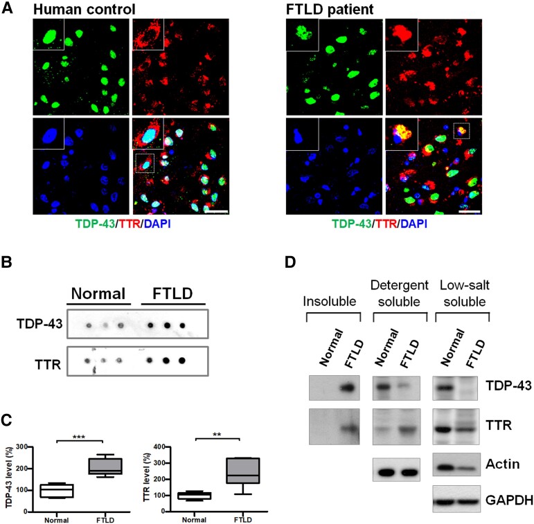
Figure 1.
TTR atypically mislocalizes to TDP-43 inclusions in human patients with FTLD-TDP. (A) The specimens from the frontal cortices were immunostained with TDP-43 (green, upper left) and TTR (red, upper right) for normal and FTLD-TDP human patients. Nuclei were counterstained with DAPI (blue, bottom left). The magnified view shown in the upper left is indicated with a white square line. Scale bar = 30 μm. (B) Dot blotting of TDP-43 and TTR in the insoluble fractions from human brains of normal and FTLD-TDP individuals. (C) Quantification of insoluble TDP-43 and TTR in the insoluble fractions from human brains of normal and FTLD-TDP individuals. Data are shown as box-and-whisker plots (min to max). The central horizontal line within the box indicates the median value. (TDP-43 level: ***P = 0.0004; TTR level: **P = 0.0036, t-test) n = 6 human brain tissue extracts. (D) Comparative analyses of brain extracts were fractionated from insoluble (urea-soluble), detergent-soluble and LS soluble fractions of normal and FTLD humans by western blotting.

