Abstract
Background
Tyrosine kinase inhibitors (TKIs) significantly improve clinical outcomes in patients with non-small cell lung cancer due to anaplastic lymphoma kinase (ALK) gene rearrangement. However, the rate of relapse with TKIs is high owing to the development of resistance mutations during treatment. Repeated biopsies during disease progression are crucial for elucidating the molecular mechanisms underlying the development of resistance to ALK inhibitors. Analysis of cell-free DNA (cfDNA) obtained from plasma is a novel approach for tumor genotyping.
Methods
In this mixed prospective and retrospective observational cohort study, we investigated the clinical feasibility of continuous quantitative monitoring of ALK-acquired mutations in plasma obtained from patients with ALK+ non-small cell lung cancer by using a highly sensitive and specific droplet digital polymerase chain reaction (ddPCR) assay. We enrolled nine patients, including three treatment-naïve patients recently diagnosed with ALK+ non-small cell lung cancer via tissue biopsy and expected to receive ALK TKIs and six patients already receiving ALK TKIs. Plasma samples were collected from these patients every 3 months. cfDNA was extracted from 66 samples during the study period, and 10 ALK mutations were simultaneously evaluated.
Results
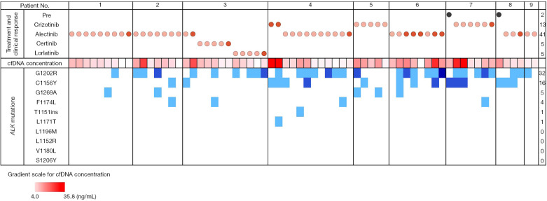
The numbers of samples showing the G1202R, C1156Y, G1269A, F1174L, T1151ins, and I1171T mutations were 32, 16, 5, 4, 1, and 1, respectively. The L1196M, L1152R, V1180L, and S1206Y mutations were not detected. Correlation analyses between progression-free survival and the time from treatment initiation (or treatment modification) to the detection of resistance mutations revealed that although resistance mutations may occur before a drug change becomes necessary, there is a duration during which the disease does not progress.
Conclusions
Our findings suggest that real-time quantitative monitoring of ALK resistance mutations during the response period could provide a time course of changes while acquiring resistance mutations. This information would be beneficial for designing an appropriate treatment strategy.
Keywords: Tumor genotyping, ALK mutations, digital polymerase chain reaction (digital PCR), resistance mutations, lung cancer
Highlight box.
Key findings
• In patients with ALK+ NSCLC, a ddPCR assay detected ALK mutations even during the period of response to ALK inhibitors.
• Our findings also suggested that the frequency of resistance mutations varies among individuals.
What is known and what is new?
• Repeated biopsies during disease progression are crucial to elucidate the molecular mechanisms underlying the development of ALK inhibitor resistance.
• Our mutation analyses showed that resistance mutations appeared before clinical disease progression.
What is the implication, and what should change now?
• Studies with large sample sizes, using a panel incorporating a larger number of genes and incorporating statistical analyses, are warranted to confirm our findings.
• In the future, multiplexed ddPCR assays may play a role in detecting multiple mutations when the input template is limited.
• This would facilitate application of the observed correlation between the time at which the mutation was detected and PFS in the clinic.
Introduction
Among all lung cancer cases, non-small cell lung cancer (NSCLC) accounts for approximately 80–85%. It is the most common cause of cancer-associated mortality in men and women globally. Anaplastic lymphoma kinase (ALK) gene rearrangement is one of the underlying mutations associated with NSCLC development (1-3) and has been identified in 3–8% of NSCLC cases (4). Since the confirmation of the role of ALK rearrangements in NSCLC, five tyrosine kinase inhibitors (TKIs), alectinib, crizotinib, ceritinib, lorlatinib, and brigatinib, have become the gold standard therapies for patients with advanced NSCLC. Although these TKIs significantly improve clinical outcomes, the rate of relapse with these drugs is high owing to the development of resistance mutations during the treatment course. ALK genetic alterations were initially described in 2007 in a Japanese subset (7%) of patients with NSCLC harboring a fusion oncogene EML4-ALK formed by the rearrangement of EML4 with ALK (5). The EML4-ALK fusion gene is a novel molecular target for cancer therapy. The incidence of ALK rearrangement is 3–13% in patients with NSCLC (6-9). Aside from EML4, ALK gene rearrangements also occur with partner genes such as KIF5B, KLC1, TFG, TPR, HIP1, STRN, DCTN1, SQSTM1, NPM1, BCL11A, and BIRC6 (4).
Repeated biopsies during disease progression are crucial to elucidate the molecular mechanisms underlying the development of resistance to ALK inhibitors (1,2,10). Different ALK mutations show different sensitivities to each TKI in vitro, and none of the TKIs share the same spectrum of activities against ALK mutants (1). Similar findings have been reported in patients. However, the pathological significance of ALK mutations occurring before disease progression has not yet been elucidated. Due to the scarcity of in vivo data, caution should be exercised when extrapolating data from in vitro experiments to predict treatment responses in humans, particularly for ALK mutations that occur before disease progression.
Examination of cfDNA obtained from plasma is a revolutionary method for tumor genotyping. Plasma sampling is minimally invasive and facilitates cfDNA collection from all metastatic sites; thus, this method can be useful for analyzing spatial heterogeneity in resistance mechanisms. Moreover, long-term real-time monitoring of genetic changes in cfDNA can overcome many limitations of tissue sampling, including repeated biopsies. Genotyping in plasma using digital polymerase chain reaction (PCR) is a reliable method for detecting mutations in patients with EGFR-mutant NSCLC. We previously reported that ALK mutations can be detected using droplet digital PCR (ddPCR) with an allele frequency of approximately 0.01% (10). This approach may be useful for identifying genetic mutations in cfDNA obtained from the plasma samples of patients undergoing ALK TKI therapy. In addition, it would allow us to examine whether the evolution of ALK mutations during treatment is associated with clinical response and evaluate the duration between the appearance of resistance mutations and tumor progression. Therefore, the objective of the present study was to investigate the clinical feasibility of the continuous quantitative monitoring of ALK-acquired mutations in plasma obtained from patients with ALK+ NSCLC by using a highly sensitive and specific ddPCR assay. We present this article in accordance with the STROBE reporting checklist (available at https://tlcr.amegroups.com/article/view/10.21037/tlcr-22-671/rc) (11).
Methods
Study design and patients
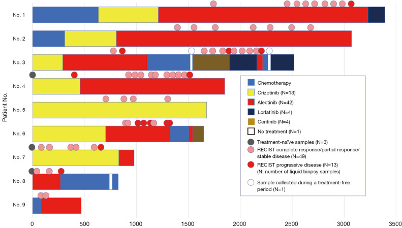
The present study was conducted at the Asahikawa Medical University Hospital, Asahikawa, Japan. In total, nine patients with ALK+ NSCLC were enrolled in the study. All patients were diagnosed with ALK+ NSCLC through reverse transcription PCR, immunohistochemistry, or fluorescence in situ hybridization. Six patients were already being treated with the ALK TKI alectinib or crizotinib [median duration of crizotinib treatment prior to study enrolment in the retrospectively enrolled patients: 524 (range, 291–1,678) days; Figure 1]. Three patients were enrolled before treatment initiation. All procedures were performed in compliance with the principles of the Declaration of Helsinki (as revised in 2013). Written informed consent was obtained from all patients before enrolment, and the study protocol was approved by the Research Ethics Committee of the Biomedical Research Institute, Asahikawa Medical University Hospital (approval No. 14106, October 23, 2014). Six patients who were retrospectively enrolled in this study visited our department between November 2015 and November 2017. Three treatment-naïve patients (approval No. 17160, December 5, 2017) were enrolled between December 2017 and November 2020. The inclusion criteria were (I) age ≥20 years; (II) patients newly diagnosed with ALK+ NSCLC, which had been confirmed by tissue biopsy, and expected to receive ALK TKIs; and (III) patients already diagnosed with ALK+ NSCLC who were being treated with ALK TKIs and had preserved tissue samples. The exclusion criteria were (I) patients with more than one type of carcinoma and (II) patients unwilling to provide written informed consent.
Figure 1.
Temporal relationship between plasma collection and treatment administration. The circles above the box indicate the timing of plasma collection and the bars represent the treatment status of the patient. The colors of the circles indicate treatment response, except that black circles indicate treatment-naïve samples and the white circle indicates a sample that was collected during a treatment-free period. Pink circles indicate clinical response (includes complete response, partial response, and stable disease). Red circles indicate disease progression. The x-axis represents the duration of treatment in days. RECIST, response evaluation criteria in solid tumors.
All patients received standard therapy, including ALK inhibitors, as part of their usual care. This study had a prospective observational design in which plasma samples were collected from all patients every 3 months during the treatment course. The treatment response of each patient was evaluated through computed tomography (CT) imaging approximately every 3 months, and additional CT scans were performed when the symptoms worsened. The standard Response Evaluation Criteria in Solid Tumors (version 1.0) was used based on the CT and other imaging findings.
Sample collection
In total, 20 mL of blood from each patient was collected into two PAXgene® Blood ccfDNA tubes (BD Biosciences, Franklin Lakes, NJ, USA). Plasma separation was performed using both tubes for each patient in accordance with the manufacturer’s protocol. Briefly, the tubes were centrifuged at room temperature using a swinging rotor at 1,900 ×g for 15 min, and the plasma layer was transferred to a separate container. One of the plasma tubes was stored for later use, if required, and the other was subjected to a second centrifugation step at 1,900 ×g for 10 min. The supernatant obtained was used for extracting cell-free DNA (cfDNA). It was stored in a deep freezer at ≥−70 ℃ until use.
Extraction of cfDNA and measurement of DNA concentration
Genetic analysis was performed between November 6, 2018 and September 30, 2020. The cfDNA was extracted from plasma using the QIAamp® Circulating Nucleic Acid Kit (Qiagen, Hilden, Germany) in accordance with the manufacturer’s protocol. The elution volume of DNA was set at 150 µL. The maximum volume of plasma used for extraction was 5 mL. The plasma volume of each patient was recorded. If the volume of plasma was >5 mL, the excess plasma was stored in a deep freezer at ≥−70 ℃. The extracted cfDNA solution was stored in a refrigerator at 2–8 ℃ until concentration measurement.
The concentration of the extracted cfDNA solution was measured the day after extraction using the QubitTM dsDNA HS Assay Kit (Thermo Fisher Scientific, Waltham, MA, USA) and a microspectrophotometer in accordance with the manufacturer’s instructions. Following concentration measurement, cfDNA solution was dispensed into 60-µL aliquots, which were stored in a freezer at −35 to −20 ℃. One aliquot (60 µL) was used for ddPCR analysis.
ddPCR analysis
ddPCR was performed as described previously (10). The ddPCR probes (LBx® Probe ALK Multi1, Multi2, and Multi3) were purchased from Riken Genesis, Tokyo, Japan. Details of the probes are provided in Table S1. ddPCR was performed using a droplet generator, thermal cycler, and droplet reader. The volume of cfDNA solution used for analysis was 8.8 µL, and one reaction was performed for each sample and probe pair. Additionally, one simultaneous reaction was performed using UltraPureTM DNase/RNase-Free Distilled Water (Thermo Fisher Scientific, Waltham, MA, USA) instead of DNA (negative control reaction) for each probe. The composition of the reaction mixture is shown in Table S2, and the PCR conditions are shown in Table S3. The amount of cfDNA contained in 8.8 µL of template DNA varied between 35.2–315.0 pg/µL. The amount of template DNA also varied between 0.7–6.3 ng/well because the digital PCR could not contain the same concentration of template DNA due to the volume of the PCR reaction solution.
After PCR, the fluorescence of the droplets was measured using a QX200TM Droplet Reader (Bio-Rad Laboratories, Hercules, CA, USA). The center of the distribution of negative droplets was checked from the histogram of 1D amplitude in panel 1 using QuantaSoftTM Software (QuantaSoft, ver.1.7, Bio-Rad Laboratories, Hercules, CA, USA). For channel 2, the threshold line was set such that all droplets were negative. The 2D amplitude images with a defined threshold line were used to confirm that no positive droplets were detected in the negative control shown in panel 1.
The reactions performed with LBx® Probe ALK Multi1 and LBx® Probe ALK Multi2 were determined to be mutation positive if the copy number (copies/µL) of the mutant gene was greater than or equal to the cutoff value of 0.25 (copies/µL) set in the ddPCR measurement conditions.
Results
Patient characteristics
The median age of the patients at baseline was 53 years, and the proportions of men and women were almost equal. The baseline characteristics of the patients are detailed in Table 1.
Table 1. Patient characteristics (n=9).
| Characteristics | Values |
|---|---|
| Sex | |
| Male | 5 [56] |
| Female | 4 [44] |
| Age at baseline (years), median [range] | 53 [37–80] |
| Smoking history | |
| Current | 1 [11] |
| Former | 2 [22] |
| Never | 6 [67] |
| Histology | |
| Adenocarcinoma | 9 [100] |
| Others | 0 [0] |
| Clinical stage | |
| IV | 9 [100] |
| Others | 0 [0] |
| No. of treatments† | |
| 1 | 1 [11] |
| 2 | 4 [44] |
| 3 | 1 [11] |
| ≥4 | 3 [33] |
| No. of ALK TKI treatments | |
| 1 | 3 [33] |
| 2 | 3 [33] |
| 3 | 2 [22] |
| 4 | 1 [11] |
| No. of cytotoxic chemotherapy‡ treatments | |
| 0 | 5 [56] |
| 1 | 3 [33] |
| 2 | 1 [11] |
Data are presented as n [%] unless otherwise stated. †, treatments include ALK TKI, chemotherapy, and immune checkpoint inhibitors; ‡, cytotoxic chemotherapy: pemetrexed +/−, cisplatin or carboplatin +/−, bevacizumab +/−, and/or pembrolizumab +/−. ALK, anaplastic lymphoma kinase; TKI, tyrosine kinase inhibitor.
Concentration of cfDNA
A total of 66 samples from the nine patients were analyzed. The median number of samples collected from each patient was 7 (range, 2–12). The plasma cfDNA concentrations of the 66 samples are shown in Figure S1. The median cfDNA concentration was 10.2 (range, 4.0–35.8) ng/µL in plasma. No samples were excluded due to technical issues or insufficient quantities.
Temporal relationship between administration of therapeutic agents and plasma collection
The course of administration of therapeutic agents (TKIs) and plasma sampling in the nine patients is summarized in Figure 1. Classification into clinical response and disease progression was conducted to evaluate the efficacy of TKIs. The clinical responses included complete response, partial response, and stable disease. The median survival time was 1,678 days [95% confidence interval (CI): 943–2,513]. Patients 2, 4, and 9 died because of disease progression. The six other patients were still undergoing treatment on the cutoff date (November 30, 2021).
ALK resistance mutations during treatment with ALK TKIs and at relapse
Figure 2 shows the ALK mutations detected in the plasma of patients during treatment with ALK TKIs or at relapse. We examined 10 resistance mutations (T1151ins, C1156Y, L1196M, G1269A, F1174L, L1152R, V1180L, I1171T, G1202R, and S1206Y). The numbers of samples showing the G1202R, C1156Y, G1269A, F1174L, T1151ins, and I1171T mutations were 32, 16, 5, 4, 1, and 1, respectively. G1202R, which is frequently detected as an acquired resistance mutation to alectinib, was detected in 20 of the 42 (48%) samples treated with alectinib in this study. Other mutations were detected in 26 (62%) of 42 samples during treatment with alectinib. Figure 2 and Figure S2 show the cfDNA concentrations in the same patients over time and across different patients. We also explored the possibility of a relationship between mutation number and cfDNA concentrations (Figure S3). However, no apparent relationship was found, and the sample size was too small to perform statistical analyses.
Figure 2.
ALK mutations during treatment with ALK TKIs and at relapse. The grid depicts ALK mutations detected in the plasma of patients on ALK TKIs or at relapse. The top box shows the status (treatment naïve or on ALK inhibitor) and clinical response when the blood was drawn. Black circles indicate treatment-naïve samples. Light red circles indicate clinical response (includes complete response, partial response, and stable disease). Red circles indicate disease progression. The numbers in the right column indicate the total number of samples in each treatment group. The middle box indicates cfDNA concentration. The darker the red color, the higher is the cfDNA concentration. Gradient for cfDNA concentration ranging from 4.0 to 35.8 ng/mL is given below. The blue boxes at the bottom indicate that an ALK mutation was detected in the plasma. The darker the blue color, the higher is the number of copies detected. The numbers in the right column indicate the total number of specific ALK mutations detected. cfDNA, cell-free DNA; ALK, anaplastic lymphoma kinase; TKIs, tyrosine kinase inhibitors.
Correlation between progression-free survival (PFS) and time from alectinib treatment initiation (or treatment modification) to detection of resistance mutations
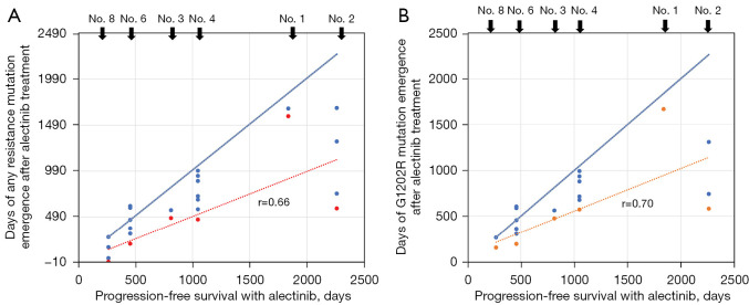
We also examined the correlation between PFS and the time from treatment initiation (or treatment modification) to the detection of resistance mutations through sensitive ddPCR. The correlation coefficient was 0.66 for the 10 resistance mutations we examined (Figure 3A). The correlation coefficient for the G1202R mutation, which is the most frequently reported alectinib resistance in vitro and in vivo, was 0.70 (Figure 3B). In addition, the resistance mutations appeared earlier than disease progression (Figure S4).
Figure 3.
Correlation between PFS and time from alectinib treatment initiation (or treatment modification) to detection of resistance mutations in six patients. (A) Correlation between PFS and detection of the 10 genetic mutations examined in this study with alectinib treatment. The correlation coefficient between the period when the resistance mutation was detected for the first time and PFS was 0.66. (B) Correlation between PFS and detection of the G1202R mutation with alectinib treatment. The correlation coefficient between the period when the resistance mutation was detected for the first time and PFS was 0.70. In five of six cases, the G1202R mutation was detected repeatedly until disease progression. The blue dots indicate the days when any of the resistance mutation was detected after treatment with alectinib, and the red dots indicate the days when they were first detected. The correlation analyses were performed using Spearman’s rank correlation coefficients. The red dotted lines represent the ‘lines of best fit’ for the red dots. The solid blue lines represent slopes with a correlation of +1 and are provided to indicate where the ‘line of best fit’ would be positioned if the clinical PFS correlated perfectly with the time period in which the resistant mutation appeared; thus, the figure indicates that the resistant mutation was detected earlier than what would be expected if clinical PFS was perfectly correlated with the appearance of the resistant mutation. PFS, progression-free survival.
Discussion
To the best of our knowledge, this study is the first to elucidate the clinical feasibility of continuous quantitative monitoring of ALK-acquired mutations in plasma through ddPCR. In previous studies, genetic analyses were conducted after the cancer became drug resistant and progressed. In the present study, genetic analyses were performed when the cancer was still responding to drugs to analyze the development of resistance mutations during treatment with ALK TKIs.
The ddPCR assay, which detects ALK mutations with high sensitivity, detected ALK mutations even during the period of response to ALK inhibitors. Moreover, ddPCR using plasma samples has several advantages over tissue biopsies. Intratumoral heterogeneity is a major obstacle to effective tumor genotyping. Intratumoral heterogeneity is characterized by the existence of distinct cell populations with different genetic profiles within the same tissue specimen harboring the tumor. Growing evidence suggests that single-tissue biopsy does not represent the entire tumor (12). For the adequate management of cancer, serial biopsies are indispensable; however, they involve additional risks for patients and could be unfeasible in certain clinical situations (13,14). Inevitably, recent research has focused on the development of noninvasive strategies for the elucidation of cancer-associated genetic features. Liquid biopsy is a novel approach that involves the use of tumor DNA fragments present in the circulation as an alternative source for tumor genotyping (15,16). This approach was used in the present study. Despite the aforementioned advantages, plasma specimens during the treatment course should be handled cautiously because the tumor volume is low owing to the treatment; additionally, plasma samples contain lower amounts of cfDNA and therefore require attention to the detection limit.
Epidermal growth factor receptor (EGFR)_T790M mutations, which occur during treatment with EGFR inhibitors in EGFR-mutated lung cancer, are associated with disease progression as indicators of resistance mutations. EGFR is dimeric or monomeric in the case of the EGFR_exon19 deletion type. However, the development of resistance mutations is associated with clinical drug resistance.
If ALK mutations increase over time, the tumor may worsen clinically. However, in the present study, we encountered cases in which ALK mutations appeared and disappeared repeatedly. ALK resistance mutations are thought to emerge during treatment with ALK inhibitors. EML4, the most frequent fusion partner of the ALK fusion genes, forms trimers (17). If a resistance mutation such as G1202R is inserted into all three dimers, then the clinical effect is expected to reflect drug sensitivity. However, if the mutation is inserted in only one of the three dimers, the other two dimers are expected to cross-phosphorylate ALK and the resistance will not be acquired (18,19). Furthermore, even mutations that are tumorigenic in the monomeric form, such as ALK_F1174L, are expected to confer resistance when double mutations such as F1174L + C1156Y are inserted at the cis position (20). Therefore, genetic analyses should be performed cautiously.
The approach used in the present study allowed us to infer that the evolution of ALK mutations during treatment is associated with clinical response. We also examined the correlation between PFS and the time from treatment initiation (or treatment modification) to the detection of resistance mutations. These findings suggest that the frequency of resistance mutations varies among individuals. Mutation analyses over time showed that resistance mutations appeared before clinical progression of the disease. Correlation analysis also suggested that although resistance mutations may appear long before a drug change becomes necessary, there is a duration when the disease does not progress. Furthermore, L1196M, L1152R, V1180L, and S1206Y, which are known resistance mutations of ALK inhibitors, were not detected. Other bypass mechanisms are also associated with TKI resistance. We did not study ALK amplification of the ALK kinase domain or activation of the bypass signaling pathway in this study as it was outside the scope of this study. Mechanisms of TKI resistance can be categorized into those associated with alterations that prevent the inhibition of the target receptor TK by TKIs, changes in tumor cell lineage, and activation of bypass +/− downstream signaling pathways that promote cell survival and proliferation (21).
The current study was limited by a small sample size and lack of statistical analyses; however, the results suggest that continuous quantitative monitoring of ALK-acquired mutations in plasma samples obtained from patients with ALK+ NSCLC using a highly sensitive and specific ddPCR assay is clinically beneficial for monitoring the emergence of resistance mutations during the treatment course. Long-term studies with large sample sizes and accompanying statistical analyses are warranted to confirm the findings of the present study and apply the observed correlation between the time at which the mutation was detected and PFS in the clinic. Our study was also limited by the use of different input amounts of cfDNA and plasma in the analyses, which could increase the chance of false negative results. We also only used one well for each sample, meaning technical replicates were not performed. In addition, digital PCR is limited by the fact that it cannot detect ALK fusion partners or cis- or trans-splicing, even when two resistance patterns are detected simultaneously. We used 10 ALK mutations for tumor monitoring in this study. Future studies should investigate the clinical utility of molecular profiling using a panel incorporating a larger number of genes (22,23). Finally, as our analysis is based on samples with low copy numbers, the fluctuations observed may be attributed to the limit of detection of ddPCR.
Conclusions
We investigated the clinical feasibility of the continuous quantitative monitoring of ALK-acquired mutations in plasma obtained from patients with ALK+ NSCLC through a highly sensitive and specific ddPCR assay. Our findings suggest that real-time quantitative monitoring of ALK resistance mutations during the response period would provide the time course of changes while acquiring resistance mutations. This information would be beneficial for designing an appropriate treatment strategy. In the future, multiplexed ddPCR assays may play a role in detecting multiple mutations when the input template is limited. Although our data indicate that the detection of genetic mutations do not correlate with clinical disease progression, the absence of a correlation may be the result of the higher sensitivity of liquid biopsy technologies, enabling the capture of molecular progression prior to the detection of disease progression by imaging.
Supplementary
The article’s supplementary files as
Acknowledgments
We thank the Northeast Japan Study Group Protocol Committee for reviewing the study protocol. Editorial support, in the form of medical writing, assembling tables and creating high-resolution images based on authors’ detailed directions, collating author comments, copyediting, fact checking, and referencing, was provided by Editage, Cactus Communications, and funded by Investigator Initiated Research support from Pfizer Japan Inc.
Funding: This study was conducted using the Investigator-Initiated Research Support from Pfizer Japan Inc. to TS. The funding source had no role in the study design; collection, analysis, and interpretation of data; writing of the report; or decision to submit the article for publication.
Ethical Statement: The authors are accountable for all aspects of the work in ensuring that questions related to the accuracy or integrity of any part of the work are appropriately investigated and resolved. The study was conducted in accordance with the Declaration of Helsinki (as revised in 2013). Written informed consent was obtained from all patients prior to the enrolment, and the study protocol was approved by the Research Ethics Committee of the Biomedical Research Institute, Asahikawa Medical University Hospital (approval No. 14106, October 23, 2014).
Footnotes
Reporting Checklist: The authors have completed the STROBE reporting checklist. Available at https://tlcr.amegroups.com/article/view/10.21037/tlcr-22-671/rc
Data Sharing Statement: Available at https://tlcr.amegroups.com/article/view/10.21037/tlcr-22-671/dss
Peer Review File: Available at https://tlcr.amegroups.com/article/view/10.21037/tlcr-22-671/prf
Conflicts of Interest: All the authors have completed the ICMJE uniform disclosure form (available at https://tlcr.amegroups.com/article/view/10.21037/tlcr-22-671/coif). TS received research funding from Pfizer Inc., Japan, and has received honoraria for lectures from Pfizer Japan, Inc., Chugal Pharmaceutical Co., Ltd., and Novartis Pharma. The other authors have no conflicts of interest to declare.
References
- 1.Gainor JF, Dardaei L, Yoda S, et al. Molecular Mechanisms of Resistance to First- and Second-Generation ALK Inhibitors in ALK-Rearranged Lung Cancer. Cancer Discov 2016;6:1118-33. 10.1158/2159-8290.CD-16-0596 [DOI] [PMC free article] [PubMed] [Google Scholar]
- 2.Recondo G, Mezquita L, Facchinetti F, et al. Diverse Resistance Mechanisms to the Third-Generation ALK Inhibitor Lorlatinib in ALK-Rearranged Lung Cancer. Clin Cancer Res 2020;26:242-55. 10.1158/1078-0432.CCR-19-1104 [DOI] [PMC free article] [PubMed] [Google Scholar]
- 3.Cognigni V, Pecci F, Lupi A, et al. The Landscape of ALK-Rearranged Non-Small Cell Lung Cancer: A Comprehensive Review of Clinicopathologic, Genomic Characteristics, and Therapeutic Perspectives. Cancers (Basel) 2022;14:4765. 10.3390/cancers14194765 [DOI] [PMC free article] [PubMed] [Google Scholar]
- 4.Fukui T, Tachihara M, Nagano T, et al. Review of Therapeutic Strategies for Anaplastic Lymphoma Kinase-Rearranged Non-Small Cell Lung Cancer. Cancers (Basel) 2022;14:1184. 10.3390/cancers14051184 [DOI] [PMC free article] [PubMed] [Google Scholar]
- 5.Soda M, Choi YL, Enomoto M, et al. Identification of the transforming EML4-ALK fusion gene in non-small-cell lung cancer. Nature 2007;448:561-6. 10.1038/nature05945 [DOI] [PubMed] [Google Scholar]
- 6.Shaw AT, Yeap BY, Mino-Kenudson M, et al. Clinical features and outcome of patients with non-small-cell lung cancer who harbor EML4-ALK. J Clin Oncol 2009;27:4247-53. 10.1200/JCO.2009.22.6993 [DOI] [PMC free article] [PubMed] [Google Scholar]
- 7.Sun Y, Ren Y, Fang Z, et al. Lung adenocarcinoma from East Asian never-smokers is a disease largely defined by targetable oncogenic mutant kinases. J Clin Oncol 2010;28:4616-20. 10.1200/JCO.2010.29.6038 [DOI] [PMC free article] [PubMed] [Google Scholar]
- 8.Inamura K, Takeuchi K, Togashi Y, et al. EML4-ALK fusion is linked to histological characteristics in a subset of lung cancers. J Thorac Oncol 2008;3:13-7. 10.1097/JTO.0b013e31815e8b60 [DOI] [PubMed] [Google Scholar]
- 9.Horn L, Pao W. EML4-ALK: honing in on a new target in non-small-cell lung cancer. J Clin Oncol 2009;27:4232-5. 10.1200/JCO.2009.23.6661 [DOI] [PMC free article] [PubMed] [Google Scholar]
- 10.Yoshida R, Sasaki T, Umekage Y, et al. Highly sensitive detection of ALK resistance mutations in plasma using droplet digital PCR. BMC Cancer 2018;18:1136. 10.1186/s12885-018-5031-0 [DOI] [PMC free article] [PubMed] [Google Scholar]
- 11.von Elm E, Altman DG, Egger M, et al. The Strengthening the Reporting of Observational Studies in Epidemiology (STROBE) statement: guidelines for reporting observational studies. Lancet 2007;370:1453-7. 10.1016/S0140-6736(07)61602-X [DOI] [PubMed] [Google Scholar]
- 12.Bettoni F, Masotti C, Habr-Gama A, et al. Intratumoral Genetic Heterogeneity in Rectal Cancer: Are Single Biopsies representative of the entirety of the tumor? Ann Surg 2017;265:e4-6. 10.1097/SLA.0000000000001937 [DOI] [PubMed] [Google Scholar]
- 13.Heitzer E, Ulz P, Geigl JB. Circulating tumor DNA as a liquid biopsy for cancer. Clin Chem 2015;61:112-23. 10.1373/clinchem.2014.222679 [DOI] [PubMed] [Google Scholar]
- 14.Crowley E, Di Nicolantonio F, Loupakis F, et al. Liquid biopsy: monitoring cancer-genetics in the blood. Nat Rev Clin Oncol 2013;10:472-84. 10.1038/nrclinonc.2013.110 [DOI] [PubMed] [Google Scholar]
- 15.Diaz LA, Jr, Bardelli A. Liquid biopsies: genotyping circulating tumor DNA. J Clin Oncol 2014;32:579-86. 10.1200/JCO.2012.45.2011 [DOI] [PMC free article] [PubMed] [Google Scholar]
- 16.Pesta M, Shetti D, Kulda V, et al. Applications of Liquid Biopsies in Non-Small-Cell Lung Cancer. Diagnostics (Basel) 2022;12:1799. 10.3390/diagnostics12081799 [DOI] [PMC free article] [PubMed] [Google Scholar]
- 17.Richards MW, O'Regan L, Roth D, et al. Microtubule association of EML proteins and the EML4-ALK variant 3 oncoprotein require an N-terminal trimerization domain. Biochem J 2015;467:529-36. 10.1042/BJ20150039 [DOI] [PubMed] [Google Scholar]
- 18.Hirai N, Sasaki T, Okumura S, et al. Monomerization of ALK Fusion Proteins as a Therapeutic Strategy in ALK-Rearranged Non-small Cell Lung Cancers. Front Oncol 2020;10:419. 10.3389/fonc.2020.00419 [DOI] [PMC free article] [PubMed] [Google Scholar]
- 19.Sampson J, Richards MW, Choi J, et al. Phase-separated foci of EML4-ALK facilitate signalling and depend upon an active kinase conformation. EMBO Rep 2021;22:e53693. 10.15252/embr.202153693 [DOI] [PMC free article] [PubMed] [Google Scholar]
- 20.Holcmann M, Amberg N, Drobits B, et al. Mouse models of receptor tyrosine kinases. In: Wheeler DL, Yarden Y. editors. Receptor tyrosine kinases: structure, functions and role in human disease. New York, NY, USA: Springer; 2015:279-438. [Google Scholar]
- 21.Cooper AJ, Sequist LV, Lin JJ. Third-generation EGFR and ALK inhibitors: mechanisms of resistance and management. Nat Rev Clin Oncol 2022;19:499-514. Erratum in: Nat Rev Clin Oncol 2022;19:744. 10.1038/s41571-022-00639-9 [DOI] [PMC free article] [PubMed] [Google Scholar]
- 22.Dietz S, Christopoulos P, Yuan Z, et al. Longitudinal therapy monitoring of ALK-positive lung cancer by combined copy number and targeted mutation profiling of cell-free DNA. EBioMedicine 2020;62:103103. 10.1016/j.ebiom.2020.103103 [DOI] [PMC free article] [PubMed] [Google Scholar]
- 23.Angeles AK, Christopoulos P, Yuan Z, et al. Early identification of disease progression in ALK-rearranged lung cancer using circulating tumor DNA analysis. NPJ Precis Oncol 2021;5:100. 10.1038/s41698-021-00239-3 [DOI] [PMC free article] [PubMed] [Google Scholar]
Associated Data
This section collects any data citations, data availability statements, or supplementary materials included in this article.
Supplementary Materials
The article’s supplementary files as


