Abstract
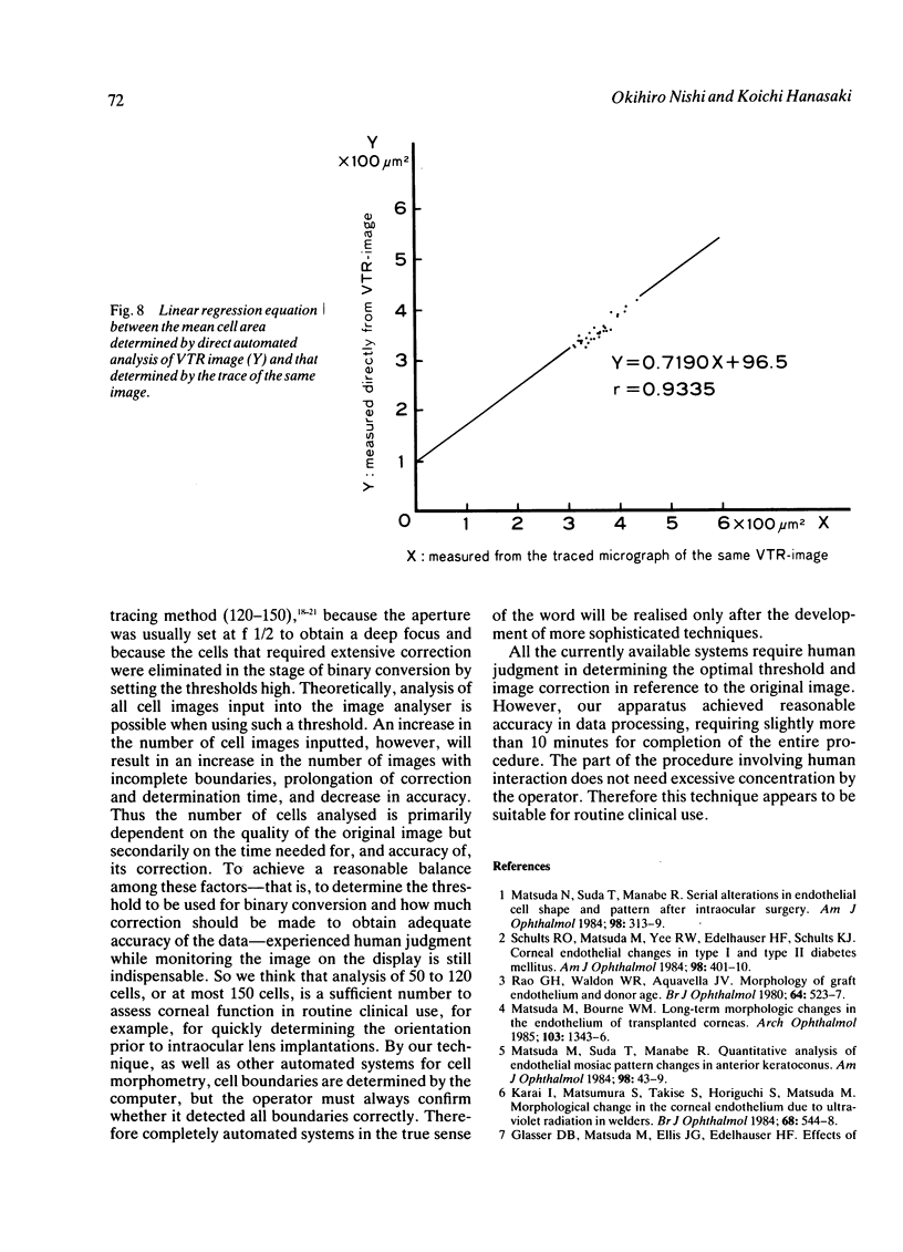
We developed an apparatus for automated morphometry of the corneal endothelium, which was photographed through a specular microscope connected to a video camera, and the images were stored on a video tape. The clearest stationary image was input into an image analyser to determine automatically the cell boundaries. Although human interaction is generally necessary, the mean time required to complete this procedure was about 13 minutes, based on the results of the 30 normal eyes, and the time needed for manual correction was about 4 minutes. The mean cell area obtained by this method correlated well (r = 0.9335) with that obtained by tracing the same images. This apparatus is clinically useful for immediately obtaining the mean cell area of corneal endothelium and will extend the application of specular microscopy to the routine clinical setting.
Full text
PDF
Images in this article
Selected References
These references are in PubMed. This may not be the complete list of references from this article.
- Glasser D. B., Matsuda M., Ellis J. G., Edelhauser H. F. Effects of intraocular irrigating solutions on the corneal endothelium after in vivo anterior chamber irrigation. Am J Ophthalmol. 1985 Mar 15;99(3):321–328. doi: 10.1016/0002-9394(85)90363-0. [DOI] [PubMed] [Google Scholar]
- Hartmann C., Köditz W. Automated morphometric endothelial analysis combined with video specular microscopy. Cornea. 1984;3(3):155–167. [PubMed] [Google Scholar]
- Hartmann C., Weingart M., Dünner P., Giraud J. P. Automatisierte Endothelmorphometrie. Fortschr Ophthalmol. 1982 Nov;79(3):261–265. [PubMed] [Google Scholar]
- Hirst L. W., Sterner R. E., Grant D. G. Automated analysis of wide-field specular photomicrographs. Cornea. 1984;3(2):83–87. [PubMed] [Google Scholar]
- Kandori T., Sawa M., Tanishima T. [Computerized morphometry of the human corneal endothelium (author's transl)]. Nippon Ganka Gakkai Zasshi. 1981 Sep 10;85(9):1204–1207. [PubMed] [Google Scholar]
- Karai I., Matsumura S., Takise S., Horiguchi S., Matsuda M. Morphological change in the corneal endothelium due to ultraviolet radiation in welders. Br J Ophthalmol. 1984 Aug;68(8):544–548. doi: 10.1136/bjo.68.8.544. [DOI] [PMC free article] [PubMed] [Google Scholar]
- Komine T., Oikawa T., Kato K. [Analysis of specular photomicrography with a computerized digitizer and an image analyzer(author's transl)]. Nippon Ganka Gakkai Zasshi. 1981 Jun 10;85(6):457–463. [PubMed] [Google Scholar]
- Laing R. A., Sanstrom M. M., Berrospi A. R., Leibowitz H. M. Changes in the corneal endothelium as a function of age. Exp Eye Res. 1976 Jun;22(6):587–594. doi: 10.1016/0014-4835(76)90003-8. [DOI] [PubMed] [Google Scholar]
- Lester J. M., McFarland J. L., Bursell S. E., Laing R. A., Brenner J. F. Automated morphometric analysis of corneal endothelial cells. Invest Ophthalmol Vis Sci. 1981 Mar;20(3):407–410. [PubMed] [Google Scholar]
- Matsuda M., Bourne W. M. Long-term morphologic changes in the endothelium of transplanted corneas. Arch Ophthalmol. 1985 Sep;103(9):1343–1346. doi: 10.1001/archopht.1985.01050090095040. [DOI] [PubMed] [Google Scholar]
- Matsuda M., Suda T., Manabe R. Quantitative analysis of endothelial mosaic pattern changes in anterior keratoconus. Am J Ophthalmol. 1984 Jul 15;98(1):43–49. doi: 10.1016/0002-9394(84)90187-9. [DOI] [PubMed] [Google Scholar]
- Matsuda M., Suda T., Manabe R. Serial alterations in endothelial cell shape and pattern after intraocular surgery. Am J Ophthalmol. 1984 Sep 15;98(3):313–319. doi: 10.1016/0002-9394(84)90321-0. [DOI] [PubMed] [Google Scholar]
- Nishi O. [Direct measurement of the corneal endothelial cell area on the negative film]. Nippon Ganka Gakkai Zasshi. 1985 Oct;89(10):1120–1124. [PubMed] [Google Scholar]
- Rao G. N., Waldron W. R., Aquavella J. V. Morphology of graft endothelium and donor age. Br J Ophthalmol. 1980 Jul;64(7):523–527. doi: 10.1136/bjo.64.7.523. [DOI] [PMC free article] [PubMed] [Google Scholar]
- Schultz R. O., Matsuda M., Yee R. W., Edelhauser H. F., Schultz K. J. Corneal endothelial changes in type I and type II diabetes mellitus. Am J Ophthalmol. 1984 Oct 15;98(4):401–410. doi: 10.1016/0002-9394(84)90120-x. [DOI] [PubMed] [Google Scholar]
- Shaw E. L., Rao G. N., Arthur E. J., Aquavella J. V. The functional reserve of corneal endothelium. Ophthalmology. 1978 Jun;85(6):640–649. doi: 10.1016/s0161-6420(78)35634-7. [DOI] [PubMed] [Google Scholar]
- Waring G. O., 3rd, Krohn M. A., Ford G. E., Harris R. R., Rosenblatt L. S. Four methods of measuring human corneal endothelial cells from specular photomicrographs. Arch Ophthalmol. 1980 May;98(5):848–855. doi: 10.1001/archopht.1980.01020030842008. [DOI] [PubMed] [Google Scholar]


