Abstract
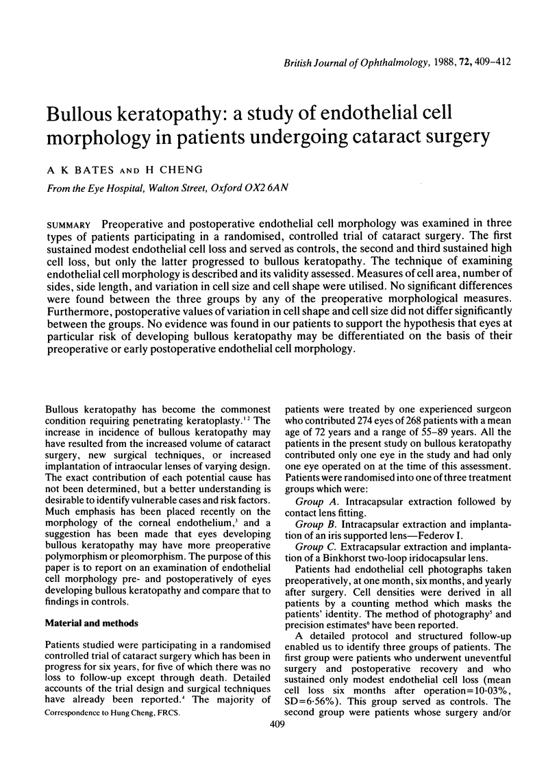
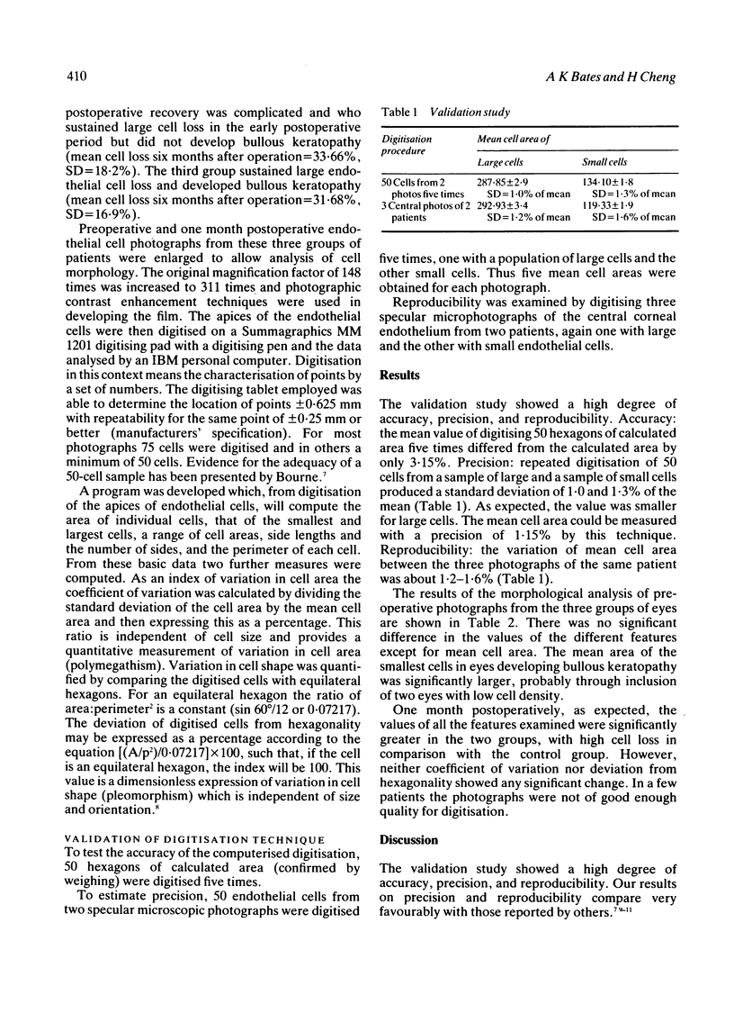
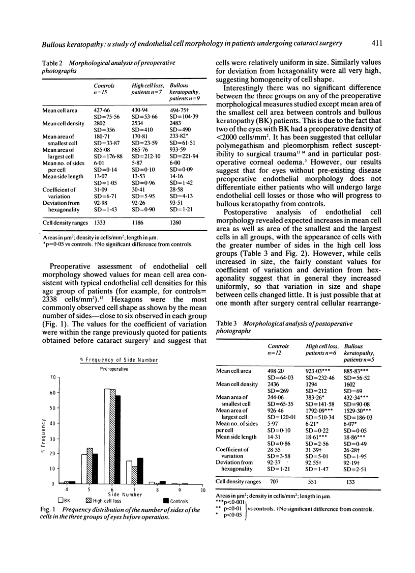
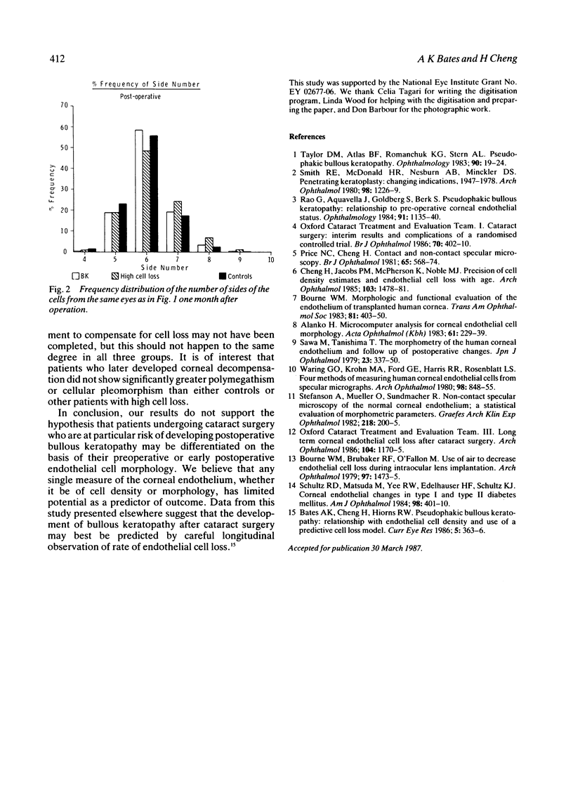
Preoperative and postoperative endothelial cell morphology was examined in three types of patients participating in a randomised, controlled trial of cataract surgery. The first sustained modest endothelial cell loss and served as controls, the second and third sustained high cell loss, but only the latter progressed to bullous keratopathy. The technique of examining endothelial cell morphology is described and its validity assessed. Measures of cell area, number of sides, side length, and variation in cell size and cell shape were utilised. No significant differences were found between the three groups by any of the preoperative morphological measures. Furthermore, postoperative values of variation in cell shape and cell size did not differ significantly between the groups. No evidence was found in our patients to support the hypothesis that eyes at particular risk of developing bullous keratopathy may be differentiated on the basis of their preoperative or early postoperative endothelial cell morphology.
Full text
PDF


Selected References
These references are in PubMed. This may not be the complete list of references from this article.
- Alanko H. I. Microcomputer analysis for corneal endothelial cell morphology. Acta Ophthalmol (Copenh) 1983 Apr;61(2):229–239. doi: 10.1111/j.1755-3768.1983.tb01416.x. [DOI] [PubMed] [Google Scholar]
- Bates A. K., Cheng H., Hiorns R. W. Pseudophakic bullous keratopathy: relationship with endothelial cell density and use of a predictive cell loss model. A preliminary report. Curr Eye Res. 1986 May;5(5):363–366. doi: 10.3109/02713688609025174. [DOI] [PubMed] [Google Scholar]
- Bourne W. M., Brubaker R. F., O'Fallon W. M. Use of air to decrease endothelial cell loss during intraocular lens implantation. Arch Ophthalmol. 1979 Aug;97(8):1473–1475. doi: 10.1001/archopht.1979.01020020135009. [DOI] [PubMed] [Google Scholar]
- Bourne W. M. Morphologic and functional evaluation of the endothelium of transplanted human corneas. Trans Am Ophthalmol Soc. 1983;81:403–450. [PMC free article] [PubMed] [Google Scholar]
- Cheng H., Jacobs P. M., McPherson K., Noble M. J. Precision of cell density estimates and endothelial cell loss with age. Arch Ophthalmol. 1985 Oct;103(10):1478–1481. doi: 10.1001/archopht.1985.01050100054017. [DOI] [PubMed] [Google Scholar]
- Price N. C., Cheng H. Contact and noncontact specular microscopy. Br J Ophthalmol. 1981 Aug;65(8):568–574. doi: 10.1136/bjo.65.8.568. [DOI] [PMC free article] [PubMed] [Google Scholar]
- Rao G. N., Aquavella J. V., Goldberg S. H., Berk S. L. Pseudophakic bullous keratopathy. Relationship to preoperative corneal endothelial status. Ophthalmology. 1984 Oct;91(10):1135–1140. [PubMed] [Google Scholar]
- Schultz R. O., Matsuda M., Yee R. W., Edelhauser H. F., Schultz K. J. Corneal endothelial changes in type I and type II diabetes mellitus. Am J Ophthalmol. 1984 Oct 15;98(4):401–410. doi: 10.1016/0002-9394(84)90120-x. [DOI] [PubMed] [Google Scholar]
- Smith R. E., McDonald H. R., Nesburn A. B., Minckler D. S. Penetrating keratoplasty: changing indications, 1947 to 1978. Arch Ophthalmol. 1980 Jul;98(7):1226–1229. doi: 10.1001/archopht.1980.01020040078009. [DOI] [PubMed] [Google Scholar]
- Stefansson A., Müller O., Sundmacher R. Non-contact specular microscopy of the normal corneal endothelium. A statistical evaluation of morphometric parameters. Graefes Arch Clin Exp Ophthalmol. 1982;218(4):200–205. doi: 10.1007/BF02150095. [DOI] [PubMed] [Google Scholar]
- Taylor D. M., Atlas B. F., Romanchuk K. G., Stern A. L. Pseudophakic bullous keratopathy. Ophthalmology. 1983 Jan;90(1):19–24. doi: 10.1016/s0161-6420(83)34607-8. [DOI] [PubMed] [Google Scholar]
- Waring G. O., 3rd, Krohn M. A., Ford G. E., Harris R. R., Rosenblatt L. S. Four methods of measuring human corneal endothelial cells from specular photomicrographs. Arch Ophthalmol. 1980 May;98(5):848–855. doi: 10.1001/archopht.1980.01020030842008. [DOI] [PubMed] [Google Scholar]


