Abstract
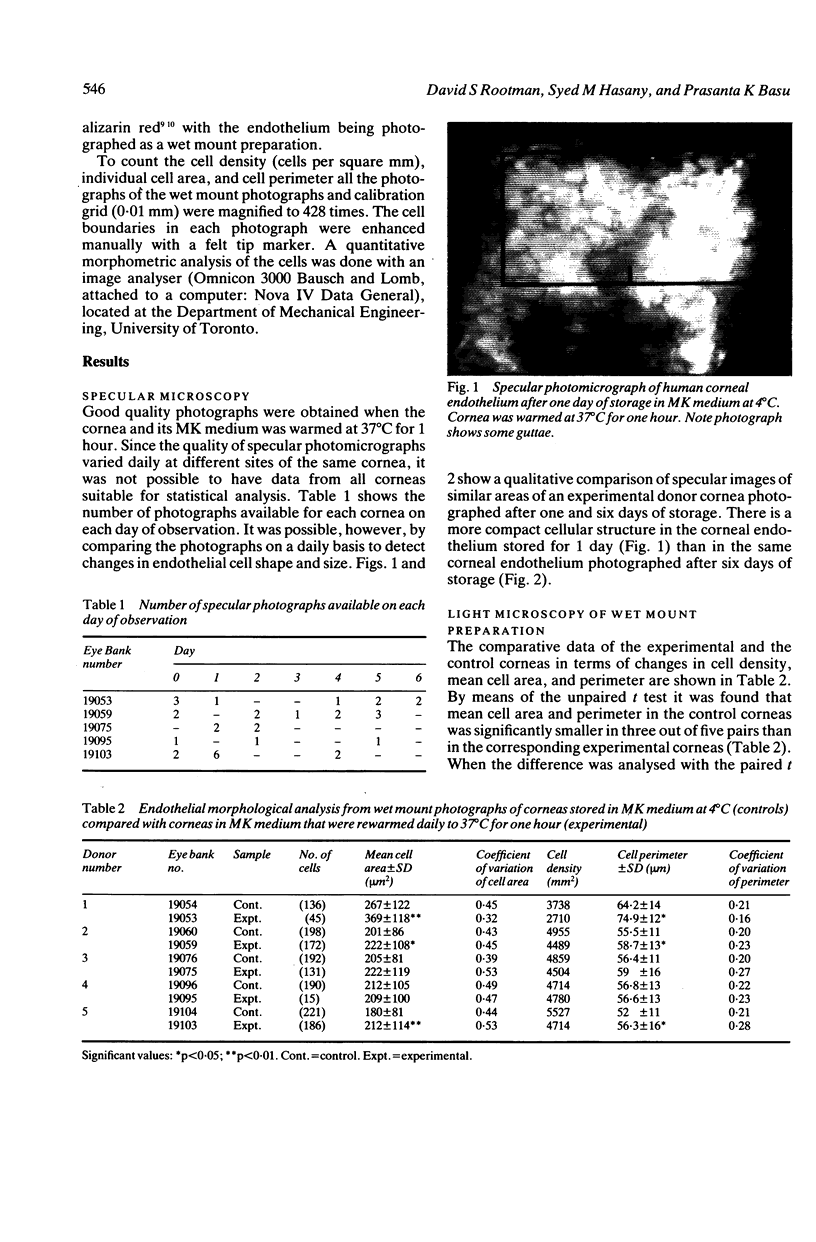
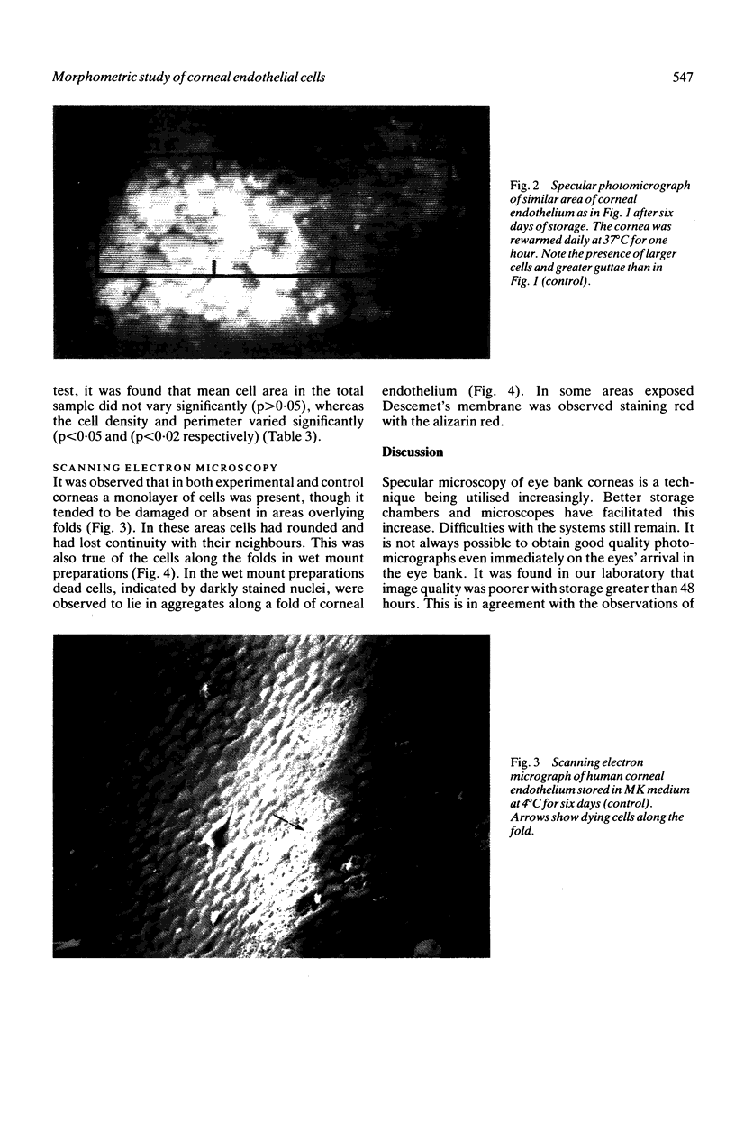
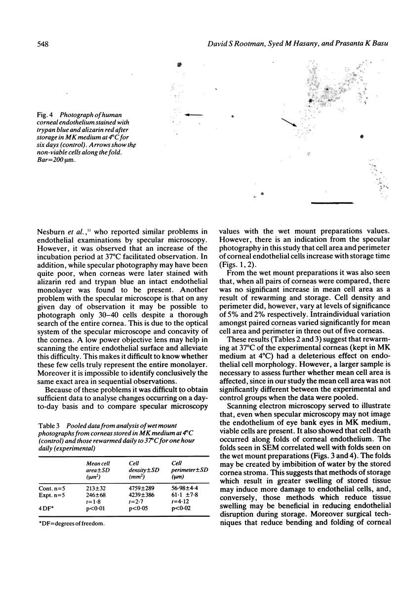
No study has yet been done to investigate the changes in endothelial cell size, perimeter, and density that may result from the warming of corneas in MK (McCarey-Kaufman) medium for specular microscopy. In the present investigation eye bank eyes were stored in MK medium at 4 degrees C and rewarmed daily for six days at 37 degrees C before specular photography of the endothelium was performed. These photographs were compared with wet mount preparations stained with trypan blue and alizarin red made from the same corneas and those stored without rewarming for six days. In addition all corneas were qualitatively analysed with the scanning electron microscope (SEM). The data from serial specular photography were insufficient to allow significant conclusions to be drawn about day to day changes in cell morphology. However, analysis of wet mount preparations revealed that cell density and perimeter varied significantly between those corneas rewarmed daily and those held in cold storage for six days. SEM studies showed an intact cell monolayer with cell loss along the folds of corneal endothelium. We therefore concluded that repeated rewarming at 37 degrees C of corneas stored in MK medium at 4 degrees has a deleterious effect on cell morphology and that folds induced by swelling of corneal tissue result in endothelial cell damage with some loss.
Full text
PDF
Images in this article
Selected References
These references are in PubMed. This may not be the complete list of references from this article.
- Basu P. K., Hasany S. M., Doane F. W., Schultes K. Can steroid reduce endothelial damage in stored corneas? Effect on cell viability and ultrastructure. Can J Ophthalmol. 1978 Jan;13(1):31–38. [PubMed] [Google Scholar]
- Bourne W. M. Examination and Photography of donor corneal endothelium. Arch Ophthalmol. 1976 Oct;94(10):1799–1800. doi: 10.1001/archopht.1976.03910040573017. [DOI] [PubMed] [Google Scholar]
- Bourne W. M., Kaufman H. E. Specular microscopy of human corneal endothelium in vivo. Am J Ophthalmol. 1976 Mar;81(3):319–323. doi: 10.1016/0002-9394(76)90247-6. [DOI] [PubMed] [Google Scholar]
- Grutzmacher R. D., Oiland D., McKillop B. R., Bunt-Milam A. H. Donor corneal endothelial striae. Am J Ophthalmol. 1986 Oct 15;102(4):508–515. doi: 10.1016/0002-9394(86)90082-6. [DOI] [PubMed] [Google Scholar]
- Hartmann C., Köditz W. Automated morphometric endothelial analysis combined with video specular microscopy. Cornea. 1984;3(3):155–167. [PubMed] [Google Scholar]
- Laing R. A., Sandstrom M. M., Leibowitz H. M. In vivo photomicrography of the corneal endothelium. Arch Ophthalmol. 1975 Feb;93(2):143–145. doi: 10.1001/archopht.1975.01010020149013. [DOI] [PubMed] [Google Scholar]
- Maurice D. M. Cellular membrane activity in the corneal endothelium of the intact eye. Experientia. 1968 Nov 15;24(11):1094–1095. doi: 10.1007/BF02147776. [DOI] [PubMed] [Google Scholar]
- Nesburn A. B., Mandelbaum S., Willey D. E., Trousdale M. D., Maguen E., Ward D. E. A specular microscopic viewing system for donor corneas. Ophthalmology. 1983 Jun;90(6):686–691. doi: 10.1016/s0161-6420(83)34513-9. [DOI] [PubMed] [Google Scholar]
- Rao G. N., Aquavella J. V., Goldberg S. H., Berk S. L. Pseudophakic bullous keratopathy. Relationship to preoperative corneal endothelial status. Ophthalmology. 1984 Oct;91(10):1135–1140. [PubMed] [Google Scholar]
- Schultz R. O., Matsuda M., Yee R. W., Edelhauser H. F., Schultz K. J. Corneal endothelial changes in type I and type II diabetes mellitus. Am J Ophthalmol. 1984 Oct 15;98(4):401–410. doi: 10.1016/0002-9394(84)90120-x. [DOI] [PubMed] [Google Scholar]
- Spence D. J., Peyman G. A. A new technique for the vital staining of the corneal endothelium. Invest Ophthalmol. 1976 Dec;15(12):1000–1002. [PubMed] [Google Scholar]
- Taylor M. J., Hunt C. J. Dual staining of corneal endothelium with trypan blue and alizarin red S: importance of pH for the dye-lake reaction. Br J Ophthalmol. 1981 Dec;65(12):815–819. doi: 10.1136/bjo.65.12.815. [DOI] [PMC free article] [PubMed] [Google Scholar]


