

Abstract
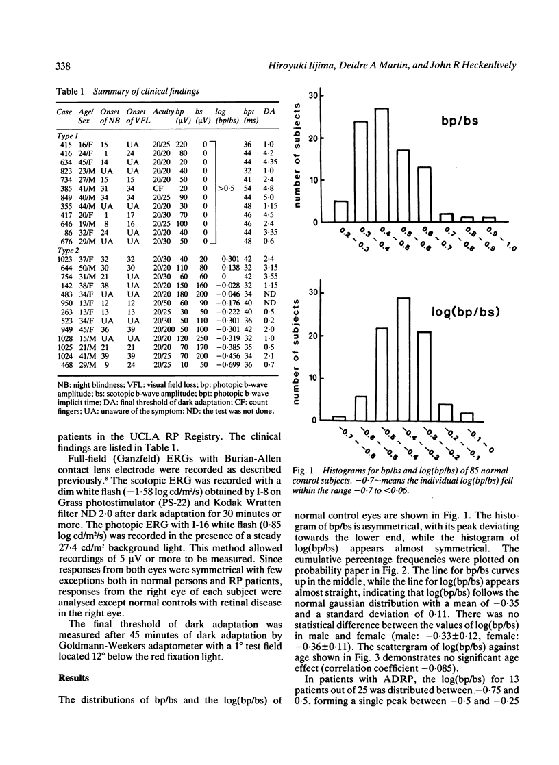
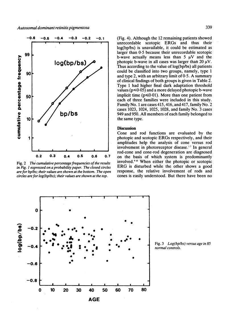
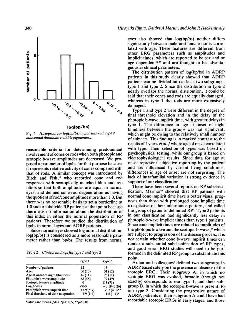
The relationship of the photopic and the scotopic b-wave amplitudes of the electroretinogram was studied in 85 normal subjects and 25 patients with autosomal dominant retinitis pigmentosa, in which one amplitude was at least 20 microvolts. The log quotient of their b-wave amplitudes--that is log of the photopic b-wave amplitude divided by the scotopic b-wave amplitude--was considered to represent the activity of cones relative to rods. The log quotient values had a normal gaussian distribution in the normal control eyes, while they formed two groups in the patients with autosomal dominant retinitis pigmentosa. In the first group (type 1), the scotopic b-wave was non-recordable while the photopic b-wave amplitude was larger than 20 microvolts in all cases, indicating that the log quotient is larger than 0.5 and that the rod system is much more severely affected than the cone system. The second group (type 2) had a log quotient smaller than 0.5 and its distribution almost overlapped the normal one, indicating more symmetrical damage in the cone and rod systems. The mean final rod threshold at 45 minutes for type 1 was significantly higher than that for type 2. The log quotient proved to be a useful index for analysing the cone and rod involvement and consequently provides a better understanding of autosomal dominant retinitis pigmentosa.
Full text
PDF
Selected References
These references are in PubMed. This may not be the complete list of references from this article.
- Arden G. B., Carter R. M., Hogg C. R., Powell D. J., Ernst W. J., Clover G. M., Lyness A. L., Quinlan M. P. Rod and cone activity in patients with dominantly inherited retinitis pigmentosa: comparisons between psychophysical and electroretinographic measurements. Br J Ophthalmol. 1983 Jul;67(7):405–418. doi: 10.1136/bjo.67.7.405. [DOI] [PMC free article] [PubMed] [Google Scholar]
- Berson E. L., Gouras P., Gunkel R. D. Progressive cone-rod degeneration. Arch Ophthalmol. 1968 Jul;80(1):68–76. doi: 10.1001/archopht.1968.00980050070010. [DOI] [PubMed] [Google Scholar]
- Birch D. G., Fish G. E. Rod ERGs in retinitis pigmentosa and cone-rod degeneration. Invest Ophthalmol Vis Sci. 1987 Jan;28(1):140–150. [PubMed] [Google Scholar]
- Birch D. G., Sandberg M. A. Dependence of cone b-wave implicit time on rod amplitude in retinitis pigmentosa. Vision Res. 1987;27(7):1105–1112. doi: 10.1016/0042-6989(87)90025-3. [DOI] [PubMed] [Google Scholar]
- Fishman G. A., Alexander K. R., Anderson R. J. Autosomal dominant retinitis pigmentosa. A method of classification. Arch Ophthalmol. 1985 Mar;103(3):366–374. doi: 10.1001/archopht.1985.01050030062023. [DOI] [PubMed] [Google Scholar]
- Heckenlively J. R. RP cone-rod degeneration. Trans Am Ophthalmol Soc. 1987;85:438–470. [PMC free article] [PubMed] [Google Scholar]
- Lyness A. L., Ernst W., Quinlan M. P., Clover G. M., Arden G. B., Carter R. M., Bird A. C., Parker J. A. A clinical, psychophysical, and electroretinographic survey of patients with autosomal dominant retinitis pigmentosa. Br J Ophthalmol. 1985 May;69(5):326–339. doi: 10.1136/bjo.69.5.326. [DOI] [PMC free article] [PubMed] [Google Scholar]
- Marmor M. F. The electroretinogram in retinitis pigmentosa. Arch Ophthalmol. 1979 Jul;97(7):1300–1304. doi: 10.1001/archopht.1979.01020020042009. [DOI] [PubMed] [Google Scholar]
- Massof R. W., Finkelstein D. Two forms of autosomal dominant primary retinitis pigmentosa. Doc Ophthalmol. 1981 Nov;51(4):289–346. doi: 10.1007/BF00143336. [DOI] [PubMed] [Google Scholar]
- Retinitis pigmentosa. A symposium on terminology and methods of examination. Ophthalmology. 1983 Feb;90(2):126–131. [PubMed] [Google Scholar]
- Weleber R. G. The effect of age on human cone and rod ganzfeld electroretinograms. Invest Ophthalmol Vis Sci. 1981 Mar;20(3):392–399. [PubMed] [Google Scholar]
- Wright C. E., Williams D. E., Drasdo N., Harding G. F. The influence of age on the electroretinogram and visual evoked potential. Doc Ophthalmol. 1985 Jun 30;59(4):365–384. doi: 10.1007/BF00159171. [DOI] [PubMed] [Google Scholar]