Abstract
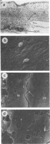
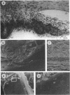
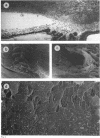
The iridocorneal angle and inner layers of the trabecular meshwork in human fetal eyes were studied by scanning electron microscopy. Tissue from 32 eyes of 17 fetuses with a gestational age from 12 to 22 weeks were investigated in order to determine the morphological changes in the cellular lining of the anterior chamber angle recess during development. The findings indicate that, although hexagonal corneal endothelial profiles extend almost to the angle apex in a few of the younger eyes examined (12-14 weeks), the lining is always perforated by a few discrete intercellular gaps (2-6 microns diameter). As development progresses it becomes clearer that the maturing meshwork is lined by uveal trabecular endothelial cells which are morphologically distinguishable from corneal endothelium. The frequency and size of the gaps between the inner uveal trabecular endothelial cells increase and are well developed by 18-20 weeks, clearly providing a route of communication between the fetal anterior chamber and the developing intercellular spaces in the primitive trabecular tissue. The implications of these observations on the 'Barkan's membrane' theory of congenital glaucoma are discussed.
Full text
PDF


Images in this article
Selected References
These references are in PubMed. This may not be the complete list of references from this article.
- ALLEN L., BURIAN H. M., BRALEY A. E. A new concept of the development of the anterior chamber angle; its relationship to developmental glaucoma and other structural anomalies. AMA Arch Ophthalmol. 1955 Jun;53(6):783–798. doi: 10.1001/archopht.1955.00930010791002. [DOI] [PubMed] [Google Scholar]
- BARKAN O. Pathogenesis of congenital glaucoma: gonioscopic and anatomic observation of the angle of the anterior chamber in the normal eye and in congenital glaucoma. Am J Ophthalmol. 1955 Jul;40(1):1–11. [PubMed] [Google Scholar]
- Barishak Y. R. The development of the angle of the anterior chamber in vertebrate eyes. Doc Ophthalmol. 1978 Aug 1;45(2):329–360. doi: 10.1007/BF00161671. [DOI] [PubMed] [Google Scholar]
- Hansson H. A., Jerndal T. Scanning electron microscopic studies on the development of the iridocorneal angle in human eyes. Invest Ophthalmol. 1971 Apr;10(4):252–265. [PubMed] [Google Scholar]
- Kupfer C. A note on the development of the anterior chamber angle. Invest Ophthalmol. 1969 Feb;8(1):69–74. [PubMed] [Google Scholar]
- MAUMENEE A. E. The pathogenesis of congenital glaucoma; a new theory. Am J Ophthalmol. 1959 Jun;47(6):827–858. doi: 10.1016/s0002-9394(14)76504-3. [DOI] [PubMed] [Google Scholar]
- McMenamin P. G. A morphological study of the inner surface of the anterior chamber angle in pre and postnatal human eyes. Curr Eye Res. 1989 Jul;8(7):727–739. doi: 10.3109/02713688909025808. [DOI] [PubMed] [Google Scholar]
- McMenamin P. G., Lee W. R. The normal anatomy of the pig tailed macaque (Macaca nemestrina) outflow apparatus with particular reference to the presence of smooth muscle. Graefes Arch Clin Exp Ophthalmol. 1982;219(5):225–232. doi: 10.1007/BF00231240. [DOI] [PubMed] [Google Scholar]
- O'Rahilly R. The prenatal development of the human eye. Exp Eye Res. 1975 Aug;21(2):93–112. doi: 10.1016/0014-4835(75)90075-5. [DOI] [PubMed] [Google Scholar]
- Pandolfi M., Astedt B. Outflow resistance in the foetal eye. Acta Ophthalmol (Copenh) 1971;49(2):344–350. doi: 10.1111/j.1755-3768.1971.tb00959.x. [DOI] [PubMed] [Google Scholar]
- Remé C., d'Epinay S. L. Periods of development of the normal human chamber angle. Doc Ophthalmol. 1981 Jul 15;51(3):241–268. doi: 10.1007/BF00143888. [DOI] [PubMed] [Google Scholar]
- Richardson T. M., Marks M. S., Ausprunk D. H., Miller M. A morphologic and morphometric analysis of the aqueous outflow system of the developing cat eye. Exp Eye Res. 1985 Jul;41(1):31–51. doi: 10.1016/0014-4835(85)90092-2. [DOI] [PubMed] [Google Scholar]
- SHAFFER R. N. Pathogenesis of congenital glaucoma; gonioscopic and microscopic anatomy. Trans Am Acad Ophthalmol Otolaryngol. 1955 May-Jun;59(3):297–308. [PubMed] [Google Scholar]
- Smelser G. K., Ozanics V. The development of the trabecular meshwork in primate eyes. Am J Ophthalmol. 1971 Jan;71(1 Pt 2):366–385. doi: 10.1016/0002-9394(71)90412-0. [DOI] [PubMed] [Google Scholar]
- Van Buskirk E. M. Clinical implications of iridocorneal angle development. Ophthalmology. 1981 Apr;88(4):361–367. doi: 10.1016/s0161-6420(81)35024-6. [DOI] [PubMed] [Google Scholar]
- Worst J. G. Congenital glaucoma. Remarks on the aspect of chamber angle, ontogenetic and pathogenetic background, and mode of action of goniotomy. Invest Ophthalmol. 1968 Apr;7(2):127–134. [PubMed] [Google Scholar]
- deLuise V. P., Anderson D. R. Primary infantile glaucoma (congenital glaucoma). Surv Ophthalmol. 1983 Jul-Aug;28(1):1–19. doi: 10.1016/0039-6257(83)90174-1. [DOI] [PubMed] [Google Scholar]


