PLOS ONE logoLink to PLOS ONE
. 2023 Aug 11;18(8):e0289892. doi: 10.1371/journal.pone.0289892

Aged gut microbiota contribute to different changes in antioxidant defense in the heart and liver after transfer to germ-free mice

Yang Hong 1, Han Dong 1, Jing Zhou 1, Ya Luo 1, Ming-Ming Yuan 1, Jia-Fei Zhan 1, Yang-Lu Liu 1, Jie-Ying Xia 1, Lei Zhang 1,*
Editor: David M Ojcius2
PMCID: PMC10420372  PMID: 37566569

Abstract

Age-associated impairment in antioxidant defense is an important cause of oxidative stress, and elderly individuals are usually associated with gut microbiota (GM) changes. Studies have suggested a potential relationship between the GM and changes in antioxidant defense in aging animals. Direct evidence regarding the impact of aging-associated shifts in GM on the antioxidant defense is lacking. The heart is a kind of postmitotic tissue, which is more prone to oxidative stress than the liver (mitotic tissue). To test and compare the influence of an aged GM on antioxidant defense changes in the heart and liver of the host, in this study, GM from young adolescent (5 weeks) or aged (20 months) mice was transferred to young adolescent (5 weeks) germ-free (GF) mice (N = 5 per group) by fecal microbiota transplantation (FMT). Four weeks after the first FMT was performed, fecal samples were collected for 16S rRNA sequencing. Blood, heart and liver samples were harvested for oxidative stress marker and antioxidant defense analysis. The results showed that mice that received young or aged microbiota showed clear differences in GM composition and diversity. Mice that received aged microbiota had a lower ratio of Bacteroidetes/Firmicutes in GM at the phylum level and an increased relative abundance of four GM genera: Akkermansia, Dubosiella, Alistipes and Rikenellaceae_RC9_gut_group. In addition, GM α-diversity scores based on the Shannon index and Simpson index were significantly higher in aged GM-treated mice. Oxidative stress marker and antioxidant defense tests showed that FMT from aged donors did not have a significant influence on malondialdehyde content in serum, heart and liver. However, the capacity of anti-hydroxyl radicals in the heart and liver, as well as the capacity of anti-superoxide anions in the liver, were significantly increased in mice with aged microbiota. FMT from aged donors increased the activities of Cu/Zn superoxide SOD (Cu/Zn-SOD), catalase (CAT) and glutathione-S-transferase in the heart, as well as the activity of Cu/Zn-SOD in the liver. Positive correlations were found between Cu/Zn-SOD activity and radical scavenging capacities. On the other hand, glutathione reductase activity and glutathione content in the liver were decreased in mice that received aged GM. These findings suggest that aged GM transplantation from hosts is sufficient to influence the antioxidant defense system of young adolescent recipients in an organ-dependent manner, which highlights the importance of the GM in the aging process of the host.

Introduction

Over recent decades, life expectancy has significantly increased globally. However, this increase has not led to a corresponding improvement in health outcomes, which is represented by an increase in numerous age-related diseases. It is well accepted that oxidative stress is an important contributor to aging, and a state of chronic oxidative stress characterizes older organisms and participates pivotally in the development of aging-related diseases [1]. Oxidative stress is the result of an elevation of reactive oxygen species (ROS), which are produced mainly in mitochondria as byproducts of normal cell respiration [1]. To remove excessive ROS, aerobic organisms have developed antioxidant defenses, which mainly rely on antioxidants, such as antioxidant enzymes and nonenzymatic glutathione (GSH) [1]. However, this critical defense has been found to decline with increasing aging, which is regarded as one major cause of the generation of oxidative stress in elderly individuals [1]. Hence, factors that undermine the antioxidant defense in elderly individuals need to be identified, which would be highly beneficial in developing interventions to promote healthy aging.

The human gut microbiota (GM), comprised of approximately 1014 microorganisms, is a complex and dynamic population that resides in the gastrointestinal tract [2]. To date, the interactions between the GM and host aging are a hotspot of research interest and have shed new light on the involvement of the GM in aging-associated decreases in antioxidant defense [3]. Generally, the composition of GM changes with aging, which leads to great GM differences between young and elderly subjects [4]. Recently, using a D-galactose-induced mouse aging model, studies showed that the alleviation of oxidative stress in aging bodies was associated with restoration of GM structure [3, 57]. In addition, Brunt et al. (2019) reported that suppression of the GM by antibiotics ameliorated age-related vascular oxidative stress and antioxidant enzyme decreases in mice [8]. To date, there are few studies concerning the direct effect of an aged GM on the antioxidant defense of the host. Colonizing young pseudo germ-free (GF) rats with the GM of aged rats by fecal microbiota transplantation (FMT), Li et al. (2020) reported that aged GM induced superoxide dismutase (SOD) enzyme activity decrease and malondialdehyde (MDA) elevation in the serum, hippocampus and medial prefrontal cortex [9]. Based on the findings above, an aged GM might modulate antioxidant defense, thus inducing oxidative stress in the host. If this is true, it would provide important evidence to explore strategies targeting GM-related alleviation in age-related oxidative stress.

As the organs requiring a high-energy source generated from mitochondrial respiration, the heart and liver are particularly prone to suffer from oxidative stress [1]. Indeed, a substantial percentage of morbidity and mortality in the elderly is attributable to oxidative stress-associated heart and liver diseases, which also cause tremendous clinical and economic burden outcomes [10, 11]. On the other hand, studies have shown that there are distinct variations in vulnerability to oxidative stress among different organs and tissues [12]. Postmitotic tissues, such as the heart, are more prone to oxidative stress than mitotic tissues (e.g., liver) due to their reduced ability to upregulate antioxidant defenses and/or repair accumulated oxidative damage [12]. Compared with the liver, a greater increase in oxidative stress has been found in the heart of aging rats [13]. We speculated that an aged GM might generate greater antioxidant defense impairment, thus leading to a higher level of oxidative stress in the heart than in the liver. To test this hypothesis, in this study, we conducted FMT from aged and young adolescent donor mice into young adolescent GF mice. Then, the impact of the aged GM on oxidative stress and antioxidant defense in the heart and liver of recipient mice was explored. FMT is the delivery of a donor microbiota to a recipient in the form of a purified fecal microbiota or a stool suspension [14]. Combined with the use of GF animals, FMT has become one of the most common techniques to analyze the effect of different types of GM on the physiological state of hosts [14]. As mentioned above, the present study aimed to explore the influences of an aged GM on the antioxidant defense of the host.

Materials and methods

All animal experiments were carried out according to ethical policies and procedures approved by the Animal Care Advisory Committee of the Sichuan Academy of Chinese Medicine Sciences (SYLL (2022)-047).

Mice

C57BL/6-GF mice (male; 4 weeks) were purchased from the Third Military Medical University (Chongqing, China). Upon arrival, the mice were transferred into flexible film isolators in the gnotobiotic facility at the Sichuan Academy of Chinese Medicine Sciences. Fecal samples were collected from each mouse to ensure the absence of bacteria in the gut according to Lee et al. (2020) [15]. After one week of acclimation, mice were exported from flexible film isolators into sterile individually ventilated cages (IVCs) for fecal transplantation [1517]. We included only males in this study for the following reasons: (1) There are differences in the GM and antioxidant response between males and females, and the gender effect is not the subject of our research [18, 19]; (2) according to the animal welfare guidelines of our institution, a single sex is suggested for a single study.

Fecal microbiota transplantation (FMT)

Donors for FMT were specific pathogen-free C57BL/6 male mice (5 weeks & 20 months; N = 10 per group). Young adolescent recipients were GF C57BL/6 male mice aged 5 weeks (N = 5 per group). Fresh fecal samples were collected from the donors, and all fecal samples from the same group were mixed, homogenized in ice-cold PBS (120 mg feces/1 ml) and centrifuged at 800 × g for 3 min at 4 °C [15]. Then, 100 μL of this supernatant was given to recipient mice by oral gavage on Days 1, 4, 7, 14, and 21 [9, 15, 20]. Recipients were individually housed in IVCs for 4 weeks [21]. The mice gavaged with fecal supernatant from aged C57BL/6 mice were referred to as the aged-FMT group, and the other group was referred to as the young-FMT group.

16S rRNA sequencing

Feces were collected from all recipient mice at the end of the experiment and immediately frozen at -80°C. DNA isolation and sequencing, as well as data analysis, were carried out at Novogene Co., Ltd. (China). Approximately 100 mg of fecal samples were used for total genome DNA extraction using TIANGEN kits. By performing 1% agarose gel electrophoresis, DNA concentration and purity were assessed.

The extracted DNA fragments were subjected to Illumina MiSeq deep sequencing. Briefly, amplification of the V4 hypervariable region of the 16S rRNA gene was accomplished by using the 515F (5’ GTGCCAGCMGCCGCGGTAA 3’) and 806R (5’ GGACTACVSGGGTATCTAAT 3’) set of primers with a barcode. PCR was performed using Phusion® High-FIdelity PCR Master Mix (New England Biolabs, Ipswich, MA) with GC buffer with the reaction conditions described by Frolinger et al. [22]. Then, the PCR products were run on agarose gel electrophoresis, purified using the TIANGEN Gel Extraction kit (TIANGEN, China) and sequenced by the Illumina NovaSeq6000 platform (Novogene Bioinformatics Technology Co., Ltd., Tianjin, China). According to the manufacturer’s instructions, sequencing library generation was accomplished by using the NEBNext Ultra DNA Library Prep Kit for Illumina (Illumina, USA), and the index codes were assigned. The Illumina paired-end reads were overlapped and merged using Flash ver. 1.2.7 after cutting off the primer sequence and the barcode. Then, high-quality clean tags were obtained according to Bokulich et al. [23]. Subsequently, chimera filtering was undertaken using VSEARCH. Chimeric sequences were removed according to Hess et al. [24] to obtain effective tags.

Based on 97% sequence similarity, the generation of the operational taxonomic unit (OTU) table was achieved by using UPARSE v.7.0.1001 software [25]. Then, a representative sequence was chosen for each OTU, annotating the taxonomic information of that unit. Subsequently, all OTUs were analyzed for abundance and diversity. The analysis of alpha (α) diversity was calculated using Mothur. A UPGMA tree was constructed on the basis of the UniFrac distance calculated by using QIIME (Version 1.9.1).

Reduction‒oxidation (Redox) state analysis

Mice were sacrificed at the end of the experiment. Mice were anesthetized with isoflurane inhalation, bled and then sacrificed by cervical dislocation. Then, the blood, heart and liver were collected. Serum was prepared by centrifuging the blood samples at 3 000 ×g for 15 min. All the samples were immediately frozen and stored at -80 °C until analysis. Heart and liver samples were homogenized on ice in 10 volumes (w/v) of ice-cold physiological saline and centrifuged at 6000 g for 20 min at 4 °C, and then supernatants were collected for analysis. The MDA content in the serum, heart and liver was investigated using the thiobarbituric acid reaction, which was accomplished by using a test kit (Nanjing Jiancheng Bioengineer Institute, Nanjing, China). Briefly, serum or supernatant samples were mixed with trichloroacetic acid and centrifuged. Subsequently, thiobarbituric acid was added to the supernatant. The mixture underwent a 40-minute heating process in water at 95 °C, resulting in the formation of a red complex between MDA and thiobarbituric acid, the absorption of which was measured by spectrophotometry at 532 nm. The Fenton reaction (Fe2++H2O2−Fe3++OH-+OH) was used to evaluate the anti-hydroxyl radical (AHR) capacity by using a test kit (Nanjing Jiancheng Bioengineer Institute, Nanjing, China), in which hydroxyl radicals (OH) are generated. Upon addition of the electron acceptor, a coloration reaction (absorbance at 550 nm) is induced by utilizing the Griess reagent. The degree of coloration is directly proportional to the concentration of hydroxyl radicals. The anti-superoxide anion (ASA) capacity was analyzed following the instructions of the test kit (Nanjing Jiancheng Bioengineer Institute, Nanjing, China). In brief, superoxide radicals (O2•−) were generated through the action of xanthine and xanthine oxidase. When an electron acceptor is introduced, a coloration reaction is initiated using the Griess reagent. The intensity of the coloration (absorbance at 550 nm) is directly proportional to the amount of superoxide anion present in the reaction. In addition, the activities of antioxidant enzymes, including Cu/Zn superoxide dismutase (Cu/Zn-SOD) and Mn superoxide dismutase (Mn-SOD), were assessed as described by Jiang et al. [26]. The reaction mixture consisted of 30 μL of tissue sample supernatant, 1.08 mmol L-1 diethylenetriaminepentaacetic acid, 50 mmol L-1 phosphate buffer (pH 7–8), 0.16 mmol L-1 xanthine solution, and 0.06 mmol L-1 nitro blue tetrazolium. Following the introduction of 0.19 U mL-1 xanthine oxidase, an alteration in absorbance at 550 nm was observed. To distinguish between SOD isoforms, the responsiveness of Cu/Zn-SOD to cyanide was employed, while Mn-SOD remained unaffected. The decomposition of hydrogen peroxide was determined to analyze catalase (CAT) activity [26]. The assay mixture comprised a total volume of 1 mL containing 100 mmol L-1 KPO4 buffer (pH 7.0), 10 mmol L-1 H2O2, and 50 μL of tissue supernatant. The reduction of H2O2 was analyzed by measuring the absorbance at 240 nm. Glutathione peroxidase (GPX) activity was detected according to the method of Jiang et al. (2012). In brief, the reaction mixture consisted of 15 μL of tissue supernatant, 40 μL of 0.25 mmol L-1 H2O2, 0.5 mmol L-1 GSH, 10 mmol L-1 sodium phosphate buffer (pH 7.0) and 1.25 mmol L-1 NaN3 in a total volume of 1 mL. After 3-min intervals, 0.5 mL of dithiobisnitrobenzoic acid was introduced. The formation of a yellow product, resulting from the reaction between GSH and dithiobisnitrobenzoic acid, was observed and measured at 412 nm. The activity of glutathione reductase (GR) was tested by measuring the rate of NADPH oxidation [27], and the main procedure was as follows: The initial reaction mixture contained 1 mM EDTA, 0.1 mM NADPH, 100 mM NaCI/Pi (pH 7.0), and 100 μL of tissue sample supernatant in a final volume of 1 mL. In this assay, the oxidation of NADPH, regardless of GR, was assessed by measuring the reduction in absorbance at 340 nm over a period of 60 seconds. Subsequently, 50 μL of 20 mM GSSG was added, and the reduction in absorbance at 340 nm was observed for an additional 120 seconds. Glutathione-S-transferase (GST) activity was analyzed according to the method of Lushchak et al. [28]. The reaction mixture consisted of 5 mmol L-1 GSH, 50 mmol L-1 KPi buffer (pH 7.0), 1 mmol L-1 1-chloro-2,4-dinitrobenzene (CDNB), 0.5 mmol L-1 EDTA, and 40 μL of tissue sample supernatant. The formation of an adduct between GSH and CDNB was observed and measured at 340 nm. The determination of GSH content was accomplished by the formation of 5-thio-2-nitrobenzoate, which was subsequently measured spectrophotometrically at a wavelength of 412 nm [29].

Statistical analysis

The redox state parameter results were expressed as the means±SDs. The differences between the two groups were analyzed with a two-tailed t test. Differences in the α-diversity of the GM between the two groups were analyzed with the Wilcox test. The correlations between antioxidant enzyme activity and radical scavenging capacity were analyzed using Pearson’s correlation analysis. P<0.05 was considered significant, and P<0.01 was considered very significant.

Results

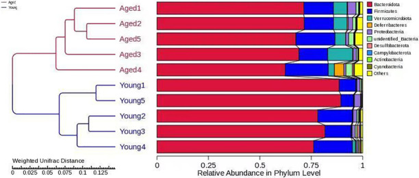
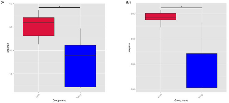
A total of 86,487 tags were obtained from 10 samples, which were clustered into 1051 OTUs. To determine the extent of similarity in gut microbial communities between the groups, UniFrac distance was calculated. The UPGMA tree built using weighted UniFrac distances revealed clustering of aged-FMT group fecal samples from young-FMT group fecal samples (Fig 1). The phylum composition of GM in each group is shown in Fig 2A. In agreement with previous results, Firmicutes and Bacteroidetes phyla covered the vast majority of the microbiota [30], and the ratio of Bacteroidetes/Firmicutes was decreased in the aged-FMT group. The GM composition was also analyzed at the genus level. The relative abundances of the genera in the two groups are shown in Fig 2B. Among all the top 10 genera, the relative abundance of the genera Akkermansia, Dubosiella, Alistipes and Rikenellaceae_RC9_gut_group was increased in the aged-FMT group. For GM diversity, α-diversity scores based on the Shannon index and Simpson index were significantly higher in the aged-FMT group (Fig 3).

Fig 1. UPGMA clustering analysis of gut microbiota based on weighted UniFrac distance.

Fig 1

The left side displays the structure of the clustering tree, while the right side shows the relative abundance of different phyla.

Fig 2. Gut microbiota composition at the level of phylum and genus.

Fig 2

(A) Relative abundance of gut microbiota at the phylum level. (B) Relative abundance of gut microbiota at the genus level. Each bar represents the relative abundance of a bacterial taxon, with the top 10 most abundant taxa being displayed.

Fig 3. α-diversity of gut microbiota.

Fig 3

(A) Shannon index. (B) Simpson index. The bottom and top boundaries of each box show the 25th and 75th percentiles, respectively. The line within each box represents the median. Ends of the whiskers indicate the maximum and minimum values. * indicates P<0.05.

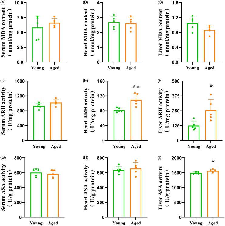
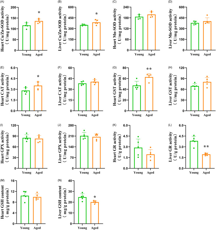
The MDA content in serum, heart and liver was not significantly different between the groups (Fig 4). As shown in Fig 4, the capacity of AHR in the heart and liver, as well as the capacity of ASA in the liver, were significantly increased in the aged-FMT group. In contrast, the capacity of AHR in serum, as well as the capacity of ASA in serum and heart, did not significantly differ between the groups. Fig 5 reveals the antioxidant indicators. FMT from aged donors increased the activities of Cu/Zn-SOD, CAT and GST in the heart, as well as the activity of Cu/Zn-SOD in the liver of the recipient mice. However, the activity of GR in the liver was significantly decreased in the aged-FMT group. In addition, the activities of Mn-SOD, GPX and GR in the heart, as well as the activities of Mn-SOD, CAT, GPX and GST in the liver, were not significantly different between the groups. FMT from aged donors did not have a significant effect on GSH content in the heart but decreased GSH content in the liver of the recipient mice (Fig 5). To uncover the potential relationship between the increased antioxidant enzyme activities and the increased radical scavenging capacities, a Pearson correlation analysis was conducted. The results only showed positive correlations between Cu/Zn-SOD activity and radical scavenging capacities (Table 1).

Fig 4. Levels of MDA, AHR and ASA in the serum, heart and liver of the recipient mice.

Fig 4

The bars represent the mean±SD (n = 5). * indicates P<0.05, and ** represents P<0.01. (A) Serum MDA content = serum malondialdehyde content; (B) Heart MDA content = heart malondialdehyde content; (C) Liver MDA content = liver malondialdehyde content; (D) Serum AHR = serum anti-hydroxyl radical capacity; (E) Heart AHR = heart anti-hydroxyl radical capacity; (F) Liver AHR = liver anti-hydroxyl radical capacity; (G) Serum ASA = serum anti-superoxide anion capacity; (H) Heart ASA = heart anti-superoxide anion capacity; (I) Liver ASA = liver anti-superoxide anion capacity.

Fig 5. Activities of antioxidant enzymes and GSH content in the heart and liver of recipient mice.

Fig 5

The bars represent the mean±SD (n = 5). * indicates P<0.05, and ** represents P<0.01. (A) Heart Cu/Zn-SOD activity = heart Cu/Zn superoxide dismutase activity; (B) Liver Cu/Zn-SOD activity = liver Cu/Zn superoxide dismutase activity; (C) Heart Mn-SOD activity = heart Mn superoxide dismutase activity; (D) Liver Mn-SOD activity = liver Mn superoxide dismutase activity; (E) Heart CAT activity = heart catalase activity; (F) Liver CAT activity = liver catalase activity; (G) Heart GST activity = heart glutathione-S-transferase activity; (H) Liver GST activity = liver glutathione-S-transferase activity; (I) Heart GPX activity = heart glutathione peroxidase activity; (J) Liver GPX activity = liver glutathione peroxidase activity; (K) Heart GR activity = heart glutathione reductase activity; (L) Liver GR activity = liver glutathione reductase activity; (M) Heart GSH content = heart glutathione content; (N) Liver GSH content = liver glutathione content.

Table 1. Correlations between Cu/Zn-SOD activity and radical scavenging capacity.

Antioxidant parameters Correlation Radical scavenging capacity
Heart Cu/Zn-SOD activity (U/mg protein) + Heart AHR (r = 0.693 p = 0.026)
Liver Cu/Zn-SOD activity (U/mg protein) + Liver AHR (r = 0.867 p = 0.01)
Liver Cu/Zn-SOD activity (U/mg protein) + Liver ASA (r = 0.886 p = 0.01)

Cu/Zn-SOD: Cu/Zn superoxide dismutase; AHR: anti-hydroxyl radical capacity; ASA: anti-superoxide anion capacity; +: positive correlation; r: Pearson’s correlation coefficient

Discussion

In recent years, the close relationship between the GM and host aging has become evident. However, the influence of age-associated shifts in the GM on host antioxidant defense remains to be determined. Antioxidant defense impairment is a major contributor to age-related oxidative stress, which is associated with various diseases in elderly individuals [1]. Here, we attempted to uncover the alterations in oxidative stress and antioxidant defense in the heart and liver of young adolescent GF mice that received GM from either young or aged adolescent donor mice. First, the GM profiles between the young and aged recipients were compared to confirm successful GM transfer. After FMT for 4 weeks, significant differences existed between the GM of the aged-FMT group and the young-FMT group, as revealed by the UPGMA clustering result. Our study showed that both the composition and diversity of the GM differed between the two groups. Firmicutes and Bacteroides are two dominant gut bacteria at the phylum level. In this study, a decreased ratio of Firmicutes to Bacteroides (F/B) was found in the aged-FMT group, which was consistent with a previous result [9]. Moreover, intergroup GM variability was also observed at the genus level. Compared with the young-FMT group, the relative abundance of the genera Akkermansia, Dubosiella, Alistipes and Rikenellaceae_RC9_gut_group was increased in the aged-FMT group. Among those four GM genera, Alistipes and Rikenellaceae_RC9_gut_group were found to be positively associated with age and some diseases [31, 32]. However, Akkermansia and Dubosiella were indicated to act as healthy microbiota in the aging process [33, 34]. Regarding diversity, the GM of mice in the aged-FMT group had higher Shannon and Simpson indices, reflecting higher species richness and evenness [33]. This result was contrary to our expectations, as a decrease in GM diversity with aging has been widely reported [3537]. However, some studies have also shown an increase in GM diversity with aging in long-living people [38]. Together with the increased Akkermansia and Dubosiella in the aged-FMT group, we speculated that the aged donor mice used in this study might represent a healthy aging group.

Oxidative stress is a major factor involved in the pathogenesis of age-associated diseases. With increasing age, oxidative stress tends to increase in a GM-associated manner [57]. The index typically used to assess the degree of oxidative stress is the product of lipid peroxidation, named MDA [1]. In this study, FMT from aged donors failed to induce MDA elevation in the serum, heart and liver of the recipients, and all the MDA contents were similar to those observed in mice under normal physiological conditions [39, 40], which indicates that there is no obvious oxidative stress in the recipient mice. To date, information regarding the influence of aged GM on oxidative stress in GF recipient mice is limited. Li et al. [9] reported that an aged GM induced elevation of serum and brain MDA content in young pseudo-GF rats. Such discrepancies might be correlated with differences in GM characteristics. At the genus level, aged FMT induced a totally different microbiota composition change in pseudo-GF rats compared with our study. In addition, the results of GM diversity change were also completely opposite. As healthy and unhealthy aging subjects have great differences in GM and physiological characteristics, our study, associated with the report of Li et al. [9], highlights the importance of considering GM characteristics in studying topics of aging-related oxidative status. Despite the insignificant difference in MDA content between the recipients, our further test showed some changes in the antioxidant system induced by aged-FMT. OH and O2•− are two free radicals that are highly involved in oxidative stress [1]. OH is considered the most reactive oxygen radical in biological systems and can react rapidly with many molecules in the cell, including proteins, lipids, DNA, and others [1]. O2•− can cause great oxidative damage within the cell due to its relatively long half-life [1]. Two indices, namely, antihydroxyl radical (AHR) capacity and antisuperoxide anion (ASA) capacity, are used to represent the total scavenging ability against OH and O2•−, respectively. In this study, we observed different responses of these two capacities to aged GM between the heart and liver, which was a proof that the heart has a reduced ability to upregulate antioxidant defense compared with the liver. Transplantation of aged-GM did not have a significant influence on ASA capacity in the heart. However, AHR capacity in the heart, as well as ASA and AHR capacity in the liver, were significantly higher in the aged-FMT group. As the MDA content was similar between the young and aged-FMT groups, the enhanced free radical scavenging ability indicates a response against increased ROS production in the aged-FMT group [41]. Studies have shown that some gut bacterial metabolites, such as small formylated peptides and lipopolysaccharides, could contribute to host ROS production [42, 43], and those metabolites were found to increase with increasing host age [44]. However, further studies are needed to test the influence of aged GM on host ROS production.

In this study, the levels of antioxidant compounds were analyzed to further determine the possible mechanism by which aged-FMT enhanced the free radical scavenging abilities in young adolescent GF recipients. Endogenous antioxidant enzymes provide the first line of cellular defense against toxic free radicals [1]. SOD, a metalloenzyme, is the first enzyme involved in responding to oxygen free radicals [1]. On the basis of the type of metal ion required as cofactor, various forms of SOD exist. Cu/Zn-SOD is widely distributed in the cytoplasm of eukaryotes, while Mn-SOD is present in mitochondria [1]. In this study, the activity of Mn-SOD was not significantly different between the groups. However, the activity of Cu/Zn-SOD in the heart and liver was improved in the aged-FMT group. CAT is essential for the detoxification of hydroxyl radicals [1]. In this study, CAT activity in the heart was higher in the aged-FMT group. Furthermore, glutathione-dependent enzymes are also able to protect cells against free radical-induced injury [1]. Our study showed that FMT from aged donors enhanced the activity of GST in the heart but had no significant effect on the activities of GPX and GR in the heart or the activities of GPX and GST in the liver. On the other hand, FMT from aged donors decreased liver GR activity in recipient mice. It is well known that GR is capable of catalyzing the reduction of oxidized GSH [1]. Similar to the GR activity, the GSH content in the liver was lower in the aged-FMT group, which might be partly attributed to the decrease in GR activity. Among all antioxidant compounds mentioned above, Pearson’s correlation analysis uncovered positive correlations between the Cu/Zn-SOD activity and the radical scavenging capacities, which indicated that the increased radical scavenging capacities were partially attributed to the increased Cu/Zn-SOD activity. Liu et al. (2022) reported that Dubosiella newyorkensis, a strain of Dubosiella, possessed a strong effect of increasing SOD in aged mice [34]. Thus, in this study, the increased Dubosiella might play an important role in shaping SOD activity, as well as radical scavenging capacity characteristics in the heart and liver. Further study is needed to test this hypothesis. Comparing the antioxidant defense changes between the heart and liver, it is evident that an aged GM had varying effects on antioxidant defense in these organs. It has been widely reported that antioxidant defense follows a mixed pattern of age-related changes, and those changes are markedly variable depending on the tissues and organs examined [18, 4547]. Our study indicated that GM is involved in these tissue- and organ-dependent antioxidant defense changes during aging.

In conclusion, the present study highlights the importance of the GM in aging-related antioxidant defense changes in the host. Transplantation of an aged GM into young adolescent GF mice did not have an impact on lipid peroxidation but enhanced free radical scavenging abilities in the heart and liver. The aged GM had different influences on antioxidant enzyme activities and GSH content between the heart and liver, which suggests an organ-dependent effect of the aged GM on host antioxidant defense. Among all the significantly changed antioxidant compounds, Cu/Zn-SOD activity was a factor that contributed to the enhanced radical scavenging abilities in aged GM recipients. Our study identified significant differences in four types of GM genera (Akkermansia, Dubosiella, Alistipes and Rikenellaceae_RC9_gut_group) between the two groups. Further work is warranted to investigate the detailed mechanisms by which the GM affects the antioxidant response in elderly individuals.

Data Availability

All data generated or analyzed during this study are included in this article.

Funding Statement

This research was financially supported by the Sichuan Provincial Science and Technology Plan Project (2022YFS0432), Basic Research Project of Sichuan Province (2022JDKY0013), Scientific and Technical Research Project of Sichuan Provincial Administration of TCM (2023MS485). The funders had no role in study design, data collection and analysis, decision to publish, or preparation of the manuscript.

References

  • 1.Liguori I, Russo G, Curcio F, Bulli G, Aran L, Della-Morte D, et al. Oxidative stress, aging, and diseases. Clin Interv Aging. 2018;13: 757–72. doi: 10.2147/CIA.S158513 [DOI] [PMC free article] [PubMed] [Google Scholar]
  • 2.DeJong EN, Surette MG, Bowdish DME. The gut microbiota and unhealthy aging: disentangling cause from consequence. Cell Host Microbe. 2020;28: 180–9. doi: 10.1016/j.chom.2020.07.013 [DOI] [PubMed] [Google Scholar]
  • 3.Zhang J, Zhao X, Jiang Y, Zhao W, Guo T, Cao Y, et al. Antioxidant status and gut microbiota change in an aging mouse model as influenced by exopolysaccharide produced by Lactobacillus plantarum YW11 isolated from Tibetan kefir. J Dairy Sci. 2017;100: 6025–41. doi: 10.3168/jds.2016-12480 [DOI] [PubMed] [Google Scholar]
  • 4.Maynard C, Weinkove D. The gut microbiota and ageing. Subcell Biochem. 2018;90: 351–71. doi: 10.1007/978-981-13-2835-0_12 [DOI] [PubMed] [Google Scholar]
  • 5.Liu XL, Zhao YC, Zhu HY, Wu M, Zheng YN, Yang M, et al. Taxifolin retards the D-galactose-induced aging process through inhibiting Nrf2-mediated oxidative stress and regulating the gut microbiota in mice. Food Funct. 2021;12: 12142–58. doi: 10.1039/d1fo01349a [DOI] [PubMed] [Google Scholar]
  • 6.Chen H, Dong L, Chen X, Ding C, Hao M, Peng X, et al. Anti-aging effect of phlorizin on D-galactose-induced aging in mice through antioxidant and anti-inflammatory activity, prevention of apoptosis, and regulation of the gut microbiota. Exp Gerontol. 2022;163: 111769. doi: 10.1016/j.exger.2022.111769 [DOI] [PubMed] [Google Scholar]
  • 7.Guo X, Dong Z, Li Q, Wan D, Zhong J, Dongzhi D, et al. Flavonoids from Rhododendron nivale Hook. f delay aging via modulation of gut microbiota and glutathione metabolism. Phytomedicine. 2022;104: 154270. doi: 10.1016/j.phymed.2022.154270 [DOI] [PubMed] [Google Scholar]
  • 8.Brunt VE, Gioscia-Ryan RA, Richey JJ, Zigler MC, Cuevas LM, Gonzalez A, et al. Suppression of the gut microbiome ameliorates age-related arterial dysfunction and oxidative stress in mice. J Physiol. 2019;597: 2361–78. doi: 10.1113/JP277336 [DOI] [PMC free article] [PubMed] [Google Scholar]
  • 9.Li Y, Ning L, Yin Y, Wang R, Zhang Z, Hao L, et al. Age-related shifts in gut microbiota contribute to cognitive decline in aged rats. Aging (Albany NY). 2020;12: 7801–17. doi: 10.18632/aging.103093 [DOI] [PMC free article] [PubMed] [Google Scholar]
  • 10.Kim IH, Kisseleva T, Brenner DA. Aging and liver disease. Curr Opin Gastroenterol. 2015;31: 184–91. doi: 10.1097/MOG.0000000000000176 [DOI] [PMC free article] [PubMed] [Google Scholar]
  • 11.Ikeda Y, Sciarretta S, Nagarajan N, Rubattu S, Volpe M, Frati G, et al. New insights into the role of mitochondrial dynamics and autophagy during oxidative stress and aging in the heart. Oxid Med Cell Longev. 2014;2014: 210934. doi: 10.1155/2014/210934 [DOI] [PMC free article] [PubMed] [Google Scholar]
  • 12.Sawada M, Sester U, Carlson JC. Superoxide radical formation and associated biochemical alterations in the plasma membrane of brain, heart, and liver during the lifetime of the rat. J Cell Biochem. 1992;48: 296–304. doi: 10.1002/jcb.240480310 [DOI] [PubMed] [Google Scholar]
  • 13.Ochoa JJ, Quiles JL, Ibáñez S, Martínez E, López-Frías M, Huertas JR, et al. Aging-related oxidative stress depends on dietary lipid source in rat postmitotic tissues. J Bioenerg Biomembr. 2003;35: 267–75. doi: 10.1023/a:1024615816839 [DOI] [PubMed] [Google Scholar]
  • 14.Burz SD, Abraham AL, Fonseca F, David O, Chapron A, Béguet-Crespel F, et al. A guide for ex vivo handling and storage of stool samples intended for fecal microbiota transplantation. Sci Rep. 2019;9: 8897. doi: 10.1038/s41598-019-45173-4 [DOI] [PMC free article] [PubMed] [Google Scholar]
  • 15.Lee J, Venna VR, Durgan DJ, Shi H, Hudobenko J, Putluri N, et al. Young versus aged microbiota transplants to germ-free mice: Increased short-chain fatty acids and improved cognitive performance. Gut Microbes. 2020;12: 1–14. doi: 10.1080/19490976.2020.1814107 [DOI] [PMC free article] [PubMed] [Google Scholar]
  • 16.Lange ME, Uwiera RRE, Inglis GD. Housing gnotobiotic mice in conventional animal facilities. Curr Protoc Mouse Biol. 2019;9: e59. doi: 10.1002/cpmo.59 [DOI] [PubMed] [Google Scholar]
  • 17.Studer N, Desharnais L, Beutler M, Brugiroux S, Terrazos MA, Menin L, et al. Functional intestinal bile acid 7α-dehydroxylation by Clostridium scindens associated with protection from Clostridium difficile infection in a gnotobiotic mouse model. Front Cell Infect Microbiol. 2016;6: 191. doi: 10.3389/fcimb.2016.00191 [DOI] [PMC free article] [PubMed] [Google Scholar]
  • 18.Carrillo MC, Kanai S, Sato Y, Kitani K. Age-related changes in antioxidant enzyme activities are region and organ, as well as sex, selective in the rat. Mech Ageing Dev. 1992;65: 187–98. doi: 10.1016/0047-6374(92)90035-c [DOI] [PubMed] [Google Scholar]
  • 19.Yoon K, Kim N. Roles of sex hormones and gender in the gut microbiota. J Neurogastroenterol Motil. 2021;27: 314–25. doi: 10.5056/jnm20208 [DOI] [PMC free article] [PubMed] [Google Scholar]
  • 20.Li X, Khan I, Xia W, Huang G, Liu L, Law BYK, et al. Icariin enhances youth-like features by attenuating the declined gut microbiota in the aged mice. Pharmacol Res. 2021;168: 105587. doi: 10.1016/j.phrs.2021.105587 [DOI] [PubMed] [Google Scholar]
  • 21.Fransen F, van Beek AA, Borghuis T, Aidy SE, Hugenholtz F, van der Gaast-de Jongh C, et al. Aged gut microbiota contributes to systemical inflammaging after transfer to germ-free mice. Front Immunol. 2017;8: 1385. doi: 10.3389/fimmu.2017.01385 [DOI] [PMC free article] [PubMed] [Google Scholar]
  • 22.Frolinger T, Sims S, Smith C, Wang J, Cheng H, Faith J, et al. The gut microbiota composition affects dietary polyphenols-mediated cognitive resilience in mice by modulating the bioavailability of phenolic acids. Sci Rep. 2019;9: 3546. doi: 10.1038/s41598-019-39994-6 [DOI] [PMC free article] [PubMed] [Google Scholar]
  • 23.Bokulich NA, Subramanian S, Faith JJ, Gevers D, Gordon JI, Knight R, et al. Quality-filtering vastly improves diversity estimates from Illumina amplicon sequencing. Nat Methods. 2013;10: 57–9. doi: 10.1038/nmeth.2276 [DOI] [PMC free article] [PubMed] [Google Scholar]
  • 24.Hess M, Sczyrba A, Egan R, Kim TW, Chokhawala H, Schroth G, et al. Metagenomic discovery of biomass-degrading genes and genomes from cow rumen. Science. 2011;331: 463–7. doi: 10.1126/science.1200387 [DOI] [PubMed] [Google Scholar]
  • 25.Cheng JE, Su P, Zhang ZH, Zheng LM, Wang ZY, Hamid MR, et al. Metagenomic analysis of the dynamical conversion of photosynthetic bacterial communities in different crop fields over different growth periods. PLoS One. 2022;17: e0262517. doi: 10.1371/journal.pone.0262517 [DOI] [PMC free article] [PubMed] [Google Scholar]
  • 26.Jiang WD, Kuang SY, Liu Y, Jiang J, Hu K, Li SH, et al. Effects of myo-inositol on proliferation, differentiation, oxidative status and antioxidant capacity of carp enterocytes in primary culture. Aquacul Nutr. 2013;19: 45–53. doi: 10.1111/j.1365-2095.2011.00934.x [DOI] [Google Scholar]
  • 27.Lora J, Alonso FJ, Segura JA, Lobo C, Márquez J, Matés JM. Antisense glutaminase inhibition decreases glutathione antioxidant capacity and increases apoptosis in Ehrlich ascitic tumour cells. Eur J Biochem. 2004;271: 4298–306. doi: 10.1111/j.1432-1033.2004.04370.x [DOI] [PubMed] [Google Scholar]
  • 28.Lushchak VI, Lushchak LP, Mota AA, Hermes-Lima M. Oxidative stress and antioxidant defenses in goldfish Carassius auratus during anoxia and reoxygenation. Am J Physiol Regul Integr Comp Physiol. 2001;280: R100–7. doi: 10.1152/ajpregu.2001.280.1.R100 [DOI] [PubMed] [Google Scholar]
  • 29.Vardi N, Parlakpinar H, Ozturk F, Ates B, Gul M, Cetin A, et al. Potent protective effect of apricot and beta-carotene on methotrexate-induced intestinal oxidative damage in rats. Food Chem Toxicol. 2008;46: 3015–22. doi: 10.1016/j.fct.2008.05.039 [DOI] [PubMed] [Google Scholar]
  • 30.Bezek K, Petelin A, Pražnikar J, Nova E, Redondo N, Marcos A, et al. Obesity measures and dietary parameters as predictors of gut microbiota phyla in healthy individuals. Nutrients. 2020;12: 2695. doi: 10.3390/nu12092695 [DOI] [PMC free article] [PubMed] [Google Scholar]
  • 31.Khannous-Lleiffe O, Willis JR, Saus E, Cabrera-Aguilera I, Almendros I, Farré R, et al. A mouse model suggests that heart failure and its common comorbidity sleep fragmentation have no synergistic impacts on the gut microbiome. Microorganisms. 2021;9. doi: 10.3390/microorganisms9030641 [DOI] [PMC free article] [PubMed] [Google Scholar]
  • 32.Fu Q, Huang H, Ding A, Yu Z, Huang Y, Fu G, et al. Portulaca oleracea polysaccharides reduce serum lipid levels in aging rats by modulating intestinal microbiota and metabolites. Front Nutr. 2022;9: 965653. doi: 10.3389/fnut.2022.965653 [DOI] [PMC free article] [PubMed] [Google Scholar]
  • 33.Langille MG, Meehan CJ, Koenig JE, Dhanani AS, Rose RA, Howlett SE, et al. Microbial shifts in the aging mouse gut. Microbiome. 2014;2: 50. doi: 10.1186/s40168-014-0050-9 [DOI] [PMC free article] [PubMed] [Google Scholar]
  • 34.Liu TH, Wang J, Zhang CY, Zhao L, Sheng YY, Tao GS, et al. Gut microbial characteristical comparison reveals potential anti-aging function of Dubosiella newyorkensis in mice. Front Endocrinol (Lausanne). 2023;14: 1133167. doi: 10.3389/fendo.2023.1133167 [DOI] [PMC free article] [PubMed] [Google Scholar]
  • 35.Nielsen CC, Gascon M, Osornio-Vargas AR, Shier C, Guttman DS, Becker AB, et al. Natural environments in the urban context and gut microbiota in infants. Environ Int. 2020;142: 105881. doi: 10.1016/j.envint.2020.105881 [DOI] [PubMed] [Google Scholar]
  • 36.Leite G, Pimentel M, Barlow GM, Chang C, Hosseini A, Wang J, et al. Age and the aging process significantly alter the small bowel microbiome. Cell Rep. 2021;36: 109765. doi: 10.1016/j.celrep.2021.109765 [DOI] [PubMed] [Google Scholar]
  • 37.Meng C, Feng S, Hao Z, Dong C, Liu H. Changes in gut microbiota composition with age and correlations with gut inflammation in rats. PLoS One. 2022;17: e0265430. doi: 10.1371/journal.pone.0265430 [DOI] [PMC free article] [PubMed] [Google Scholar]
  • 38.De la Cuesta-Zuluaga J, Kelley ST, Chen Y, Escobar JS, Mueller NT, Ley RE, et al. Age-and sex-dependent patterns of gut microbial diversity in human adults. mSystems. 2019;4. doi: 10.1128/mSystems.00261-19 [DOI] [PMC free article] [PubMed] [Google Scholar]
  • 39.Li YJ, Wang PH, Chen C, Zou MH, Wang DW. Improvement of mechanical heart function by trimetazidine in db/db mice. Acta Pharmacol Sin. 2010;31: 560–9. doi: 10.1038/aps.2010.31 [DOI] [PMC free article] [PubMed] [Google Scholar]
  • 40.Xu H, Zhang L, Xu D, Deng W, Yang W, Tang F, et al. Knockout of calpain-1 protects against high-fat diet-induced liver dysfunction in mouse through inhibiting oxidative stress and inflammation. Food Sci Nutr. 2021;9: 367–74. doi: 10.1002/fsn3.2002 [DOI] [PMC free article] [PubMed] [Google Scholar]
  • 41.Chen D, Zhang Z, Yao H, Liang Y, Xing H, Xu S. Effects of atrazine and chlorpyrifos on oxidative stress-induced autophagy in the immune organs of common carp (Cyprinus carpio L.). Fish Shellfish Immunol. 2015;44: 12–20. doi: 10.1016/j.fsi.2015.01.014 [DOI] [PubMed] [Google Scholar]
  • 42.Kabanov DS, Vwedenskaya OY, Fokina MA, Morozova EM, Grachev SV, Prokhorenko IR. Impact of CD14 on reactive oxygen species production from human leukocytes primed by Escherichia coli lipopolysaccharides. Oxid Med Cell Longev. 2019;2019: 6043245. doi: 10.1155/2019/6043245 [DOI] [PMC free article] [PubMed] [Google Scholar]
  • 43.Karlsson A, Markfjäll M, Strömberg N, Dahlgren C. Escherichia coli-induced activation of neutrophil NADPH-oxidase: lipopolysaccharide and formylated peptides act synergistically to induce release of reactive oxygen metabolites. Infect Immun. 1995;63: 4606–12. doi: 10.1128/iai.63.12.4606-4612.1995 [DOI] [PMC free article] [PubMed] [Google Scholar]
  • 44.Jia Q, Xie Y, Lu C, Zhang A, Lu Y, Lv S, et al. Endocrine organs of cardiovascular diseases: Gut microbiota. J Cell Mol Med. 2019;23: 2314–23. doi: 10.1111/jcmm.14164 [DOI] [PMC free article] [PubMed] [Google Scholar]
  • 45.Carbone MC, Tatone C, Delle Monache S, Marci R, Caserta D, Colonna R, et al. Antioxidant enzymatic defences in human follicular fluid: characterization and age-dependent changes. Mol Hum Reprod. 2003;9: 639–43. doi: 10.1093/molehr/gag090 [DOI] [PubMed] [Google Scholar]
  • 46.Ceballos-Picot I, Nicole A, Clément M, Bourre JM, Sinet PM. Age-related changes in antioxidant enzymes and lipid peroxidation in brains of control and transgenic mice overexpressing copper-zinc superoxide dismutase. Mutat Res. 1992;275: 281–93. doi: 10.1016/0921-8734(92)90032-k [DOI] [PubMed] [Google Scholar]
  • 47.Sohal RS, Arnold LA, Sohal BH. Age-related changes in antioxidant enzymes and prooxidant generation in tissues of the rat with special reference to parameters in two insect species. Free Radic Biol Med. 1990;9: 495–500. doi: 10.1016/0891-5849(90)90127-5 [DOI] [PubMed] [Google Scholar]

Decision Letter 0

David M Ojcius

2 Jun 2023

PONE-D-23-09106Aged Versus Young Gut Microbiota Transplants to Young Germ-free C57BL/6 Mice: Changes in the Reduction-Oxidation (Redox) State in the Heart and LiverPLOS ONE

Dear Dr. Zhang,

Thank you for submitting your manuscript to PLOS ONE. After careful consideration, we feel that it has merit but does not fully meet PLOS ONE’s publication criteria as it currently stands. Therefore, we invite you to submit a revised version of the manuscript that addresses the points raised during the review process.

Both reviewers request experimental clarifications, rationale for some study design, and major revisions of text. Please note that a revised manuscript will be sent back to the same reviewers.

Please submit your revised manuscript by Jul 17 2023 11:59PM. If you will need more time than this to complete your revisions, please reply to this message or contact the journal office at plosone@plos.org. When you're ready to submit your revision, log on to https://www.editorialmanager.com/pone/ and select the 'Submissions Needing Revision' folder to locate your manuscript file.

Please include the following items when submitting your revised manuscript:

  • A rebuttal letter that responds to each point raised by the academic editor and reviewer(s). You should upload this letter as a separate file labeled 'Response to Reviewers'.

  • A marked-up copy of your manuscript that highlights changes made to the original version. You should upload this as a separate file labeled 'Revised Manuscript with Track Changes'.

  • An unmarked version of your revised paper without tracked changes. You should upload this as a separate file labeled 'Manuscript'.

If you would like to make changes to your financial disclosure, please include your updated statement in your cover letter. Guidelines for resubmitting your figure files are available below the reviewer comments at the end of this letter.

If applicable, we recommend that you deposit your laboratory protocols in protocols.io to enhance the reproducibility of your results. Protocols.io assigns your protocol its own identifier (DOI) so that it can be cited independently in the future. For instructions see: https://journals.plos.org/plosone/s/submission-guidelines#loc-laboratory-protocols. Additionally, PLOS ONE offers an option for publishing peer-reviewed Lab Protocol articles, which describe protocols hosted on protocols.io. Read more information on sharing protocols at https://plos.org/protocols?utm_medium=editorial-email&utm_source=authorletters&utm_campaign=protocols.

We look forward to receiving your revised manuscript.

Kind regards,

David M. Ojcius

Academic Editor

PLOS ONE

Journal Requirements:

When submitting your revision, we need you to address these additional requirements.

1. Please ensure that your manuscript meets PLOS ONE's style requirements, including those for file naming. The PLOS ONE style templates can be found at 

https://journals.plos.org/plosone/s/file?id=wjVg/PLOSOne_formatting_sample_main_body.pdf and 

https://journals.plos.org/plosone/s/file?id=ba62/PLOSOne_formatting_sample_title_authors_affiliations.pdf

2. We suggest you thoroughly copyedit your manuscript for language usage, spelling, and grammar. If you do not know anyone who can help you do this, you may wish to consider employing a professional scientific editing service. 

Whilst you may use any professional scientific editing service of your choice, PLOS has partnered with both American Journal Experts (AJE) and Editage to provide discounted services to PLOS authors. Both organizations have experience helping authors meet PLOS guidelines and can provide language editing, translation, manuscript formatting, and figure formatting to ensure your manuscript meets our submission guidelines. To take advantage of our partnership with AJE, visit the AJE website (http://learn.aje.com/plos/) for a 15% discount off AJE services. To take advantage of our partnership with Editage, visit the Editage website (www.editage.com) and enter referral code PLOSEDIT for a 15% discount off Editage services.  If the PLOS editorial team finds any language issues in text that either AJE or Editage has edited, the service provider will re-edit the text for free.

Upon resubmission, please provide the following:

The name of the colleague or the details of the professional service that edited your manuscript

A copy of your manuscript showing your changes by either highlighting them or using track changes (uploaded as a *supporting information* file)

A clean copy of the edited manuscript (uploaded as the new *manuscript* file)

3. We note that the grant information you provided in the ‘Funding Information’ and ‘Financial Disclosure’ sections do not match. 

When you resubmit, please ensure that you provide the correct grant numbers for the awards you received for your study in the ‘Funding Information’ section.

4. Thank you for stating the following financial disclosure: 

"The funders had no role in study design, data collection and analysis, decision to publish, or preparation of the manuscript."

At this time, please address the following queries:

a) Please clarify the sources of funding (financial or material support) for your study. List the grants or organizations that supported your study, including funding received from your institution. 

b) State what role the funders took in the study. If the funders had no role in your study, please state: “The funders had no role in study design, data collection and analysis, decision to publish, or preparation of the manuscript.”

c) If any authors received a salary from any of your funders, please state which authors and which funders.

d) If you did not receive any funding for this study, please state: “The authors received no specific funding for this work.”

Please include your amended statements within your cover letter; we will change the online submission form on your behalf.

5. Thank you for stating the following in the Acknowledgments Section of your manuscript: 

"This research was supported by the Basic Research Project of Sichuan Province (2022JDKY0013)."

We note that you have provided funding information that is not currently declared in your Funding Statement. However, funding information should not appear in the Acknowledgments section or other areas of your manuscript. We will only publish funding information present in the Funding Statement section of the online submission form. 

Please remove any funding-related text from the manuscript and let us know how you would like to update your Funding Statement. Currently, your Funding Statement reads as follows: 

"The funders had no role in study design, data collection and analysis, decision to publish, or preparation of the manuscript."

Please include your amended statements within your cover letter; we will change the online submission form on your behalf.

6. PLOS requires an ORCID iD for the corresponding author in Editorial Manager on papers submitted after December 6th, 2016. Please ensure that you have an ORCID iD and that it is validated in Editorial Manager. To do this, go to ‘Update my Information’ (in the upper left-hand corner of the main menu), and click on the Fetch/Validate link next to the ORCID field. This will take you to the ORCID site and allow you to create a new iD or authenticate a pre-existing iD in Editorial Manager. Please see the following video for instructions on linking an ORCID iD to your Editorial Manager account: https://www.youtube.com/watch?v=_xcclfuvtxQ

7. Your ethics statement should only appear in the Methods section of your manuscript. If your ethics statement is written in any section besides the Methods, please move it to the Methods section and delete it from any other section. Please ensure that your ethics statement is included in your manuscript, as the ethics statement entered into the online submission form will not be published alongside your manuscript. 

[Note: HTML markup is below. Please do not edit.]

Reviewers' comments:

Reviewer's Responses to Questions

Comments to the Author

1. Is the manuscript technically sound, and do the data support the conclusions?

The manuscript must describe a technically sound piece of scientific research with data that supports the conclusions. Experiments must have been conducted rigorously, with appropriate controls, replication, and sample sizes. The conclusions must be drawn appropriately based on the data presented.

Reviewer #1: Partly

Reviewer #2: Yes

**********

2. Has the statistical analysis been performed appropriately and rigorously?

Reviewer #1: Yes

Reviewer #2: I Don't Know

**********

3. Have the authors made all data underlying the findings in their manuscript fully available?

The PLOS Data policy requires authors to make all data underlying the findings described in their manuscript fully available without restriction, with rare exception (please refer to the Data Availability Statement in the manuscript PDF file). The data should be provided as part of the manuscript or its supporting information, or deposited to a public repository. For example, in addition to summary statistics, the data points behind means, medians and variance measures should be available. If there are restrictions on publicly sharing data—e.g. participant privacy or use of data from a third party—those must be specified.

Reviewer #1: Yes

Reviewer #2: No

**********

4. Is the manuscript presented in an intelligible fashion and written in standard English?

PLOS ONE does not copyedit accepted manuscripts, so the language in submitted articles must be clear, correct, and unambiguous. Any typographical or grammatical errors should be corrected at revision, so please note any specific errors here.

Reviewer #1: No

Reviewer #2: Yes

**********

5. Review Comments to the Author

Please use the space provided to explain your answers to the questions above. You may also include additional comments for the author, including concerns about dual publication, research ethics, or publication ethics. (Please upload your review as an attachment if it exceeds 20,000 characters)

Reviewer #1: Aged Versus Young Gut Microbiota Transplants to Young Germ-free C57BL/6 Mice: Changes in the Reduction-Oxidation (Redox) State in the Heart and Liver

The authors present an interesting manuscript looking at FMT of aged microbiome into you and aged germ free mice.

There are several issues with the manuscript that will require a lot of work.

Major

Why was aged microbiome used as FMT material? There is no rationale or translational validity to this experiment that I can see.

There is no clear hypothesis that I can see.

The study is underpowered, was a power calculation performed?

The grammar throughout needs revision.

Show graphs rather than tables for the redox experiments.

Include individual data points.

The authors need to discuss why they looked at the heart and the liver, there is not rationale introduced in the introduction.

Please give a rationale for why only male mice were used.

Were the mice germ free on purchase?

What was the rationale for the time of administration of the FMT, was it administered at the same time every day?

line 91, OUT spelt wrong.

What was the statistical method for analysing microbiome?

Line 98 is confusing, 4th week of what?

How was the serum compared?

More details of the methods for redox analysis are needed

Reviewer #2: The study investigates the effects of gut microbiota on the redox state in two important organs such as heart and liver. In particular, they used FMT mouse-to-mouse approach using two different types of donors: aged and young. The recipients of the FMT were young germ-free mice. They evaluated the engraftment of the gut microbiota after the FMT and also the redox state in heart and liver. The work is interesting. However, the paper could be improved doing some major corrections. Taken together, the paper could be acceptable for publication only if the comments are addressed, as needs a big change. Please read my comments:

1. Title: In my opinion, the title is too long. For example, use only Germ-Free and indicate that you use C57BL/6 in the body of the paper. Choose between Reduction-Oxidation or Redox, as sound redundant. Perhaps, you can create a more informative title rather than a descriptive title.

2. Abstract: The abstract needs more work as is not clear on the objectives, the methods and the results are not clear. For example, in the line 9 the acronym GM is not clear if refers to age gut microbiota or just gut microbiota, so this makes a bit confusing the rest of the reading. Another example is that the reason of why the study was performed is not clear.

3. Introduction:

a. Lines 32-34, this refers to FMT studies? What are the characteristics of the aging in rat brain and mice intestine?

b. Line 49: Why is important or interesting to evaluate Redox state in heart and liver?

c. Line 53: As the term reduction-oxidation has been associated with the acronym redox, use that throughout the manuscript, as can be confusing to read it all the time.

4. Material and Methods:

a. Mice: Just wondering why only males were used in the study? Is there a prevalence in alteration of the redox state in a male population rather than the females? You purchase the mice at 4 weeks old and then performed the experiments at 5 weeks old, which to my understanding correspond to adolescence period, so you might clarify that you are doing and intervention in the adolescence and the readouts are in adulthood. The term “young” is not clear and is important to mention that the intervention was performed during adolescence as many processes are still on going.

b. FMT: Is the FMT procedure performed as one-to-one mouse?

c. Redox state analysis: Can the isoflurane influence the redox state in the tissue analysed? Performing only cervical dislocation was not an option? In general, this section needs more attention, a brief explanation on how each analysis was performed would improve the paper.

5. Results:

a. Figures: Please provide a more elaborated figure legend for ach figure.

b. Figure 3: Elaborate the legend in terms of what * means, what are the error bars SD or SEM? Add more info to the letters used like A) Shannon, B) Simpson. This will help the reader to better understand the data.

c. Redox data: Perhaps having the data in graphs instead of table might help the reader to visualise the findings. As there are quite a lot of results, this can be clustered in panels.

In general, the figures need to be made in a better image quality. Ignore these comments if the image quality was affected during the PDF exportation process.

6. Discussion: The begging of the discussion need rephrasing as is confusing.

a. Line 167: What about the MDA levels in GF mice? Is this parameter already altered in the model? Please add discussion around this.

b. Line 172: Please elaborate on how and what future work should be performed.

c. Line 174: O2 and OH terms has not been used before so can be confusing to the reader to interpret the provided discussion. Please clarify this throughout the manuscript.

d. Line 179: Please elaborate regarding the importance or relevance of the increased rate of free radicals in the aged-FMT, what this mean?

e. Lines 198-200: Is the gut microbiota involved in this process? In general, there is a very little information about the gut microbiota in the manuscript and why this type of intervention was chosen. In the introduction should be more info about the microbiota and why the FMT approach was used.

**********

6. PLOS authors have the option to publish the peer review history of their article (what does this mean?). If published, this will include your full peer review and any attached files.

If you choose “no”, your identity will remain anonymous but your review may still be made public.

Do you want your identity to be public for this peer review? For information about this choice, including consent withdrawal, please see our Privacy Policy.

Reviewer #1: Yes: Gerard Moloney

Reviewer #2: No

**********

[NOTE: If reviewer comments were submitted as an attachment file, they will be attached to this email and accessible via the submission site. Please log into your account, locate the manuscript record, and check for the action link "View Attachments". If this link does not appear, there are no attachment files.]

While revising your submission, please upload your figure files to the Preflight Analysis and Conversion Engine (PACE) digital diagnostic tool, https://pacev2.apexcovantage.com/. PACE helps ensure that figures meet PLOS requirements. To use PACE, you must first register as a user. Registration is free. Then, login and navigate to the UPLOAD tab, where you will find detailed instructions on how to use the tool. If you encounter any issues or have any questions when using PACE, please email PLOS at figures@plos.org. Please note that Supporting Information files do not need this step.

PLoS One. 2023 Aug 11;18(8):e0289892. doi: 10.1371/journal.pone.0289892.r002

Author response to Decision Letter 0


19 Jul 2023

Response to reviewer #1

Dear reviewer,

Thank you very much for your review and kind suggestions. Also, we take great appreciation for your kindness that giving us an opportunity to revise. We have seriously revised our manuscript according to your advising. The detail responses list as follows.

1: Why was aged microbiome used as FMT material? There is no rationale or translational validity to this experiment that I can see.

Answer: Thank you very much for your kind suggestion. According to your comment, the reason that an aged microbiota was used as FMT material has been added in Instruction, page 4, line 54-68:

“To date, the interactions between the GM and host aging are a hotspot of research interest and have shed new light on the involvement of the GM in aging-associated decreases in antioxidant defense [3]. Generally, the composition of GM changes with aging, which leads to great GM differences between young and elderly subjects [4]. Recently, using a D-galactose-induced mouse aging model, studies showed that the alleviation of oxidative stress in aging bodies was associated with restoration of GM structure [3, 5-7]. In addition, Brunt et al. (2019) reported that suppression of the GM by antibiotics ameliorated age-related vascular oxidative stress and antioxidant enzyme decreases in mice [8]. To date, there are few studies concerning the direct effect of an aged GM on the antioxidant defense of the host. Colonizing young pseudogerm-free (GF) rats with the GM of aged rats by fecal microbiota transplantation (FMT), Li et al. (2020) reported that aged GM induced superoxide dismutase (SOD) enzyme activity decrease and malondialdehyde (MDA) elevation in the serum, hippocampus and medial prefrontal cortex [9]. Based on the findings above, an aged GM might modulate antioxidant defense, thus inducing oxidative stress in the host. If this is true, it would provide important evidence to explore strategies targeting GM-related alleviation in age-related oxidative stress. ”

2: There is no clear hypothesis that I can see.

Answer: Thank you very much for your kind suggestion. According to your comment, we have added some hypothesizes in the Instruction. The detail changes are listed as follows.

Instruction, page 4, line 66-67:

The hypothesis that an aged microbiota could influence the antioxidant defense and oxidative stress of the host has been added: “Based on the findings above, an aged GM might modulate antioxidant defense, thus inducing oxidative stress in the host.”

Instruction, page 4, line 77-79:

The hypothesis that an aged microbiota might induce different influences on antioxidant defense and oxidative stress between the heart and liver has been added: “We speculated that an aged GM might generate greater antioxidant defense impairment, thus leading to a higher level of oxidative stress in the heart than in the liver.”

3. The study is underpowered, was a power calculation performed?

Answer: Thanks very much for your kind review. We very appreciate your precious suggestion. According to your advice, we have added some calculations. The detail changes are listed as follows.

The data of gut microbiota composition at genus level has been added in Results, page 10, line 201-204:“The GM composition was also analyzed at the genus level. The relative abundances of the genera in the two groups are shown in Fig. 2B. Among all the top 10 genera, the relative abundance of the genera Akkermansia, Dubosiella, Alistipes and Rikenellaceae_RC9_gut_group was increased in the aged-FMT group.”

The correlations between increased antioxidant enzymes and increased radical scavenging capacities were analyzed and results have been added (Results, page 11, line 231-234): “To uncover the potential relationship between the increased antioxidant enzyme activities and the increased radical scavenging capacities, a Pearson correlation analysis was conducted. The results only showed positive correlations between Cu/Zn-SOD activity and radical scavenging capacities (Table 1).”

4. The grammar throughout needs revision.

Answer: Thanks very much for your kind review. According to your advice, our manuscript has been edited by the American Journal Experts (AJE).

5. Show graphs rather than tables for the redox experiments.

Answer: Thank you very much for your kind suggestion. According to your comment, we have presented the redox data in the form of graphs.

6. Include individual data points.

Answer: Thank you very much for your kind comments. According to your suggestion, the individual data points have been added in graphs.

7. The authors need to discuss why they looked at the heart and the liver, there is not rationale introduced in the introduction.

Answer: Thank you very much for your kind comments. According to your suggestion, the reason that the heart and liver were investigated has been added in Instruction, page 4, line 69-79: “As the organs requiring a high-energy source generated from mitochondrial respiration, the heart and liver are particularly prone to suffer from oxidative stress [1]. Indeed, a substantial percentage of morbidity and mortality in the elderly is attributable to oxidative stress-associated heart and liver diseases, which also cause tremendous clinical and economic burden outcomes [10,11]. On the other hand, studies have shown that there are distinct variations in vulnerability to oxidative stress among different organs and tissues [12]. Postmitotic tissues, such as the heart, are more prone to oxidative stress than mitotic tissues (e.g., liver) due to their reduced ability to upregulate antioxidant defenses and/or repair accumulated oxidative damage [12]. Compared with the liver, a greater increase in oxidative stress has been found in the heart of aging rats [13]. We speculated that an aged GM might generate greater antioxidant defense impairment, thus leading to a higher level of oxidative stress in the heart than in the liver.”

8. Please give a rationale for why only male mice were used.

Answer: Thank you very much for your kind comments. According to your suggestion, the reason that only male mice were used has been added in Materials and Methods, page 5, line 97-101: “We included only males in this study for the following reasons: (1) There are differences in the GM and antioxidant response between males and females, and the gender effect is not the subject of our research [18,19]; (2) according to the animal welfare guidelines of our institution, a single sex is suggested for a single study.”

9. Were the mice germ free on purchase?

Answer: The germ free mice were purchased, and the information was provided in Materials and Methods, page 5, line 92-93: “C57BL/6-GF mice (male; 4 weeks) were purchased from the Third Military Medical University (Chongqing, China).”

10. What was the rationale for the time of administration of the FMT, was it administered at the same time every day?

Answer: Thanks very much for your kind review. The timing of fecal microbiota transplantation (FMT) is based on methods used in other studies. Currently, there exists some variability in the timing of FMT across different studies; however, they generally adhere to the following pattern: during the first week, it is more frequent, around 3 times. From the second week onwards, it is done once or twice a week. The exact time for FMT administration was not mentioned in most reported studies. We conducted FMT at 8:00 am according to Wen et al. (2021).

11. line 91, OUT spelt wrong.

Answer: Thanks very much for your kind review. We were very sorry for the wrong spelt, and the word has been corrected accordingly.

Materials and Methods, page 7, line 131:

“OUT” has been changed to “OTU”.

12. What was the statistical method for analysing microbiome?

Answer: Thanks very much for your kind review. According to your comment, the statistical method for analysing microbiome was added in Statistical Analysis, page 9, line 190-191: “Differences in the α-diversity of the GM between the two groups were analyzed with the Wilcox test.”

13. Line 98 is confusing, 4th week of what?

Answer: Thanks very much for your kind review. According to your comment, the sentence “Mice were sacrificed at the end of forth week” have been changed to “Mice were sacrificed at the end of the experiment.” in Materials and Methods, page 7, line 138.

14. How was the serum prepared?

Answer: Thanks very much for your kind review. According to your comment, the method for serum preparation has been added in Materials and Methods, page 7, line 140: “Serum was prepared by centrifuging the blood samples at 3 000 ×g for 15 min.”

15. More details of the methods for redox analysis are needed.

Answer: Thank you very much for your kind suggestion. According to your comment, the details of the methods for redox analysis have been added in Materials and Methods, page 7-9, line 143-187: “The MDA content in the serum, heart and liver was investigated using the thiobarbituric acid reaction, which was accomplished by using a test kit (Nanjing Jiancheng Bioengineer Institute, Nanjing, China). Briefly, serum or supernatant samples were mixed with trichloroacetic acid and centrifuged. Subsequently, thiobarbituric acid was added to the supernatant. The mixture underwent a 40-minute heating process in water at 95 ℃, resulting in the formation of a red complex between MDA and thiobarbituric acid, the absorption of which was measured by spectrophotometry at 532 nm. The Fenton reaction (Fe2++H2O2-Fe3++OH-+OH•) was used to evaluate the anti-hydroxyl radical (AHR) capacity by using a test kit (Nanjing Jiancheng Bioengineer Institute, Nanjing, China), in which hydroxyl radicals (OH•) are generated. Upon addition of the electron acceptor, a coloration reaction (absorbance at 550 nm) is induced by utilizing the Griess reagent. The degree of coloration is directly proportional to the concentration of hydroxyl radicals. The anti-superoxide anion (ASA) capacity was analyzed following the instructions of the test kit (Nanjing Jiancheng Bioengineer Institute, Nanjing, China). In brief, superoxide radicals (O2•−) were generated through the action of xanthine and xanthine oxidase. When an electron acceptor is introduced, a coloration reaction is initiated using the Griess reagent. The intensity of the coloration (absorbance at 550 nm) is directly proportional to the amount of superoxide anion present in the reaction. In addition, the activities of antioxidant enzymes, including Cu/Zn superoxide dismutase (Cu/Zn-SOD) and Mn superoxide dismutase (Mn-SOD), were assessed as described by Jiang et al. (2012) [26]. The reaction mixture consisted of 30 µL of tissue sample supernatant, 1.08 mmol L-1 diethylenetriaminepentaacetic acid, 50 mmol L-1 phosphate buffer (pH 7-8), 0.16 mmol L-1 xanthine solution, and 0.06 mmol L-1 nitro blue tetrazolium. Following the introduction of 0.19 U mL-1 xanthine oxidase, an alteration in absorbance at 550 nm was observed. To distinguish between SOD isoforms, the responsiveness of Cu/Zn-SOD to cyanide was employed, while Mn-SOD remained unaffected. The decomposition of hydrogen peroxide was determined to analyze catalase (CAT) activity [26]. The assay mixture comprised a total volume of 1 mL containing 100 mmol L-1 KPO4 buffer (pH 7.0), 10 mmol L-1 H2O2, and 50 μL of tissue supernatant. The reduction of H2O2 was analyzed by measuring the absorbance at 240 nm. Glutathione peroxidase (GPX) activity was detected according to the method of Jiang et al. (2012). In brief, the reaction mixture consisted of 15 μL of tissue supernatant, 40 μL of 0.25 mmol L-1 H2O2, 0.5 mmol L-1 GSH, 10 mmol L-1 sodium phosphate buffer (pH 7.0) and 1.25 mmol L-1 NaN3 in a total volume of 1 mL. After 3-min intervals, 0.5 mL of dithiobisnitrobenzoic acid was introduced. The formation of a yellow product, resulting from the reaction between GSH and dithiobisnitrobenzoic acid, was observed and measured at 412 nm. The activity of glutathione reductase (GR) was tested by measuring the rate of NADPH oxidation [27], and the main procedure was as follows: The initial reaction mixture contained 1 mM EDTA, 0.1 mM NADPH, 100 mM NaCI/Pi (pH 7.0), and 100 µL of tissue sample supernatant in a final volume of 1 mL. In this assay, the oxidation of NADPH, regardless of GR, was assessed by measuring the reduction in absorbance at 340 nm over a period of 60 seconds. Subsequently, 50 µL of 20 mM GSSG was added, and the reduction in absorbance at 340 nm was observed for an additional 120 seconds. Glutathione-S-transferase (GST) activity was analyzed according to the method of Lushchak et al. (2011) [28]. The reaction mixture consisted of 5 mmol L-1 GSH, 50 mmol L-1 KPi buffer (pH 7.0), 1 mmol L-1 1-chloro-2,4-dinitrobenzene (CDNB), 0.5 mmol L-1 EDTA, and 40 µL of tissue sample supernatant. The formation of an adduct between GSH and CDNB was observed and measured at 340 nm. The determination of GSH content was accomplished by the formation of 5-thio-2-nitrobenzoate, which was subsequently measured spectrophotometrically at a wavelength of 412 nm [29].”

Reference:

Wen, X., Wang, H. G., Zhang, M. N., Zhang, M. H., Wang, H., & Yang, X. Z. (2021). Fecal microbiota transplantation ameliorates experimental colitis via gut microbiota and T-cell modulation. World Journal of Gastroenterology, 27(21), 2834.

Response to reviewer #2

Dear reviewer,

Thank you very much for your review and kind suggestions. Also, we take great appreciation for your kindness that giving us an opportunity to revise. We have seriously revised our manuscript according to your advising. The detail responses list as follows.

1. Title: In my opinion, the title is too long. For example, use only Germ-Free and indicate that you use C57BL/6 in the body of the paper. Choose between Reduction-Oxidation or Redox, as sound redundant. Perhaps, you can create a more informative title rather than a descriptive title.

Answer: Thank you very much for your kind suggestion. According to your comment, the tile “Aged Versus Young Gut Microbiota Transplants to Young Germ-free C57BL/6 Mice: Changes in the Reduction-Oxidation (Redox) State in the Heart and Liver” has been changed to “Aged Gut Microbiota Contribute to Different Changes in Antioxidant Defense in the Heart and Liver after Transfer to Germ-free Mice”.

2. Abstract: The abstract needs more work as is not clear on the objectives, the methods and the results are not clear. For example, in the line 9 the acronym GM is not clear if refers to age gut microbiota or just gut microbiota, so this makes a bit confusing the rest of the reading. Another example is that the reason of why the study was performed is not clear.

Answer: Thanks very much for your kind review. According to your suggestion, we have revised the abstract carefully. The final version of abstract is as follows.

Age-associated impairment in antioxidant defense is an important cause of oxidative stress, and elderly individuals are usually associated with gut microbiota (GM) changes. Studies have suggested a potential relationship between the GM and changes in antioxidant defense in aging animals. Direct evidence regarding the impact of aging-associated shifts in GM on the antioxidant defense is lacking. The heart is a kind of postmitotic tissue, which is more prone to oxidative stress than the liver (mitotic tissue). To test and compare the influence of an aged GM on antioxidant defense changes in the heart and liver of the host, in this study, GM from young adolescent (5 weeks) or aged (20 months) mice was transferred to young adolescent (5 weeks) germ-free (GF) mice (N=5 per group) by fecal microbiota transplantation (FMT). Four weeks after the first FMT was performed, fecal samples were collected for 16S rRNA sequencing. Blood, heart and liver samples were harvested for oxidative stress marker and antioxidant defense analysis. The results showed that mice that received aged or young microbiota showed clear differences in GM composition and diversity. Mice that received aged microbiota had a lower ratio of Bacteroidetes/Firmicutes in GM at the phylum level and an increased relative abundance of four GM genera: Akkermansia, Dubosiella, Alistipes and Rikenellaceae_RC9_gut_group. In addition, GM α-diversity scores based on the Shannon index and Simpson index were significantly higher in aged GM-treated mice. Oxidative stress marker and antioxidant defense tests showed that FMT from aged donors did not have a significant influence on malondialdehyde content in serum, heart and liver. However, the capacity of anti-hydroxyl radicals in the heart and liver, as well as the capacity of anti-superoxide anions in the liver, were significantly increased in mice with aged microbiota. FMT from aged donors increased the activities of Cu/Zn superoxide SOD (Cu/Zn-SOD), catalase (CAT) and glutathione-S-transferase in the heart, as well as the activity of Cu/Zn SOD in the liver. Positive correlations were found between Cu/Zn-SOD activity and radical scavenging capacities. On the other hand, glutathione reductase activity and glutathione content in the liver were decreased in mice that received aged GM. These findings suggest that aged GM transplantation from hosts is sufficient to influence the antioxidant defense system of young adolescent recipients in an organ-dependent manner, which highlights the importance of the GM in the aging process of the host.

3. a. Lines 32-34, this refers to FMT studies? What are the characteristics of the aging in rat brain and mice intestine?

Answer: Thank you very much for your kind review. Both of these two studies have used FMT method to investigate effects of aged GM on aging characteristics in rat brain and mouse intestine (Li et al. 2020; Fransen et al. 2017). Li et al. (2020) showed that the aged GM FMT increased pro-inflammatory cytokines and oxidative stress (increased MDA content, decreased SOD activity) in the brain of young pseudo-germ free rats. Fransen et al. (2017) reported that aged GM promoted inflammation in the small intestine of GF mice. Due to the revision of our manuscript, the information of Fransen et al. (2017) was removed from our manuscript, and the information of Li et al. (2020) has been revised to: “Colonizing young pseudogerm-free (GF) rats with the GM of aged rats by fecal microbiota transplantation (FMT), Li et al. (2020) reported that aged GM induced superoxide dismutase (SOD) enzyme activity decrease and malondialdehyde (MDA) elevation in the serum, hippocampus and medial prefrontal cortex” in Introduction, page 4, line 63-66.

4. Line 49: Why is important or interesting to evaluate Redox state in heart and liver?

Answer: Thank you very much for your kind comments. According to your suggestion, the reason that the heart and liver were investigated has been added in Instruction, page 4, line 69-79: “As the organs requiring a high-energy source generated from mitochondrial respiration, the heart and liver are particularly prone to suffer from oxidative stress [1]. Indeed, a substantial percentage of morbidity and mortality in the elderly is attributable to oxidative stress-associated heart and liver diseases, which also cause tremendous clinical and economic burden outcomes [10, 11]. On the other hand, studies have shown that there are distinct variations in vulnerability to oxidative stress among different organs and tissues [12]. Postmitotic tissues, such as the heart, are more prone to oxidative stress than mitotic tissues (e.g., liver) due to their reduced ability to upregulate antioxidant defenses and/or repair accumulated oxidative damage [12]. Compared with the liver, a greater increase in oxidative stress has been found in the heart of aging rats [13]. We speculated that an aged GM might generate greater antioxidant defense impairment, thus leading to a higher level of oxidative stress in the heart than in the liver.”

5. Line 53: As the term reduction-oxidation has been associated with the acronym redox, use that throughout the manuscript, as can be confusing to read it all the time.

Answer: Thank you very much for your kind comments. Due to the revision of our manuscript, the term “reduction-oxidation” has only been used twice in the text. The second “reduction-oxidation” has been abbreviated as “redox” in Statistical Analysis, page 9, line 189.

6. Mice: Just wondering why only males were used in the study? Is there prevalence in alteration of the redox state in a male population rather than the females? You purchase the mice at 4 weeks old and then performed the experiments at 5 weeks old, which to my understanding correspond to adolescence period, so you might clarify that you are doing and intervention in the adolescence and the readouts are in adulthood. The term “young” is not clear and is important to mention that the intervention was performed during adolescence as many processes are still on going.

(1) Mice: Just wondering why only males were used in the study?

Answer: Thank you very much for your kind review. According to your comments, the reason that only male mice were used has been added in Materials and Methods, page 5, line 97-101: “We included only males in this study for the following reasons: (1) There are differences in the GM and antioxidant response between males and females, and the gender effect is not the subject of our research [18,19]; (2) according to the animal welfare guidelines of our institution, a single sex is suggested for a single study.”

(2) Is there prevalence in alteration of the redox state in a male population rather than the females?

Answer: Thank you very much for your kind review. Studies have demonstrated that aging had significant influences on redox state in both males and females. Besides, those influences were apparently sex-dependent. For example, Carrillo et al. (1992) found sex-dependent pattern of age-related changes of antioxidant enzymes in rat liver, including CAT, Cu/Zn-SOD and Mn-SOD (Table 1). In our study, we didn’t investigate the gender effect, which would be important and interesting topics in future studying.

Table 1. A sex-dependent pattern of age-related changes of antioxidant enzymes in rats

Antioxidant enzymes Gender Young rat Old rat

CAT male High activity Low activity

female Low activity High activity

Cu/Zn SOD male Low activity High activity

female No significant difference No significant difference

Mn SOD male High activity Low activity

female Low activity High activity

(3) You purchase the mice at 4 weeks old and then performed the experiments at 5 weeks old, which to my understanding correspond to adolescence period, so you might clarify that you are doing and intervention in the adolescence and the readouts are in adulthood. The term “young” is not clear and is important to mention that the intervention was performed during adolescence as many processes are still on going.

Answer: Thank you very much for your kind suggestion. According to your comments, all the terms “young” have been changed to “young adolescent”. The change spots are as follows.

Abstract, page 2, line 18;

Abstract, page 2, line 19;

Abstract, page 3, line 37;

Introduction, page 4, line 80;

Materials and Methods, page 6, line 104;

Discussion, page 12, line 260-262;

Discussion, page 14, line 314;

Discussion, page 16, line 341.

7. FMT: Is the FMT procedure performed as one-to-one mouse?

Answer: Thank you very much for your kind review. The FMT procedure is performed according to the method reported by Lee et al. (2020), and it is not in a one-to-one way. We have added the information in Materials and Methods, page 6, line 105-106: “and all fecal samples from the same group were mixed”.

8. Redox state analysis: Can the isoflurane influence the redox state in the tissue analysed? Performing only cervical dislocation was not an option? In general, this section needs more attention, a brief explanation on how each analysis was performed would improve the paper.

(1) Can the isoflurane influence the redox state in the tissue analysed? Performing only cervical dislocation was not an option?

Answer: Thank you very much for your kind review. So far, performing ocervical dislocation with or without isoflurane is both used frequently in studies on redox state. We chose this method according to Tsai et al. (2018). Besides, the blood and tissue samples were collected very quickly after anesthesia, so redox state indexes should not be influenced.

(2) A brief explanation on how each analysis was performed would improve the paper.

Answer: Thank you very much for your kind suggestion. According to your comments, the explanation on how each analysis was performed has been added in Materials and Methods, page 7-9, line 143-187: “The MDA content in the serum, heart and liver was investigated using the thiobarbituric acid reaction, which was accomplished by using a test kit (Nanjing Jiancheng Bioengineer Institute, Nanjing, China). Briefly, serum or supernatant samples were mixed with trichloroacetic acid and centrifuged. Subsequently, thiobarbituric acid was added to the supernatant. The mixture underwent a 40-minute heating process in water at 95 ℃, resulting in the formation of a red complex between MDA and thiobarbituric acid, the absorption of which was measured by spectrophotometry at 532 nm. The Fenton reaction (Fe2++H2O2-Fe3++OH-+OH•) was used to evaluate the anti-hydroxyl radical (AHR) capacity by using a test kit (Nanjing Jiancheng Bioengineer Institute, Nanjing, China), in which hydroxyl radicals (OH•) are generated. Upon addition of the electron acceptor, a coloration reaction (absorbance at 550 nm) is induced by utilizing the Griess reagent. The degree of coloration is directly proportional to the concentration of hydroxyl radicals. The anti-superoxide anion (ASA) capacity was analyzed following the instructions of the test kit (Nanjing Jiancheng Bioengineer Institute, Nanjing, China). In brief, superoxide radicals (O2•−) were generated through the action of xanthine and xanthine oxidase. When an electron acceptor is introduced, a coloration reaction is initiated using the Griess reagent. The intensity of the coloration (absorbance at 550 nm) is directly proportional to the amount of superoxide anion present in the reaction. In addition, the activities of antioxidant enzymes, including Cu/Zn superoxide dismutase (Cu/Zn-SOD) and Mn superoxide dismutase (Mn-SOD), were assessed as described by Jiang et al. (2012) [26]. The reaction mixture consisted of 30 µL of tissue sample supernatant, 1.08 mmol L-1 diethylenetriaminepentaacetic acid, 50 mmol L-1 phosphate buffer (pH 7-8), 0.16 mmol L-1 xanthine solution, and 0.06 mmol L-1 nitro blue tetrazolium. Following the introduction of 0.19 U mL-1 xanthine oxidase, an alteration in absorbance at 550 nm was observed. To distinguish between SOD isoforms, the responsiveness of Cu/Zn-SOD to cyanide was employed, while Mn-SOD remained unaffected. The decomposition of hydrogen peroxide was determined to analyze catalase (CAT) activity [26]. The assay mixture comprised a total volume of 1 mL containing 100 mmol L-1 KPO4 buffer (pH 7.0), 10 mmol L-1 H2O2, and 50 μL of tissue supernatant. The reduction of H2O2 was analyzed by measuring the absorbance at 240 nm. Glutathione peroxidase (GPX) activity was detected according to the method of Jiang et al. (2012). In brief, the reaction mixture consisted of 15 μL of tissue supernatant, 40 μL of 0.25 mmol L-1 H2O2, 0.5 mmol L-1 GSH, 10 mmol L-1 sodium phosphate buffer (pH 7.0) and 1.25 mmol L-1 NaN3 in a total volume of 1 mL. After 3-min intervals, 0.5 mL of dithiobisnitrobenzoic acid was introduced. The formation of a yellow product, resulting from the reaction between GSH and dithiobisnitrobenzoic acid, was observed and measured at 412 nm. The activity of glutathione reductase (GR) was tested by measuring the rate of NADPH oxidation [27], and the main procedure was as follows: The initial reaction mixture contained 1 mM EDTA, 0.1 mM NADPH, 100 mM NaCI/Pi (pH 7.0), and 100 µL of tissue sample supernatant in a final volume of 1 mL. In this assay, the oxidation of NADPH, regardless of GR, was assessed by measuring the reduction in absorbance at 340 nm over a period of 60 seconds. Subsequently, 50 µL of 20 mM GSSG was added, and the reduction in absorbance at 340 nm was observed for an additional 120 seconds. Glutathione-S-transferase (GST) activity was analyzed according to the method of Lushchak et al. (2011) [28]. The reaction mixture consisted of 5 mmol L-1 GSH, 50 mmol L-1 KPi buffer (pH 7.0), 1 mmol L-1 1-chloro-2,4-dinitrobenzene (CDNB), 0.5 mmol L-1 EDTA, and 40 µL of tissue sample supernatant. The formation of an adduct between GSH and CDNB was observed and measured at 340 nm. The determination of GSH content was accomplished by the formation of 5-thio-2-nitrobenzoate, which was subsequently measured spectrophotometrically at a wavelength of 412 nm [29].”

9. Figures: Please provide a more elaborated figure legend for each figure.

Answer: Thank you very much for your kind suggestion. According to your comment, we have provided more elaborated figure legends. They are as follows.

Results, page 10, line 211-212:

“The left side displays the structure of the clustering tree, while the right side shows the relative abundance of different phyla. ” has been added.

Results, page 10, line 213-216.

“Relative abundance of gut microbiota at the phylum level.” has been changed to “Gut microbiota composition at the level of phylum and genus (A) Relative abundance of gut microbiota at the phylum level. (B) Relative abundance of gut microbiota at the genus level. Each bar represents the relative abundance of a bacterial taxon, with the top 10 most abundant taxa being displayed.”

Results, page 11, line 217-220.

“α-diversity of gut microbiota. (A) Shannon index. (B) Simpson index.” has been changed to “α-diversity of gut microbiota. (A) Shannon index. (B) Simpson index. The bottom and top boundaries of each box show the 25th and 75th percentiles, respectively. The line within each box represents the median. Ends of the whiskers indicate the maximum and minimum values. * indicates P<0.05.”

10. Figure 3: Elaborate the legend in terms of what * means, what are the error bars SD or SEM? Add more info to the letters used like A) Shannon, B) Simpson. This will help the reader to better understand the data.

Answer: Thank you very much for your kind suggestion. According to your comments, the explanation for Figure 3 has been added in Results, page 11, line 217-220: “The bottom and top boundaries of each box show the 25th and 75th percentiles, respectively. The line within each box represents the median. Ends of the whiskers indicate the maximum and minimum values. * indicates P<0.05.”

11. Redox data: Perhaps having the data in graphs instead of table might help the reader to visualise the findings. As there are quite a lot of results, this can be clustered in panels.

Answer: Thank you very much for your kind suggestion. According to your comment, we have presented the redox data in the form of graphs.

12. In general, the figures need to be made in a better image quality. Ignore these comments if the image quality was affected during the PDF exportation process.

Answer: Thank you very much for your kind review. As you speculated, the figures quality was affected during the PDF exportation process. Thanks again.

13. Discussion: The beginning of the discussion need rephrasing as is confusing.

Answer: Thank you very much for your kind suggestion. According to your comment, we have rephrasing the beginning of the Discussion page 12, line 256-262: “In recent years, the close relationship between the GM and host aging has become evident. However, the influence of age-associated shifts in the GM on host antioxidant defense remains to be determined. Antioxidant defense impairment is a major contributor to age-related oxidative stress, which is associated with various diseases in elderly individuals [1]. Here, we attempted to uncover the alterations in oxidative stress and antioxidant defense in the heart and liver of young adolescent GF mice that received GM from either aged or young adolescent donor mice. First, the GM profiles between the young and aged recipients were compared to confirm successful GM transfer.”

14. Line 167: What about the MDA levels in GF mice? Is this parameter already altered in the model? Please add discussion around this.

Answer: Thank you very much for your kind review. We did not measure MDA levels in GF mice. However, values for MDA content in this study were similar to those observed in mice under normal physiological conditions. We have added the discussion around this in Discussion page 13, line 283-286: “In this study, FMT from aged donors failed to induce MDA elevation in the serum, heart and liver of the recipients, and all the MDA contents were similar to those observed in mice under normal physiological conditions [40, 41], which indicates that there is no obvious oxidative stress in the recipient mice.”

15. Line 172: Please elaborate on how and what future work should be performed.

Answer: Thank you very much for your kind suggestion. Due to the inclusion of data on GM composition at the genus level, we have discussed the reason for the discrepancy between the two studies. The details are as follows.

“Such discrepancies might be correlated with differences in GM characteristics. At the genus level, aged FMT induced a totally different microbiota composition change in pseudo-GF rats compared with our study. In addition, the results of GM diversity change were also completely opposite. As healthy and unhealthy aging subjects have great differences in GM and physiological characteristics, our study, associated with the report of Li et al. (2020) [9], highlights the importance of considering GM characteristics in studying topics of aging-related oxidative status.” has been added in Discussion page 13-14, line 288-294.

16. Line 174: O2 and OH terms has not been used before so can be confusing to the reader to interpret the provided discussion. Please clarify this throughout the manuscript.

Answer: Thank you very much for your kind suggestion. According to your comment, the interpretation on OH• and O2•− has been added in Discussion page 14, line 296-301: “OH• is considered the most reactive oxygen radical in biological systems and can react rapidly with many molecules in the cell, including proteins, lipids, DNA, and others [1]. O2•− can cause great oxidative damage within the cell due to its relatively long half-life [1]. Two indices, namely, antihydroxyl radical (AHR) capacity and antisuperoxide anion (ASA) capacity, are used to represent the total scavenging ability against OH• and O2•−, respectively. ”

17. Line 179: Please elaborate regarding the importance or relevance of the increased rate of free radicals in the aged-FMT, what this mean?

Answer: Thank you very much for your kind review. Oxidative stress is the result of an imbalance between ROS (including OH• and O2•−) production and clearance. An excessive of ROS induces lipid peroxidation, resulting in an elevation of MDA content. In our study, the ROS scavenging capacity was increased by aged-FM. If the level of ROS production is similar between the young and aged-FMT group, there should be a lower level of MDA content in aged-FMT group. However, intergoup MDA content was similar. Thus, it can be deduced that there was an elevation of ROS generation in aged-FMT group. Studies showed that some gut bacteria metabolites, such as small formylated peptides and lipopolysaccharides, could contribute to host ROS production, and those metabolites were found to increase with increasing host age. However, further studies are needed to test the influence of aged GM on host ROS production. Thanks again for your kind review.

18. Lines 198-200: Is the gut microbiota involved in this process? In general, there is a very little information about the gut microbiota in the manuscript and why this type of intervention was chosen. In the introduction should be more info about the microbiota and why the FMT approach was used.

(1) Lines 198-200: Is the gut microbiota involved in this process?

Answer: Thank you very much for your kind review. It has been widely reported that the antioxidant defense followed a mixed pattern of age-related changes, and those changes were markedly variable depending on tissues and organs examined. In our study, the GM is the independent variable. Thus, it can be concluded that GM is involved in this organ-dependent antioxidant defense change. The speculation has been added in Discussion page 15, line 338-339: “Our study indicated that GM is involved in these tissue- and organ-dependent antioxidant defense changes during aging.”

(2) In general, there is a very little information about the gut microbiota in the manuscript and why this type of intervention was chosen. In the introduction should be more info about the microbiota and why the FMT approach was used.

Answer: Thank you very much for your kind suggestion. According to your comment, information about the microbiota has been added in the Introduction page 3-4, line 53-68: “The human gut microbiota (GM), comprised of approximately 1014 microorganisms, is a complex and dynamic population that resides in the gastrointestinal tract [2]. To date, the interactions between the GM and host aging are a hotspot of research interest and have shed new light on the involvement of the GM in aging-associated decreases in antioxidant defense [3]. Generally, the composition of GM changes with aging, which leads to great GM differences between young and elderly subjects [4]. Recently, using a D-galactose-induced mouse aging model, studies showed that the alleviation of oxidative stress in aging bodies was associated with restoration of GM structure [3, 5-7]. In addition, Brunt et al. (2019) reported that suppression of the GM by antibiotics ameliorated age-related vascular oxidative stress and antioxidant enzyme decreases in mice [8]. To date, there are few studies concerning the direct effect of an aged GM on the antioxidant defense of the host. Colonizing young pseudogerm-free (GF) rats with the GM of aged rats by fecal microbiota transplantation (FMT), Li et al. (2020) reported that aged GM induced superoxide dismutase (SOD) enzyme activity decrease and malondialdehyde (MDA) elevation in the serum, hippocampus and medial prefrontal cortex [9]. Based on the findings above, an aged GM might modulate antioxidant defense, thus inducing oxidative stress in the host. If this is true, it would provide important evidence to explore strategies targeting GM-related alleviation in age-related oxidative stress.”

Besides, information about why the FMT approach was used has been added in the Introduction page 4-5, line 82-85: “FMT is the delivery of a donor microbiota to a recipient in the form of a purified fecal microbiota or a stool suspension [14]. Combined with the use of GF animals, FMT has become one of the most common techniques to analyze the effect of different types of GM on the physiological state of hosts [14].”

Reference:

[1] Fransen F, van Beek AA, Borghuis T, Aidy SE, Hugenholtz F, van der Gaast-de Jongh C, et al. Aged gut microbiota contributes to systemical inflammaging after transfer to germ-free mice. Frontiers in immunology. 2017;8: 1385.

[2] Li Y, Ning L, Yin Y, Wang R, Zhang Z, Hao L, et al. Age-related shifts in gut microbiota contribute to cognitive decline in aged rats. Aging. 2020;12: 7801-17.

[3] Carrillo, M. C., Kanai, S., Sato, Y., & Kitani, K. (1992). Age-related changes in antioxidant enzyme activities are region and organ, as well as sex, selective in the rat. Mechanisms of ageing and development, 65(2-3), 187-198.

[4] Tsai, M. S., Wang, Y. H., Lai, Y. Y., Tsou, H. K., Liou, G. G., Ko, J. L., & Wang, S. H. (2018). Kaempferol protects against propacetamol-induced acute liver injury through CYP2E1 inactivation, UGT1A1 activation, and attenuation of oxidative stress, inflammation and apoptosis in mice. Toxicology letters, 290, 97-109.

[5] Lee J, Venna VR, Durgan DJ, Shi H, Hudobenko J, Putluri N, et al. Young versus aged microbiota transplants to germ-free mice: Increased short-chain fatty acids and improved cognitive performance. Gut microbes. 2020;12: 1-14.

Attachment

Submitted filename: Response to editor 719.docx

Decision Letter 1

David M Ojcius

28 Jul 2023

Aged Gut Microbiota Contribute to Different Changes in Antioxidant Defense in the Heart and Liver after Transfer to Germ-free Mice

PONE-D-23-09106R1

Dear Dr. Zhang,

We’re pleased to inform you that your manuscript has been judged scientifically suitable for publication and will be formally accepted for publication once it meets all outstanding technical requirements.

Within one week, you’ll receive an e-mail detailing the required amendments. When these have been addressed, you’ll receive a formal acceptance letter and your manuscript will be scheduled for publication.

An invoice for payment will follow shortly after the formal acceptance. To ensure an efficient process, please log into Editorial Manager at http://www.editorialmanager.com/pone/, click the 'Update My Information' link at the top of the page, and double check that your user information is up-to-date. If you have any billing related questions, please contact our Author Billing department directly at authorbilling@plos.org.

If your institution or institutions have a press office, please notify them about your upcoming paper to help maximize its impact. If they’ll be preparing press materials, please inform our press team as soon as possible -- no later than 48 hours after receiving the formal acceptance. Your manuscript will remain under strict press embargo until 2 pm Eastern Time on the date of publication. For more information, please contact onepress@plos.org.

Kind regards,

David M. Ojcius

Academic Editor

PLOS ONE

Reviewers' comments:

Reviewer's Responses to Questions

Comments to the Author

1. If the authors have adequately addressed your comments raised in a previous round of review and you feel that this manuscript is now acceptable for publication, you may indicate that here to bypass the “Comments to the Author” section, enter your conflict of interest statement in the “Confidential to Editor” section, and submit your "Accept" recommendation.

Reviewer #1: All comments have been addressed

Reviewer #2: All comments have been addressed

**********

2. Is the manuscript technically sound, and do the data support the conclusions?

The manuscript must describe a technically sound piece of scientific research with data that supports the conclusions. Experiments must have been conducted rigorously, with appropriate controls, replication, and sample sizes. The conclusions must be drawn appropriately based on the data presented.

Reviewer #1: Yes

Reviewer #2: Yes

**********

3. Has the statistical analysis been performed appropriately and rigorously?

Reviewer #1: Yes

Reviewer #2: Yes

**********

4. Have the authors made all data underlying the findings in their manuscript fully available?

The PLOS Data policy requires authors to make all data underlying the findings described in their manuscript fully available without restriction, with rare exception (please refer to the Data Availability Statement in the manuscript PDF file). The data should be provided as part of the manuscript or its supporting information, or deposited to a public repository. For example, in addition to summary statistics, the data points behind means, medians and variance measures should be available. If there are restrictions on publicly sharing data—e.g. participant privacy or use of data from a third party—those must be specified.

Reviewer #1: Yes

Reviewer #2: Yes

**********

5. Is the manuscript presented in an intelligible fashion and written in standard English?

PLOS ONE does not copyedit accepted manuscripts, so the language in submitted articles must be clear, correct, and unambiguous. Any typographical or grammatical errors should be corrected at revision, so please note any specific errors here.

Reviewer #1: Yes

Reviewer #2: Yes

**********

6. Review Comments to the Author

Please use the space provided to explain your answers to the questions above. You may also include additional comments for the author, including concerns about dual publication, research ethics, or publication ethics. (Please upload your review as an attachment if it exceeds 20,000 characters)

Reviewer #1: The authors have done a very good job of anwering all of the questions I had. Once the amendments are finalised, this paper is ready for publication.

Reviewer #2: The authors of the manuscript titled "Aged Gut Microbiota Contribute to Different Changes in Antioxidant Defense in the Heart and Liver after Transfer to Germ-free Mice" addressed all my comments. I believe the paper is good to be published.

**********

7. PLOS authors have the option to publish the peer review history of their article (what does this mean?). If published, this will include your full peer review and any attached files.

If you choose “no”, your identity will remain anonymous but your review may still be made public.

Do you want your identity to be public for this peer review? For information about this choice, including consent withdrawal, please see our Privacy Policy.

Reviewer #1: Yes: Gerard Moloney

Reviewer #2: No

**********

Acceptance letter

David M Ojcius

1 Aug 2023

PONE-D-23-09106R1

Aged Gut Microbiota Contribute to Different Changes in Antioxidant Defense in the Heart and Liver after Transfer to Germ-free Mice

Dear Dr. Zhang:

I'm pleased to inform you that your manuscript has been deemed suitable for publication in PLOS ONE. Congratulations! Your manuscript is now with our production department.

If your institution or institutions have a press office, please let them know about your upcoming paper now to help maximize its impact. If they'll be preparing press materials, please inform our press team within the next 48 hours. Your manuscript will remain under strict press embargo until 2 pm Eastern Time on the date of publication. For more information please contact onepress@plos.org.

If we can help with anything else, please email us at plosone@plos.org.

Thank you for submitting your work to PLOS ONE and supporting open access.

Kind regards,

PLOS ONE Editorial Office Staff

on behalf of

Dr. David M. Ojcius

Academic Editor

PLOS ONE

Associated Data

    This section collects any data citations, data availability statements, or supplementary materials included in this article.

    Supplementary Materials

    Attachment

    Submitted filename: Response to editor 719.docx

    Data Availability Statement

    All data generated or analyzed during this study are included in this article.


    Articles from PLOS ONE are provided here courtesy of PLOS

    RESOURCES