Abstract
Stevia (Stevia rebaudiana Bertoni) is an aromatic plant known for its high sweetening power ascribed to its glycosides. Stevia also contains several bioactive compounds showing antioxidant, antiproliferative, antimicrobial, and anti-inflammatory activities. Since inflammation and oxidative stress play critical roles in the pathogenesis of many diseases, stevia emerges as a promising natural product that could support human health. In this study we set out to investigate the way stevia affects oxidative stress markers (e.g., SOD, CAT, GPx, GSH, MDA) in diseased rats administered stevia leaf extracts or glycosides. To this end, we performed an inclusive literature search, following PRISMA guidelines, and recruited multivariate meta-analysis and meta-regression to synthesize all available data on experimental animal models encountering (a) healthy, (b) diseased, and (c) stevia-treated diseased rats. From the 184 articles initially retrieved, 24 satisfied the eligibility criteria, containing 104 studies. Our results demonstrate that regardless of the assay employed, stevia leaf extracts restored all oxidative stress markers to a higher extent compared to pure glycosides. Meta-regression analysis revealed that results from SOD, CAT, GSH, and TAC assays are not statistically significantly different (p = 0.184) and can be combined in meta-analysis. Organic extracts from stevia leaves showed more robust antioxidant properties compared to aqueous or hydroalcoholic ones. The restoration of oxidative markers ranged from 65% to 85% and was exhibited in all tested tissues. Rats with diabetes mellitus were found to have the highest restorative response to stevia leaf extract administration. Our results suggest that stevia leaf extract can act protectively against various diseases through its antioxidant properties. However, which of each of the multitude of stevia compounds contribute to this effect, and to what extent, awaits further investigation.
Keywords: stevia, antioxidant, meta-analysis, animal model
1. Introduction
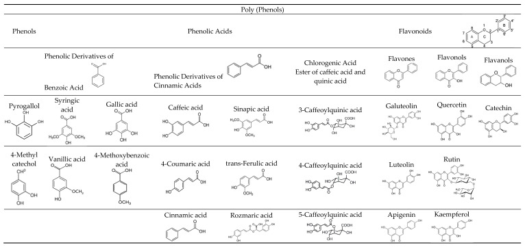
Stevia rebaudiana Bertoni is a perennial shrub of the family Asteraceae which is endemic to northeastern Paraguay but also found in the nearby regions of Brazil and Argentina [1,2]. Stevia is mainly known for the high content of steviol glycosides in its leaves that are utilized as a non-sucrose and calorie-free sweetener in a variety of food products. Glycosides are organic compounds that consist of two parts, one part is a carbohydrate called glycone, which is connected via a glycoside bond to another part, the aglycone, a non-sugar group [3]. The best-known steviol glycosides are stevioside and rebaudioside A (RebA), which are the most abundant glycosides of the plant [4,5,6]. Additionally, other diterpene glycosides such as rebaudioside B, C, D, E, and F, steviolbioside, and dulcoside A also exist in leaves, but at significant lower concentrations. In addition to the sweet compounds, stevia leaves contain carbohydrates, lipids, dietary fibers, essential oils, water-soluble vitamins, minerals, and phenolic compounds [7,8]. Recent studies have shown several benefits of stevia leaf consumption on human health. Because of the high content of various phytoconstituents, stevia leaves appear to have a broad range of biological activities such as antidiabetic, antihypertensive, antimicrobial, anti-inflammatory, anti-tumor, and antioxidant activities [9,10]. Antioxidant activity in plants is most often due to a high content of polyphenols. The main polyphenols of stevia are phenols, phenolic acids, and flavonoids [11,12], as shown in (Figure 1). The main phenols are pyrogallol and 4-methylcatechol, while the main phenolic acids are derivatives of benzoic acid (syringic, vanillic, gallic, and 4-methoxybenzoic), cinnamic acids (caffeic, 4-coumaric, sinapic, trans-ferulic, and rosmaric), and chlorogenic acid (esters of caffeic and quinic acids) [13]. Flavonoids found in stevia leaves belong to three main groups, i.e., flavones (galuteolin, luteolin, apigenin), flavonols (quercetin, rutin, kaempferol), and flavanols (catechin) [14,15,16].
Figure 1.
The main polyphenols found in Stevia rebaudiana (Bertoni) leaves.
Oxidative stress in animals occurs when there is an imbalance between the production of reactive oxygen species (ROS) and the ability of the organism to neutralize or repair the resulting damage. ROS are highly reactive molecules that can damage cells and their components, such as DNA, proteins, and lipids. Some common ROS are the superoxide anion (O2•), hydrogen peroxide (H2O2), and the hydroxyl radical (•OH). Exposure of an animal body to high levels of ROS can lead to tissue damage and ultimately to the development of various degenerative diseases, such as cancer, cardiovascular disease, and neurodegenerative diseases [17]. Humans have several antioxidant defenses to counteract the harmful effects of ROS, including enzymes such as superoxide dismutase (SOD), catalase (CAT), and glutathione peroxidase (GPx), as well as other non-enzymatic antioxidant molecules such as glutathione (GSH) and vitamins C and E [18,19,20].
SOD catalyzes the conversion of the superoxide anion into hydrogen peroxide [21] (2O2− + 2H+ → H2O2). Hydrogen peroxide is a substrate for CAT and GPx; CAT metabolizes hydrogen peroxide into harmless water and oxygen [22] (2H2O2 → 2H2O + O2), while GPx uses GSH to convert hydrogen peroxide and organic hydroperoxides into less harmful compounds, i.e., oxidized glutathione (GSSG) and water (2GSH + H2O2→ GSSG + 2H2O). GSH is a tripeptide molecule composed of three amino acids: glutamic acid, cysteine, and glycine. In its reduced form, GSH contains a thiol (-SH) group, and when GSH donates an electron to neutralize a free radical, it becomes oxidized and forms a disulfide bond (-S-S-) with another GSH molecule [23].
Lipid peroxidation is a process in which free radicals attack and damage lipids in cell membranes, leading to the production of reactive lipid peroxidation products such as malondialdehyde (MDA) and other harmful byproducts. MDA is considered a marker of lipid peroxidation and oxidative stress [24,25]. Lipid peroxidation can be triggered by a variety of factors, including oxidative stress, inflammation, and exposure to toxins or radiation. The process entails the attack of a free radical on a polyunsaturated fatty acid (PUFA) in the cell membrane [26], and the production of a peroxyl-radical lipid which is converted to MDA. MDA has been shown to have several toxic effects on the body, including DNA damage and alteration of proteins and enzymes, and it has been linked to a number of health problems, including inflammation, cancer, and cardiovascular disease [25].
Antioxidants are molecules that can contribute to the protection of cells from oxidative stress by neutralizing free radicals (ROS). Some common natural antioxidants are vitamins A, C, and E, as well as minerals such as selenium and zinc. Other natural antioxidants include phytochemicals, such as flavonoids and polyphenols, which are found in fruits, vegetables, and herbs [27]. Research results suggest that a diet high in antioxidants may protect against the harmful effects of oxidative stress [17,28,29]. The study of antioxidants and of plant extracts rich in antioxidants has emerged as an important and very potent research area on the role of oxidative stress in health and disease; it can provide qualitative and quantitative determination of their antioxidant capacity that can lead to the development of new natural-based treatments for modern-lifestyle diseases.
Studies in animal models have been used to investigate how diseases affect numerous markers of animal oxidation status. It has been reported that animal experimental models of various diseases are characterized by decreased levels of most enzymatic and non-enzymatic antioxidant markers, and by increased levels of MDA [19,24]; however, discrepancies among reported results do occur. The objective of the present study is to statistically combine all of the available data in the literature and determine the effect of stevia leaf extracts on oxidative stress markers in tissues of rats that have been infected with a disease and then (or in parallel) administered stevia extracts, mainly orally. The present meta-analysis is an effort to quantitatively synthesize all of the available data, uncover interchangeable methods, and summarize all of the existing evidence on the antioxidant impact of differently prepared stevia leaf extracts on several tissues of animals suffering from various diseases.
2. Materials and Methods
2.1. Literature Search Strategy and Eligibility Criteria
An all-inclusive literature search in the PubMed database (https://pubmed.ncbi.nlm.nih.gov/ (accessed on 26 January 2023) was carried out to retrieve all potential research articles exploring the antioxidant impacts of stevia, using the keywords ‘Stevia’, ‘antioxidant’, and ‘animal’ with their combinations and derivatives. The search was performed using the preferred reporting items for systematic reviews and meta-analyses (PRISMA) guidelines (http://www.prisma-statement.org/ (accessed on 26 January 2023)) along with the advice for best practices [30,31]. Screening of the reference lists of the included studies was also performed to incorporate all possible relevant publications. To eliminate publication bias and the implications of the grey literature, articles in various languages were taken into consideration [32]. Eligible criteria for inclusion in the meta-analysis were (a) intervention animal studies, (b) studies aimed at evaluating the effects of stevia leaves or stevia glycosides on various diseases, (c) use of control animals. Unrelated articles, in vitro studies, studies conducted on humans, observational studies, and reviews were excluded. We also excluded studies that did not provide sufficient information or data necessary for the analysis in order to ensure the reliability and validity of the results.
2.2. Data Extraction and Antioxidant Markers
Initially, titles and abstracts of the articles were screened, and relevant articles were further examined following the inclusion and exclusion criteria. The search results were assessed by two separate researchers (MP and PK), any discrepancies were discussed with GB and PB and decided upon by consensus. Upon reading all articles, it was found that the determination of oxidative stress was performed mainly with six assays measuring enzymatic activity, and levels of oxidative stress was expressed quantitatively in units of the six oxidation markers. The majority of the studies reported data for the following assays: (a) superoxide dismutase (SOD), (b) catalase (CAT), (c) glutathione peroxidase (GPx), (d) reduced glutathione (GSH), (e) malondialdehyde (MDA), and (f) total antioxidant capacity (TAC) [33].
All the above-mentioned methods by which oxidative stress can be estimated fall into three main categories [34]: (a) assays that measure the activity of antioxidant enzymes, (b) assays that measure lipid peroxidation, and (c) assays that measure total antioxidant capacity (TAC).
Assays used to measure antioxidant enzyme activity include SOD, CAT, and GPx. The activity of SOD [35] can be measured using a colorimetric assay that detects the amount of hydrogen peroxide produced by the action of SOD using formazan dye as an indicator. Formazan dye is measured colorimetrically at 560 nm. Superoxide anions react with formazan salts to produce a dye which can be detected colorimetrically. The greater the activity of SOD in the sample, the less formazan dye is produced. The activity of CAT [36,37] can be measured using a colorimetric assay that detects the rate of decomposition of hydrogen peroxide. The technique involves the reduction of dichromate in acetic acid to chromic acetate in the presence of hydrogen peroxide, which forms an impermanent intermediate perchromic acid. The amount of chromic acetate produced in the reaction is in direct proportion to the concentration of hydrogen peroxide employed. The chromic acetate produced is measured colorimetrically at 570 nm (Cr2 O7 −2+ 7H2O2 → 2CrO8 −3 + 5H2O + 4H+ followed by 2CrO8 −3 + 6CH3COO− + 6H + → 2Cr(CH3 COO)3 + 14H2O). The activity of GPx can be measured using a colorimetric assay that detects the amount of GSSG produced [38], which is coupled to the oxidation of NADPH to NADP+. The decrease in NADPH is proportional to the GPx activity and is monitored spectrophotometrically at 340 nm. When GSH reacts with 5,5′-dithiobis(2-nitrobenzoic acid) (DTNB) a yellow product is formed proportional to the GSH concentration that can be measured colorimetrically according to Ellman’s assay [39].
The extent of lipid peroxidation can be measured using a colorimetric assay that detects the amount of malondialdehyde (MDA) produced because of lipid peroxidation [25]. MDA reacts with thiobarbirutic acid (TBA), and the product is detected by the absorbance at 532 nm.
The total antioxidant capacity (TAC) assay [20] is an assay that measures the overall ability of a substance or biological sample to neutralize free radicals. It measures either the combination of both small molecule antioxidants and proteins, or the presence of small molecules alone that are present in the sample. This assay can be performed using various methods and, in the studies included herein, it was measured colorimetrically. The ferric reducing antioxidant power (FRAP) assay [40] is based on the ability of antioxidants to reduce ferric ion Fe+3 to ferrous ion Fe+2 in a redox reaction, the Fe+2 then reacts with the colorimetric reagent TPTZ (2,4,6-tripyridyl-s-triazine, and iron(III) chloride hexahydrate) to produce a complex which can be measured spectrophotometrically at 593 nm. In one study [41], TAC was indirectly measured by determining the residual H2O2 through the conversion of 3,5, dichloro dicloro-2-hydroxy benzensulphonate to a colored product. The measurement of this colored product can be measured spectrophotometrically at 500–510 nm.
Oxidative stress markers data were used to determine the effect of differently prepared stevia leaf extracts on different tissues of variously diseased animals.
In the studies included herein, dried leaves of stevia were ground to powder. The solvents used for the extraction included water or organic solvents (ethanol, acetone, or methanol). For hydroalcoholic extracts, solutions containing 70% to 80% methanol or ethanol were used. Generally, extractions took place with maceration and incubation to various temperatures [42,43] for different times (five min to 24 h) or using the Soxhlet extraction technique [44]. Subsequently, the extraction products were filtered and evaporated to complete dryness under reduced pressure using vacuum rotary evaporation. The resulting powder of the extracts was diluted with either distilled water or saline solutions for oral administration. The glycosides used in the studies reported herein were purchased ensuring that purities were >96%. In one study [45], the isolation and purification of the glycosides were performed using a diode array detector (JASCO HPLC system).
Data extraction was performed in a predetermined Microsoft Excel® sheet. From each study, the following information was extracted: first author’s last name, publication year, country, type of assay determining oxidative stress, treatment, number of experimental animals, tissue of rats, type of disease, and the type of stevia leaf extracts or steviol glycosides. Antioxidant marker data were divided into three groups as follows: ‘control’, consisting of healthy normal rats; ‘case’, consisting of diseased rats; and ‘stevia’, the group with the diseased rats that received stevia extracts.
2.3. Statistical Analysis
The primary outcome of this meta-analysis was the standardized mean differences in oxidative stress marker estimates between the three animal groups, which is referred to as Cohen’s d. Secondary outcomes included the stratification analysis of oxidative stress markers’ performance in different tissues of rats, types of diseases, and types of stevia extracts. The model we used was based on the standard model of multivariate meta-analysis method [46,47,48]. We were interested in comparing the mean difference of the oxidative stress marker estimates between the three animal groups, measured in study i = 1,2,…k. The mean differences in oxidative stress marker estimates were used to determine the contrasts ‘control’ vs. ‘case’, ‘stevia’ vs. ‘case’, and ‘stevia’ vs. ‘control’, and were estimated using:
| (1) |
where X1i, X2i, and X0i are the means of the measured values of the oxidative stress markers in the ‘control’, ‘case’, and ‘stevia’ groups, respectively. Spooled,i is the pooled standard deviation in study i, given by:
| (2) |
with n1i, n2i, and n0i being the sample size of each group and S1i, S2i, and S0i the standard deviation of the measured values for each group.
The variance of the effect size estimates d1i and d2i is estimated by:
| (3) |
and the covariance between the estimates of d1i and d2i is
| (4) |
with ni = n0i + n1i+ n2i being the total sample size of the study. The calculation of the within-studies covariance is very important since it plays a crucial role in the multivariate method.
Several statistical methods have been proposed to handle the issues of small sample sizes efficiently and to calculate accurate p-values and confidence intervals. The d can be corrected using the so-called Hedges’ g, which generates an unbiased estimate (the standardized mean difference d has the tendency to overestimate the absolute value in small samples), and the meta-analysis was conducted as proposed in [49]. In the multivariate random-effects meta-analysis, we assume that gi = (g1i, g2i) is distributed following a multivariate normal distribution around the true means, according to the marginal model:
| (5) |
By Σi we denote the within-studies covariance matrix:
| (6) |
The diagonal elements of Σi are the study-specific estimates of the variance (Equation (3)), whereas the off-diagonal elements correspond to the pairwise within-studies covariances (Equation (4)), for instance, . It is important to note that the elements of Σi are considered known quantities. In contrast, by C we denote the between-studies covariance matrix, which is estimated during the fitting process:
| (7) |
The particular model takes into account the within-studies covariance and the between-studies covariance of the random terms, which is estimated in the model fitting procedure. A major advantage of the multivariate meta-analysis model is that it can accommodate studies reporting only one of the parameters of interest, resulting in borrowing strength from external studies. Another advantage is that with the estimated variance–covariance matrix, it can be used to perform global tests for the effect sizes where the estimates of both g1 and g2 influence all outcomes (‘control’ vs. ‘case’, ‘stevia’ vs. case’, and ‘stevia’ vs. ‘control’).
Moreover, to determine whether different oxidative stress markers can be combined in a meta-analysis, meta-regression analysis [50,51] was also used. This allowed us to assess whether studies that used different markers tended to have comparable data [46,52,53]. Multivariate meta-analysis and meta-regression analysis were performed using the statistical software package Stata13 [54]. In all tests, p ≤ 0.05 was used as the decision rule for significance testing. Meta-analysis was performed when two or more studies were available.
3. Results
3.1. Study Selection and Characteristics
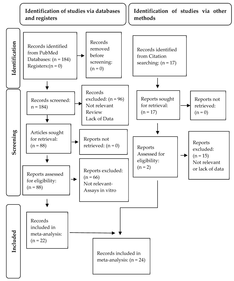
A thorough literature search for antioxidant activity of stevia resulted in 184 articles. After screening of titles and abstracts, and compliance with the PRISMA guidelines (http://www.prisma-statement.org/ (accessed on 26 January 2023)) (Figure 2), 22 articles were found to satisfy the eligibility criteria [41,42,44,45,55,56,57,58,59,60,61,62,63,64,65,66,67,68,69,70,71,72]. Seventeen records were additionally retrieved from screening lists of references, and two of them [37,73] were enrolled in the meta-analysis, making a total of 24 articles, which contained 104 studies in total.
Figure 2.
Flow diagram of systematic review to retrieve the selected studies for meta-analysis in accordance with the PRISMA statement.
All the selected studies evaluated the effects of stevia extracts or stevia glycosides on rats as experimental models. The included studies predominantly reported data on SOD (21 studies), CAT (17 studies), and GPx (4 studies) activity, the content of GSH (27 studies), the assessment of TAC (2 studies), and the content of MDA (33 studies), all of which are shown in Table 1. Data on oxidative stress markers concerned 13 rat tissues, including liver, kidney, pancreas, heart, serum, plasma, skeletal muscles, brain, ovary, colon, duodenum, jejunum, and ileum. Antioxidant activity was investigated in 13 types of diseases, comprising diabetes mellitus, liver diseases, renal disorders, ulcerative colitis, metabolic syndrome, polycystic ovary syndrome, and epilepsy. Four types of stevia extracts were tested for their effect after administration: aqueous, organic, hydroalcoholic, and fractions of methanolic extracts. Hence, overall, our data enrolled 15 studies with aqueous extracts, 19 studies with organic extracts, 40 studies with hydroalcoholic extracts and only 1 with methanolic fractions of extract. In addition, 20 studies were performed with stevioside, 5 studies with rebaudioside A, and 1 study with extracted sweeteners from stevia leaves. All the selected studies investigated the effects of oral administration of stevia extracts or steviol glycosides in rats as a treatment, except for two articles in which administration was performed intraperitoneally. In three of the articles [61,71,72], the use of stevia was investigated as a pre-treatment prior to the induction of a disease, while in all other studies, stevia was administered after the disease had been induced.
Table 1.
Characteristics of the 104 studies included in the meta-analysis.
| Author | Year | Country | Assay | After Treatment (A) Pre-Treatment (P) with Stevia | # Controls | Control Value | Control SD | # Cases |
Case Value | Case SD | # Stevia |
Stevia Value | Stevia SD | Extract/Compound | Type of Tissue Tested | Type of Disease |
|---|---|---|---|---|---|---|---|---|---|---|---|---|---|---|---|---|
| Mostafa et al. [72] | 2020 | Egypt | SOD | 80 mg/kg/day orally, 1 week (A) |
10 | 3.6 | 0.11 | 10 | 1.6 | 0.1 | 10 | 2.66 | 0.09 | Aqueous | Colon | Ulcerative colitis |
| Mehmood et al. [64] | 2019 | China | SOD | 400 mg/kg/day orally, 8 weeks (A) |
8 | 118 | 6.1 | 8 | 53 | 2.8 | 8 | 107 | 12 | Hydroalcoholic | Duodenum | Hyperuricemia |
| Mehmood et al. [64] | 2019 | China | SOD | 400 mg/kg/day orally, 8 weeks (A) |
8 | 70 | 7.9 | 8 | 30 | 7 | 8 | 51 | 12 | Hydroalcoholic | Jejunum | Hyperuricemia |
| Mehmood et al. [64] | 2019 | China | SOD | 400 mg/kg/day orally, 8 weeks (A) |
8 | 55 | 8.1 | 8 | 21 | 4.5 | 8 | 40 | 4.5 | Hydroalcoholic | Ileum | Hyperuricemia |
| Mehmood et al. [63] | 2020 | China | SOD | 200 mg/kg/day orally, 4 weeks (A) |
8 | 118 | 4.9 | 8 | 91 | 5.9 | 8 | 115 | 5 | Hydroalcoholic | Serum | Hyperuricemia |
| El-Mesallamy et al. [45] | 2018 | Egypt | SOD | 200 mg/kg/day orally, 4 weeks (A) |
10 | 48 | 4 | 10 | 25 | 7.5 | 10 | 41 | 4 | Hydroalcoholic | Skeletal muscles | Diabetes mellitus |
| El-Mesallamy et al. [45] | 2018 | Egypt | SOD | 2 mg/kg/day orally, 4 weeks (A) |
10 | 48 | 4 | 10 | 25 | 7.5 | 10 | 30 | 4 | Stevioside | Skeletal muscles | Diabetes mellitus |
| Latha et al. [70] | 2017 | India | SOD | 500 mg/kg/day orally, 1 week (A) |
8 | 21 | 4.24 | 8 | 4 | 4.52 | 8 | 16.5 | 2.54 | Hydroalcoholic | Liver | Acute liver injury |
| Latha et al. [70] | 2017 | India | SOD | 250 mg/kg/day orally, 1 week (A) |
8 | 21 | 4.24 | 8 | 4 | 4.52 | 8 | 17 | 4.52 | Stevioside | Liver | Acute liver injury |
| Moselhy et al. [66] | 2016 | Saudi Arabia | SOD | 200 mg/kg/day orally, 2 weeks (A) |
10 | 0.25 | 0.0023 | 10 | 0.18 | 0.03 | 10 | 0.21 | 0.029 | Organic | Liver | Hepatotoxic |
| Perumal et al. [67] | 2016 | India | SOD | 100 mg/kg/day orally, 3 weeks (A) |
6 | 10.15 | 1.05 | 6 | 4.63 | 0.95 | 6 | 6.75 | 1.25 | Hydroalcoholic | Liver | Diabetes mellitus |
| Perumal et al. [67] | 2016 | India | SOD | 100 mg/kg/day orally, 3 weeks (A) |
6 | 11.04 | 1.3 | 6 | 4.63 | 1.69 | 6 | 6.74 | 1.18 | Hydroalcoholic | Kidney | Diabetes mellitus |
| Shivanna et al. [71] | 2012 | India | SOD | NR orally, 4 weeks (P) | 10 | 2.41 | 0.91 | 10 | 1.21 | 0.05 | 10 | 2.72 | 0.52 | Fraction methanol | Liver | Diabetes mellitus |
| Myint et al. [73] | 2020 | China | SOD | 12 mg/kg/day orally, 6 weeks (A) |
6 | 74.53 | 1.82 | 6 | 36.61 | 1.44 | 6 | 39.23 | 1.28 | Rebaudioside A | Liver | Diabetes mellitus |
| Myint et al. [73] | 2020 | China | SOD | 10 mg/kg/day orally, 6 weeks (A) |
6 | 74.53 | 1.82 | 6 | 36.61 | 1.44 | 6 | 44.46 | 1.28 | Stevioside | Liver | Diabetes mellitus |
| Singh et al. [37] | 2013 | India | SOD | 300 mg/kg/day orally, 3 weeks (A) |
7 | 50.5 | 32.80 | 7 | 37.5 | 18.79 | 7 | 50 | 10.85 | Organic | Liver | Diabetes mellitus |
| Singh et al. [37] | 2013 | India | SOD | 300 mg/kg/day orally, 3 weeks (A) |
7 | 190.2 | 50.27 | 7 | 40 | 19.84 | 7 | 10 | 14.02 | Organic | Pancreas | Diabetes mellitus |
| Singh et al. [37] | 2013 | India | SOD | 300 mg/kg/day orally, 3 weeks (A) |
7 | 55 | 9.26 | 7 | 25 | 9.26 | 7 | 9 | 9.26 | Organic | Kidney | Diabetes mellitus |
| El-Hadary et al. [60] | 2021 | Egypt | SOD | 300 mg/kg/day orally, 8 weeks (A) |
10 | 54.3 | 2.3 | 10 | 47.2 | 1.2 | 10 | 58.8 | 0.9 | Hydroalcoholic | Liver | Diabetes mellitus |
| Morsi et al. [65] | 2022 | Egypt | SOD | 300 mg/kg/day orally, 4 weeks (A) |
7 | 36.25 | 1.25 | 7 | 21.25 | 3 | 7 | 29 | 1.75 | Glycosides-sweetener | Ovary | Polycystic ovary syndrome |
| Deenadayalan et al. [59] | 2021 | India | SOD | 20 mg/kg/day orally, 45 days (A) |
6 | 32.5 | 3.06 | 6 | 13 | 1.84 | 6 | 20 | 1.84 | Stevioside | Skeletal muscles | Diabetes mellitus |
| Mostafa et al. [72] | 2020 | Egypt | CAT | 80 mg/kg/day orally, 1 week (P) | 10 | 7.95 | 0.11 | 10 | 3.9 | 0.08 | 10 | 6.6 | 0.09 | Aqueous | Colon | Ulcerative colitis |
| Elsaid et al. [61] | 2019 | Egypt | CAT | 200 mg/kg/day orally, 5 weeks (P) | 12 | 34.66 | 3.14 | 12 | 14 | 2.36 | 12 | 27.66 | 3.72 | Hydroalcoholic | Kidney | Renal ischemia/reperfusion |
| Abdallah et al. [55] | 2022 | Egypt | CAT | 500 mg/kg/day orally, 1 week (A) | 7 | 36 | 0.5 | 7 | 21 | 5 | 7 | 30 | 0.5 | Organic | Liver | Liver disease |
| Abdallah et al. [55] | 2022 | Egypt | CAT | 250 mg/kg/day orally, 1 week (A) | 7 | 36 | 0.5 | 7 | 21 | 5 | 7 | 35 | 5 | Stevioside | Liver | Liver disease |
| Mehmood et al. [63] | 2020 | China | CAT | 200 mg/kg/day orally, 4 weeks (A) | 8 | 17.5 | 1.5 | 8 | 8 | 1.1 | 8 | 10.5 | 1.2 | Hydroalcoholic | Serum | Hyperuricemia |
| Moselhy et al. [66] | 2016 | Saudi Arabia | CAT | 200 mg/kg/day orally, 2 weeks (A) | 10 | 0.89 | 0.07 | 10 | 0.32 | 0.05 | 10 | 0.76 | 0.05 | Organic | Liver | Hepatotoxic |
| El Nashar et al. [41] | 2022 | Egypt | CAT | 200 mg/kg/day orally, 4 weeks (P and A) | 10 | 67.67 | 31.53 | 10 | 52.08 | 29.6 | 10 | 95.25 | 35.45 | Organic | Brain | Epilepsy |
| Hussein et al. [62] | 2020 | Egypt | CAT | 400 mg/kg/day orally, 4 weeks (A) | 8 | 17 | 0.45 | 8 | 7 | 0.19 | 8 | 22.5 | 0.98 | Hydroalcoholic | Heart | Diabetes mellitus |
| Shivanna et al. [71] | 2012 | India | CAT | NR orally, 4 weeks (P) | 10 | 1.02 | 0.05 | 10 | 0.52 | 0.07 | 10 | 0.78 | 0.17 | Fraction methanol | Liver | Diabetes mellitus |
| Assaei et al. [44] | 2016 | Iran | CAT | 400 mg/kg/day orally, 4 weeks (A) | 10 | 29.4 | 8.85 | 10 | 9.9 | 6.96 | 10 | 33.7 | 6.33 | Aqueous | Pancreas | Diabetes mellitus |
| Deenadayalan et al. [59] | 2021 | India | CAT | 20 mg/kg/day orally, 45 days (A) | 6 | 11.5 | 3.06 | 6 | 6.25 | 3.18 | 6 | 8.75 | 1.83 | Stevioside | Skeletal muscles | Diabetes mellitus |
| Perumal et al. [67] | 2016 | India | CAT | 100 mg/kg/day orally, 3 weeks (A) | 6 | 42.8 | 6.2 | 6 | 25.8 | 3.72 | 6 | 35.69 | 6.42 | Hydroalcoholic | Liver | Diabetes mellitus |
| Perumal et al. [67] | 2016 | India | CAT | 100 mg/kg/day orally, 3 weeks (A) | 6 | 34.04 | 5.27 | 6 | 23.56 | 1.54 | 6 | 25.3 | 1.71 | Hydroalcoholic | Kidney | Diabetes mellitus |
| El-Mesallamy et al. [45] | 2018 | Egypt | CAT | 200 mg/kg/day orally, 4 weeks (A) | 10 | 90 | 1 | 10 | 55 | 3 | 10 | 80 | 4 | Hydroalcoholic | Skeletal muscles | Diabetes mellitus |
| El-Mesallamy et al. [45] | 2018 | Egypt | CAT | 2 mg/kg/day orally, 4 weeks (A) | 10 | 90 | 1 | 10 | 55 | 3 | 10 | 72 | 4 | Stevioside | Skeletal muscles | Diabetes mellitus |
| Latha et al. [70] | 2017 | India | CAT | 500 mg/kg/day orally, 1 week (A) | 8 | 0.87 | 0.73 | 8 | 0.21 | 0.03 | 8 | 0.51 | 0.31 | Hydroalcoholic | Liver | Acute liver injury |
| Latha et al. [70] | 2017 | India | CAT | 250 mg/kg/day orally, 1 week (A) | 8 | 0.87 | 0.73 | 8 | 0.21 | 0.03 | 8 | 0.3 | 0.28 | Stevioside | Liver | Acute liver injury |
| El-Mesallamy et al. [45] | 2018 | Egypt | GPx | 200 mg/kg/day orally, 4 weeks (A) | 10 | 515 | 2 | 10 | 280 | 4 | 10 | 480 | 7 | Hydroalcoholic | Skeletal muscles | Diabetes mellitus |
| El-Mesallamy et al. [45] | 2018 | Egypt | GPx | 2 mg/kg/day Orally, 4 weeks (A) |
10 | 515 | 2 | 10 | 280 | 4 | 10 | 450 | 3.5 | Stevioside | Skeletal muscles | Diabetes mellitus |
| Deenadayalan et al. [59] | 2021 | India | GPx | 20 mg/kg/day orally, 45 days (A) | 6 | 26 | 6.12 | 6 | 14 | 3.68 | 6 | 20 | 3.68 | Stevioside | Skeletal muscles | Diabetes mellitus |
| El-Hadary et al. [60] | 2021 | Egypt | GPx | 300 mg/kg/day orally, 8 weeks (A) | 10 | 165.6 | 0.7 | 10 | 137.8 | 1.4 | 10 | 175.8 | 5.3 | Hydroalcoholic | Liver | Diabetes mellitus |
| Mostafa et al. [72] | 2020 | Egypt | GSH | 80 mg/kg/day orally, 1 week (A) | 10 | 5.7 | 0.09 | 10 | 2.2 | 0.05 | 10 | 4.9 | 0.03 | Aqueous | Colon | Ulcerative colitis |
| Abdel-Aal et al. [56] | 2021 | Egypt | GSH | 400 mg/kg/day orally, 3 weeks (A) | 8 | 22 | 3.11 | 8 | 3 | 0.70 | 8 | 12.5 | 0.70 | Aqueous | Liver | Diabetes mellitus |
| Abdel-Aal et al. [56] | 2021 | Egypt | GSH | 400 mg/kg/day orally, 3 weeks (A) | 8 | 22 | 1.41 | 8 | 7 | 0.71 | 8 | 17 | 1.27 | Aqueous | Kidney | Diabetes mellitus |
| Hussein et al. [62] | 2020 | Egypt | GSH | 400 mg/kg/day orally, 4 weeks (A) | 8 | 11.25 | 0.4 | 8 | 3.1 | 0.23 | 8 | 11.7 | 0.97 | Hydroalcoholic | Heart | Diabetes mellitus |
| Mehmood et al. [64] | 2019 | China | GSH | 400 mg/kg/day orally, 8 weeks (A) | 8 | 125 | 37.5 | 8 | 40 | 7.5 | 8 | 100 | 27 | Hydroalcoholic | Duodenum | Hyperuricemia |
| Mehmood et al. [64] | 2019 | China | GSH | 400 mg/kg/day orally, 8 weeks (A) | 8 | 100 | 27 | 8 | 40 | 2 | 8 | 80 | 17.5 | Hydroalcoholic | Jejunum | Hyperuricemia |
| Mehmood et al. [64] | 2019 | China | GSH | 400 mg/kg/day orally, 8 weeks (A) | 8 | 100 | 8 | 8 | 35 | 10 | 8 | 84 | 10 | Hydroalcoholic | Ileum | Hyperuricemia |
| Elsaid et al. [61] | 2019 | Egypt | GSH | 200 mg/kg/day orally, 5 weeks (P) | 12 | 8.65 | 0.57 | 12 | 3.38 | 0.36 | 12 | 7.17 | 0.64 | Hydroalcoholic | Kidney | Renal ischemia/reperfusion |
| Ramos-Tovar et al. [43] | 2019 | Mexico | GSH | 100 mg/kg/day orally, 12 weeks (A) | 8 | 10 | 1.98 | 8 | 3.75 | 1.95 | 8 | 8.55 | 1.27 | Aqueous | Liver | Liver cirrhosis |
| Casas-Grajales et al. [58] | 2019 | Mexico | GSH | 20 mg/kg/twice daily intraperitoneally, 8 weeks (A) | 8 | 13 | 0.85 | 8 | 10 | 0.28 | 8 | 13.5 | 1.13 | Stevioside | Liver | Liver fibrosis |
| Casas-Grajales et al. [57] | 2019 | Mexico | GSH | 20 mg/kg/twice daily intraperitoneally, 8 weeks (A) |
8 | 13.3 | 0.85 | 8 | 10 | 0.28 | 8 | 12.2 | 0.57 | Rebaudioside A | Liver | Liver fibrosis |
| Ramos-Tovar et al. [42] | 2018 | Mexico | GSH | 100 mg/kg/day orally, 10 weeks (A) | 8 | 5.6 | 1.41 | 8 | 3 | 0.28 | 8 | 4.5 | 1.56 | Aqueous | Liver | Liver cirrhosis |
| Ramos-Tovar et al. [68] | 2018 | Mexico | GSH | 100 mg/kg/day orally, 1 week (A) |
8 | 11.5 | 1.27 | 8 | 6 | 2.55 | 8 | 11 | 2.55 | Aqueous | Liver | Liver cirrhosis |
| Latha et al. [70] | 2017 | India | GSH | 500 mg/kg/day orally, 1 week (A) |
8 | 230 | 14.14 | 8 | 95 | 28.29 | 8 | 212 | 98.99 | Hydroalcoholic | Liver | Acute Liver injury |
| Latha et al. [70] | 2017 | India | GSH | 250 mg/kg/day orally, 1 week (A) |
8 | 230 | 14.14 | 8 | 95 | 28.28 | 8 | 181 | 14.14 | Stevioside | Liver | Acute Liver injury |
| Perumal et al. [67] | 2016 | India | GSH | 100 mg/kg/day orally, 3 weeks (A) |
6 | 43.4 | 7.20 | 6 | 11.83 | 8.23 | 6 | 21.4 | 6.74 | Hydroalcoholic | Liver | Diabetes mellitus |
| Perumal et al. [67] | 2016 | India | GSH | 100 mg/kg/day orally, 3 weeks (A) |
6 | 41.4 | 9.48 | 6 | 19.04 | 5.07 | 6 | 27.43 | 8.01 | Hydroalcoholic | Kidney | Diabetes mellitus |
| Shivanna et al. [71] | 2012 | India | GSH | NR orally, 4 weeks (P) | 10 | 24.58 | 0.51 | 10 | 13.58 | 0.4 | 10 | 21.11 | 0.51 | Fraction methanol | Plasma | Diabetes mellitus |
| Myint et al. [73] | 2020 | China | GSH | 12 mg/kg/day orally, 6 weeks (A) |
6 | 56.75 | 1.27 | 6 | 35.78 | 1.26 | 6 | 36.6 | 1.24 | Rebaudioside A | Liver | Diabetes mellitus |
| Myint et al. [73] | 2020 | China | GSH | 10 mg/kg/day orally, 6 weeks (A) |
6 | 56.75 | 1.27 | 6 | 35.78 | 1.26 | 6 | 39.89 | 1.24 | Stevioside | Liver | Diabetes mellitus |
| Singh et al. [37] | 2013 | India | GSH | 300 mg/kg/day orally, 3 weeks(A) |
7 | 25.2 | 10.05 | 7 | 7.1 | 6.09 | 7 | 28.1 | 19.31 | Organic | Liver | Diabetes mellitus |
| Singh et al. [37] | 2013 | India | GSH | 300 mg/kg/day orally, 3 weeks (A) |
7 | 7.5 | 6.09 | 7 | 4.1 | 1.32 | 7 | 22.1 | 10.05 | Organic | Pancreas | Diabetes mellitus |
| Singh et al. [37] | 2013 | India | GSH | 300 mg/kg/day orally, 3 weeks (A) |
7 | 22.4 | 8.73 | 7 | 3.5 | 0.26 | 7 | 18.1 | 8.73 | Organic | Kidney | Diabetes mellitus |
| El-Hadary et al. [60] | 2021 | Egypt | GSH | 300 mg/kg/day orally, 8 weeks (A) |
10 | 80.8 | 0.9 | 10 | 57.2 | 1.7 | 10 | 81.6 | 1.9 | Hydroalcoholic | Liver | Diabetes mellitus |
| Abdallah et al. [55] | 2022 | Egypt | GSH | 500 mg/kg/day orally, 1 week (A) |
7 | 50 | 1 | 7 | 42.5 | 2.5 | 7 | 48 | 1.5 | Organic | Liver | Liver disease |
| Abdallah et al. [55] | 2022 | Egypt | GSH | 250 mg/kg/day orally, 1 week (A) |
7 | 50 | 1 | 7 | 42.5 | 2.5 | 7 | 51 | 2 | Stevioside | Liver | Liver disease |
| Deenadayalan et al. [59] | 2021 | India | GSH | 20 mg/kg/day orally, 45 days (A) |
6 | 13.25 | 2.45 | 6 | 6 | 2.21 | 6 | 9 | 1.22 | Stevioside | Skeletal muscles | Diabetes mellitus |
| Ranjbar et al. [40] | 2020 | Iran | TAC | 400 mg/kg/day orally, 14 weeks (A) |
10 | 0.36 | 0.16 | 10 | 0.19 | 0.19 | 10 | 0.28 | 0.13 | Hydroalcoholic | Serum | Metabolic syndrome |
| El Nashar et al. [41] | 2022 | Egypt | TAC | 200 mg/kg/day orally, 4 weeks (P and A) | 10 | 4.65 | 1.28 | 10 | 1.91 | 1.27 | 10 | 6.3 | 2.63 | Organic | Brain | Epilepsy |
| Abdel-Aal et al. [56] | 2021 | Egypt | MDA | 400 mg/kg/day orally, 3 weeks (A) |
8 | 0.2 | 0.06 | 8 | 0.58 | 0.06 | 8 | 0.24 | 0.04 | Aqueous | Liver | Diabetes mellitus |
| Abdel-Aal et al. [56] | 2021 | Egypt | MDA | 400 mg/kg/day orally, 3 weeks (A) |
8 | 0.21 | 0.02 | 8 | 0.46 | 0.03 | 8 | 0.24 | 0.21 | Aqueous | Kidney | Diabetes mellitus |
| Ranjbar et al. [40] | 2020 | Iran | MDA | 400 mg/kg/day orally, 14 weeks (A) |
10 | 34 | 17.39 | 10 | 45 | 23.72 | 10 | 38 | 12.33 | Hydroalcoholic | Serum | Metabolic syndrome |
| Hussein et al. [62] | 2020 | Egypt | MDA | 400 mg/kg/day orally, 4 weeks (A) |
8 | 1.49 | 0.03 | 8 | 9.9 | 0.26 | 8 | 2 | 0.13 | Hydroalcoholic | Heart | Diabetes mellitus |
| Mehmood et al. [64] | 2019 | China | MDA | 400 mg/kg/day orally, 8 weeks(A) |
8 | 1.4 | 0.1 | 8 | 5.75 | 0.9 | 8 | 1.75 | 0.49 | Hydroalcoholic | Duodenum | Hyperuricemia |
| Mehmood et al. [64] | 2019 | China | MDA | 400 mg/kg/day orally, 8 weeks (A) |
8 | 1.2 | 0.25 | 8 | 4.4 | 0.79 | 8 | 1.2 | 0.95 | Hydroalcoholic | Jejunum | Hyperuricemia |
| Mehmood et al. [64] | 2019 | China | MDA | 400 mg/kg/day orally, 8 weeks (A) |
8 | 1.1 | 0.37 | 8 | 4.1 | 1.21 | 8 | 1.5 | 0.49 | Hydroalcoholic | Ileum | Hyperuricemia |
| Mehmood et al. [63] | 2020 | China | MDA | 200 mg/kg/day orally, 4 weeks (A) |
8 | 4.6 | 1.95 | 8 | 9.15 | 1 | 8 | 5.75 | 1.25 | Hydroalcoholic | Serum | Hyperuricemia |
| Elsaid et al. [61] | 2019 | Egypt | MDA | 200 mg/kg/day orally, 5 weeks (A) |
12 | 1.89 | 0.31 | 12 | 5.2 | 1.07 | 12 | 2.92 | 0.16 | Hydroalcoholic | Kidney | Renal ischemia/reperfusion |
| Ramos-Tovar et al. [43] | 2019 | Mexico | MDA | 100 mg/kg/day orally, 12 weeks (A) |
8 | 0.19 | 0.03 | 8 | 0.29 | 0.06 | 8 | 0.2 | 0.04 | Aqueous | Liver | Liver cirrhosis |
| Casas-Grajales et al. [58] | 2019 | Mexico | MDA | 20 mg/kg/twice daily intraperitoneally, 8 weeks (A) |
8 | 0.2 | 0.03 | 8 | 0.67 | 0.09 | 8 | 0.4 | 0.07 | Stevioside | Liver | Liver fibrosis |
| Casas-Grajales et al. [57] | 2019 | Mexico | MDA | 20 mg/kg twice daily intraperitoneally, 8 weeks (A) |
8 | 0.22 | 0.06 | 8 | 0.67 | 0.11 | 8 | 0.32 | 0.09 | Rebaudioside A | Liver | Liver fibrosis |
| El-Mesallamy et al. [45] | 2018 | Egypt | MDA | 200 mg/kg/day orally, 4 weeks (A) |
10 | 4 | 1.1 | 10 | 11.5 | 0.9 | 10 | 6 | 0.95 | Hydroalcoholic | Skeletal muscles | Diabetes mellitus |
| El-Mesallamy et al. [45] | 2018 | Egypt | MDA | 2 mg/kg/day orally, 4 weeks (A) |
10 | 4 | 1.1 | 10 | 11.5 | 0.9 | 10 | 8 | 0.95 | Stevioside | Skeletal muscles | Diabetes mellitus |
| Ramos-Tovar et al. [42] | 2018 | Mexico | MDA | 100 mg/kg/day orally, 10 weeks (A) |
8 | 0.17 | 0.03 | 8 | 0.34 | 0.09 | 8 | 0.24 | 0.11 | Aqueous | Liver | Liver cirrhosis |
| Ramos-Tovar et al. [68] | 2018 | Mexico | MDA | 100 mg/kg/day orally, 1 week (A) |
8 | 0.10 | 0.03 | 8 | 0.31 | 0.03 | 8 | 0.15 | 0.06 | Aqueous | Liver | Liver cirrhosis |
| Latha et al. [70] | 2017 | India | MDA | 500 mg/kg/day orally, 1 week (A) |
8 | 35 | 2.83 | 8 | 160 | 19.8 | 8 | 85 | 5.66 | Hydroalcoholic | Liver | Acute liver injury |
| Latha et al. [70] | 2017 | India | MDA | 250 mg/kg/day orally, 1 week (A) |
8 | 35 | 2.83 | 8 | 160 | 19.78 | 8 | 115 | 2.83 | Stevioside | Liver | Acute liver injury |
| Moselhy et al. [66] | 2016 | Saudi Arabia | MDA | 200 mg/kg/day orally, 2 weeks (A) |
10 | 5.11 | 0.14 | 10 | 10.14 | 0.37 | 10 | 6.94 | 0.47 | Organic | Liver | Hepatotoxic |
| Perumal et al. [67] | 2016 | India | MDA | 100 mg/kg/day orally, 3 weeks (A) |
6 | 0.9 | 0.22 | 6 | 2.33 | 0.56 | 6 | 1.06 | 0.42 | Hydroalcoholic | Liver | Diabetes mellitus |
| Perumal et al. [67] | 2016 | India | MDA | 100 mg/kg/day orally, 3 weeks (A) |
6 | 0.53 | 0.220 | 6 | 2.07 | 1.13 | 6 | 1.49 | 0.61 | Hydroalcoholic | Kidney | Diabetes mellitus |
| Assaei et al. [44] | 2016 | Iran | MDA | 400 mg/kg/day orally, 4 weeks (A) |
10 | 0.4 | 0.13 | 10 | 1.4 | 0.25 | 10 | 0.45 | 0.13 | Aqueous | Pancreas | Diabetes mellitus |
| Shivanna et al. [71] | 2012 | India | MDA | NR, 4 weeks (P) | 10 | 0.06 | 0.01 | 10 | 0.16 | 0.03 | 10 | 0.07 | 0.01 | Fraction methanol | Liver | Diabetes mellitus |
| Myint et al. [73] | 2020 | China | MDA | 12 mg/kg/day orally, 6 weeks (A) |
6 | 7.12 | 0.11 | 6 | 12.63 | 0.32 | 6 | 12.43 | 0.23 | Rebaudioside A | Liver | Diabetes mellitus |
| Myint et al. [73] | 2020 | China | MDA | 10 mg/kg/day orally, 6 weeks (A) |
6 | 7.12 | 0.11 | 6 | 12.63 | 0.32 | 6 | 11.89 | 0.23 | Stevioside | Liver | Diabetes mellitus |
| Singh et al. [37] | 2013 | India | MDA | 300 mg/kg/day orally, 3 weeks (A) |
7 | 25 | 66.14 | 7 | 410 | 66.14 | 7 | 10.2 | 1.32 | Organic | Liver | Diabetes mellitus |
| Singh et al. [37] | 2013 | India | MDA | 300 mg/kg/day orally, 3 weeks (A) |
7 | 11.1 | 2.64 | 7 | 75.2 | 47.62 | 7 | 4.8 | 1.32 | Organic | Pancreas | Diabetes mellitus |
| Singh et al. [37] | 2013 | India | MDA | 30 mg/kg/day orally, 3 weeks (A) |
7 | 50.2 | 31.75 | 7 | 415 | 165.36 | 7 | 11 | 1.32 | Organic | Kidney | Diabetes mellitus |
| El-Hadary et al. [60] | 2021 | Egypt | MDA | 300 mg/kg/day orally, 8 weeks (A) |
10 | 5.33 | 0.2 | 10 | 11.9 | 0.5 | 10 | 4.9 | 0.1 | Hydroalcoholic | Liver | Diabetes mellitus |
| Morsi et al. [65] | 2022 | Egypt | MDA | 300 mg/kg/day orally, 4 weeks (A) |
7 | 50 | 1.5 | 7 | 160 | 2 | 7 | 76 | 1.5 | Glycosides-sweetener | Ovary | Polycystic ovary syndrome |
| Abdallah et al. [55] | 2022 | Egypt | MDA | 500 mg/kg/day orally, 1 week (A) |
7 | 4.1 | 0.5 | 7 | 10.9 | 1.75 | 7 | 4.8 | 0.8 | Organic | Liver | Liver disease |
| Abdallah et al. [55] | 2022 | Egypt | MDA | 250 mg/kg/day orally, 1 week (A) |
7 | 4.1 | 0.5 | 7 | 10.9 | 1.75 | 7 | 5 | 1 | Stevioside | Liver | Liver disease |
| El Nashar et al. [41] | 2022 | Egypt | MDA | 200 mg/kg/day orally, 4 weeks (P and A) | 10 | 50.76 | 5.58 | 10 | 81.79 | 5.82 | 10 | 42.68 | 14.6 | Organic | Brain | Epilepsy |
SOD: superoxide dismutase; CAT: catalase; GPx: glutathione peroxidase; GSH: reduced glutathione; TAC: total antioxidant capacity; MDA: malondialdehyde; A: administration after disease had been established; P: administration prior to disease establishment; NR: not reported, #: number of.
The administration of the substance varied in terms of the doses employed, with doses ranging from 2 mg/kg to 300 mg/kg for pure glycosides and from 80 mg/kg to 500 mg/kg for leaf extracts. Additionally, the period of treatment varied from one to twelve weeks. It should be noted that the studies were conducted in six countries: Mexico, Egypt, Saudi Arabia, Iran, China, and India. The characteristics of the selected studies that were encountered in the meta-analysis are presented in Table 1.
3.2. Bioactive Compounds from Stevia Leaf Extracts Exert Significantly Higher Antioxidant Activity Compared to Stevia Glycosides
To assess the effect of stevia whole leaf extracts or steviol glycosides on diseased animals, the differences of oxidative stress marker measurements between control and cases were enrolled in a multivariate meta-analysis for the assays SOD, CAT, GPx, GSH, TAC, and MDA. If stevia could reverse the effect of the ‘disease’ in case animals, in terms of oxidative stress markers, then the differences ‘ ‘control–case’ and ‘stevia case’ would be almost the same; consequently, the difference ‘stevia – control’ in assays’ values would be very low and perhaps non-significant.
Overall, the meta-analysis results for each separate assay (SOD, CAT, GPx, GSH, CAT, MDA) showed that differences for the contrasts ‘control–case’ were a bit higher than the differences ‘stevia–case’ (for the MDA assay absolute values were considered). The differences ‘stevia–control’ were significantly lower, and importantly, in the GPx assay, this difference was not even statistically significant (difference −6.03, p-value 0.22) (Table 2), supporting our previous hypothesis. The above data suggest that stevia administration can, at least partially, restore the oxidation markers of diseased rat tissues.
Table 2.
Results of the multivariate meta-analysis for the assays superoxide dismutase (SOD), catalase activity (CAT), glutathione peroxidase (GPx), reduced glutathione (GSH), total antioxidant activity (TAC), and malondialdehyde (MDA), and stratification according to leaf extract and glycosides. Listed information includes differences between groups with the 95% confidence intervals.
| SOD | CAT | GPx | GSH | TAC | SOD | CAT | ||||||||||||||||||
|---|---|---|---|---|---|---|---|---|---|---|---|---|---|---|---|---|---|---|---|---|---|---|---|---|
| Difference | Coef. | p-value | 95% CI | # Studies |
Coef. | p-value | 95% CI | # Studies |
Coef. | p-value | 95% CI | # Studies |
Coef. | p-value | 95% CI | # Studies |
Coef. | p-value | 95% CI | # Studies |
Coef. | p-value | 95% CI | # Studies |
| Overall | ||||||||||||||||||||||||
| control–case | 6.09 | 0.00 | 3.99, 8.18 | 21 | 6.61 | 0.00 | 3.24, 9.99 | 17 | 31.70 | 0.05 | −0.16, 63.56 | 4 | 7.06 | 0.00 | 4.46, 9.67 | 27 | 1.25 | 0.00 | 0.46, 2.03 | 2 | −6.25 | 0.00 | −7.87, −4.63 | 33 |
| stevia–case | 2.73 | 0.00 | 1.66, 3.79 | 21 | 4.65 | 0.00 | 2.02, 7.28 | 17 | 25.67 | 0.03 | 2.47, 48.88 | 4 | 4.92 | 0.00 | 2.87, 6.98 | 27 | 1.45 | 0.12 | −0.39, 3.27 | 2 | −4.66 | 0.00 | −6.05, −3.27 | 33 |
| stevia–control | −3.36 | 0.00 | −5.24, −1.48 | 21 | 1.96 | 0.01 | −3.51, −0.41 | 17 | −6.03 | 0.22 a | −15.62, 3.56 | 4 | −2.14 | 0.00 | −3.33, −0.95 | 27 | 0.19 | 0.79 a | −1.24, 1.62 | 2 | 1.56 | 0.00 | 0.72, 2.46 | 33 |
| Leaf extract | ||||||||||||||||||||||||
| control–case | 4.72 | 0.00 | 3.08, 6.37 | 15 | 7.66 | 0.00 | 3.01, 12.33 | 13 | 28.55 | 0.16 | −11.17, 68.28 | 2 | 7.67 | 0.00 | 3.72, 11.61 | 20 | 1.25 | 0.00 | 0.46, 2.03 | 2 | −5.49 | 0.00 | −7.15, 0.83 | 25 |
| stevia–case | 2.77 | 0.00 | 1.39, 4.15 | 15 | 5.73 | 0.00 | 1.97, 9.5 | 13 | 26.54 | 0.08 | −2.99, 56.06 | 2 | 5.89 | 0.00 | 2.79, 8.99 | 20 | 1.45 | 0.12 | −0.39, 3.27 | 2 | −4.78 | 0.00 | −6.37, −3.19 | 25 |
| stevia–control | −1.95 | 0.00 | −3.18, −0.72 | 15 | −1.93 | 0.08 a | −4.07, 0.19 | 13 | −2.02 | 0.70 a | −12.30, 8.27 | 2 | −1.77 | 0.00 | −2.92, −0.62 | 20 | 0.19 | 0.79 a | −1.24, 1.62 | 2 | 0.71 | 0.00 | 0.24, 1.17 | 25 |
| Glycosides | ||||||||||||||||||||||||
| control–case | 11.04 | 0.00 | 3.83, 18.25 | 6 | 4.49 | 0.06 | −0.09, 9.08 | 4 | 36.52 | 0.29 | −31.09, 104.13 | 2 | 7.42 | 0.00 | 3.41, 11.43 | 7 | −10.28 | 0.00 | −15.02, −5.55 | 8 | ||||
| stevia–case | 2.67 | 0.00 | 1.54, 3.81 | 6 | 2.43 | 0.05 | 0.03, 4.83 | 4 | 26.13 | 0.30 | −23.34, 75.60 | 2 | 2.99 | 0.00 | 1.84, 4.13 | 7 | −3.51 | 0.00 | −4.53, −2.49 | 8 | ||||
| stevia–control | −8.36 | 0.01 | −15.34, −1.39 | 6 | −2.06 | 0.11 a | −4.58, 0.45 | 4 | −10.39 | 0.26 a | −28.55, 7.77 | 2 | −4.43 | 0.06 a | −9.07, 0.21 | 7 | 6.77 | 0.01 | 1.27, 12.27 | 8 | ||||
p-values with a represent non-statistically significant differences between ‘stevia’ and ‘control’ groups; #: number of.
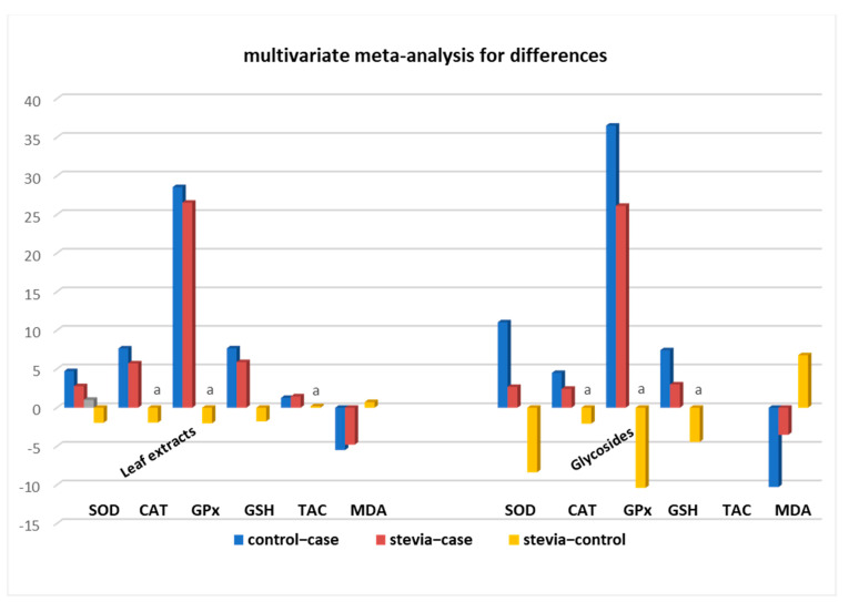
Stratification analysis of all of the assays’ measurements according to the administered leaf extracts or compounds revealed that with stevia glycosides the differences ‘control–case’ were much higher (in absolute values) than the differences ‘stevia–case’ (SOD: 11.04 vs. 2.67, GSH: 7.42 vs. 2.99, and MDA: −10.28 vs. −3.51) suggesting that stevia glycosides cannot restore antioxidant activity to the extent that whole leaf extracts can (Table 2 and Figure 3). This is further verified by the fact that ‘stevia–control’ differences (for SOD, GSH, and MDA assays) were much higher (absolute values) than the ‘stevia–case’ differences in the ‘glycosides’ datasets (Table 2). In the CAT and GPx assays, all of the differences were not statistically significant, perhaps due to the limited number of studies meaning that they could not provide a robust result. Importantly, significant restoration of antioxidant activity of diseased animals was observed with administration of whole leaf extracts in studies performed with CAT and GPx assays, as suggested by the non-statistically significant differences in the ‘stevia–control’ values (p-values 0.08 and 0.70), respectively. Moreover, the fact that significant restoration of antioxidant activity by stevia leaf extracts was seen with the SOD, GSH, TAC, and MDA assays to 59%, 77%, 111%, and 87%, respectively, further supports our findings that whole stevia leaf extracts can better restore oxidation markers in experimental animals compared with pure stevia glycosides, which showed restoration ability ranging from 24% (SOD) to 71% (GPx) (Table 2 and Figure 3).
Figure 3.
Stevia whole leaf extracts can better restore antioxidant markers of diseased animal tissues compared to pure steviol glycosides. a: non-statistically significant difference between compared groups.
Moreover, meta-regression analysis for the datasets from the SOD, GSH, TAC, and MDA assays testing leaf extracts and glycosides was also employed and revealed a statistically significant dependence of the results on the type of treatment (leaf extract or glycosides) with a p-value = 0.0023. Taken together, the results above indicate that, in all assays, treatment with whole leaf extract of stevia exhibited a remarkable restorative potential as opposed to treatment with isolated glycosides. Consequently, further analyses were performed with whole leaf extracts.
3.3. Datasets from SOD, CAT, GSH, and TAC Assays Can Be Combined for Meta-Analysis: Meta-Regression Analysis
We next wondered whether we could synthesize the results of all experiments, despite the different values each exhibited, in order to perform multivariate meta-analysis on the combined data from all assays. Towards this, multivariate meta-regression analysis for leaf extract datasets on the differences ‘control–case’ and ‘stevia–case’ from all assays was executed and revealed a relationship between the assay method and their outcomes (p = 0.000), with GPx and MDA results being the assays with statistically significant differences compared to the rest of them (coefficient = 6.14, p = 0.005; and coefficient = −10.95, p = 0.000, respectively, and constant = 5.31 for the difference ‘control–case’). Subsequently, meta-regression analysis of leaf extract datasets on the differences ‘control–case’ and ‘stevia–case’ for the rest of the assays, i.e., SOD, CAT, GSH, and TAC revealed the absence of any relationship between assay results and the type of assay (p = 0.184). As a result, datasets on the differences ‘control–case’ and ‘stevia–case’ from the above four assays can be treated as coming from the same source and can be combined in consequent meta-analyses.
3.4. Stratification Meta-Analysis for Datasets from Leaf Extracts
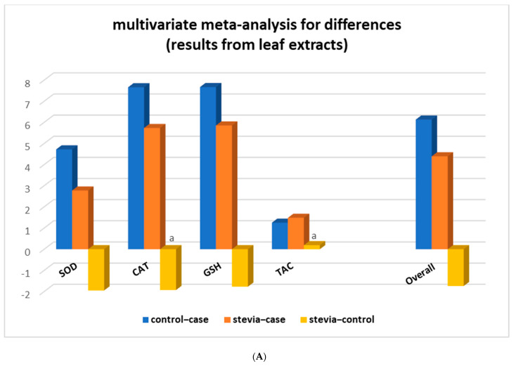
Multivariate meta-analysis was then performed with the combined leaf extract datasets from four assays (SOD, CAT, GSH, and TAC) comprising 50 studies in which rats had only leaf extracts administered. Analysis showed that in all assays, the ‘stevia–control’ differences were lower than ‘control–case’ and ‘stevia–case’, and in the CAT and TAC assays, they were not even statistically significant (Figure 4A and Table 3).
Figure 4.
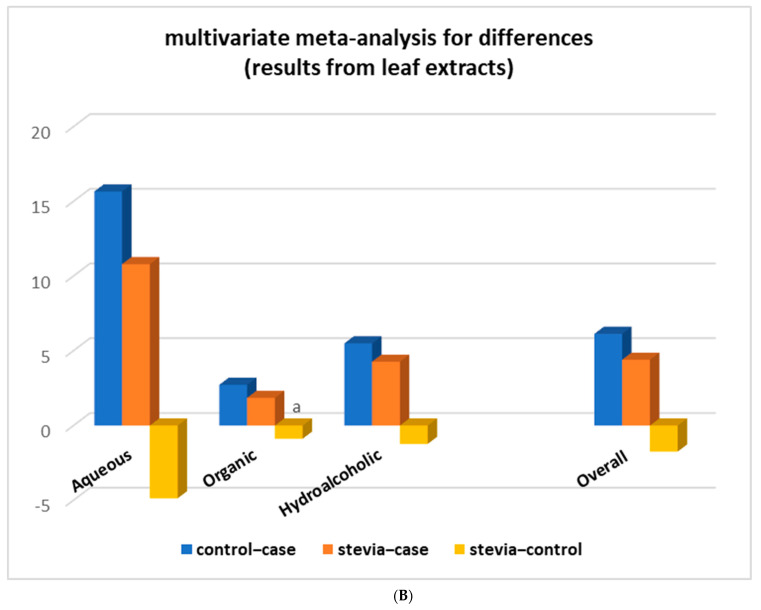
Stratification of the meta-analysis according to (A) type of assay and (B) type of leaf extract. a: non-statistically significant difference between compared groups. Overall: results from combined SOD, CAT, GSH, and TAC assays.
Table 3.
Results of the multivariate meta-analysis for the assays superoxide dismutase (SOD), catalase activity (CAT), reduced glutathione (GSH), and total antioxidant capacity (TAC) together and separated according to leaf extract, and stratification according to different types of extract. Listed information includes differences between groups with the 95% confidence intervals.
| SOD, CAT, GSH, TAC | SOD | CAT | GSH | TAC | ||||||||||||||||
|---|---|---|---|---|---|---|---|---|---|---|---|---|---|---|---|---|---|---|---|---|
| Difference | Coef. | p-value | 95% CI | # Studies | Coef. | p-value | 95% CI | # Studies | Coef. | p-value | 95% CI | # Studies | Coef. | p-value | 95% CI | # Studies | Coef. | p-value | 95% CI | # Studies |
| Combined leaf extract | ||||||||||||||||||||
| control−case | 6.13 | 0.00 | 4.42, 7.84 | 50 | 4.72 | 0.00 | 3.08, 6.37 | 15 | 7.66 | 0.00 | 3.01, 12.33 | 13 | 7.67 | 0.00 | 3.72, 11.61 | 20 | 1.25 | 0.00 | 0.46, 2.03 | 2 |
| stevia–case | 4.39 | 0.00 | 3.03, 5.74 | 50 | 2.77 | 0.00 | 1.39, 4.15 | 15 | 5.73 | 0.00 | 1.97, 9.5 | 13 | 5.85 | 0.00 | 2.79, 8.99 | 20 | 1.48 | 0.12 | −0.39, 3.27 | 2 |
| stevia–-control | −1.74 | 0.00 | −2.46, −1.02 | 50 | −1.95 | 0.00 | −3.18, −0.72 | 15 | −1.93 | 0.07 a | −4.07, 0.19 | 13 | −1.77 | 0.00 | −2.92, −0.62 | 20 | 0.19 | 0.79 a | −1.24, 1.62 | 2 |
| Aqueous | ||||||||||||||||||||
| control–case | 15.66 | 0.00 | 3.92, 27.40 | 9 | 22.47 | 0.26 | −17.18, 62.12 | 2 | 13.53 | 0.09 | −2.03, 29.07 | 6 | ||||||||
| stevia–case | 10.79 | 0.01 | 2.17, 19.40 | 9 | 15.70 | 0.22 | −9.34, 40.75 | 2 | 9.78 | 0.11 | −2.34, 21.91 | 6 | ||||||||
| stevia–control | −4.87 | 0.00 | −8.34, −1.41 | 9 | −6.76 | 0.36 a | −21.39, 7.86 | 2 | −3.74 | 0.04 | −7.30, −0.17 | 6 | ||||||||
| Organic | ||||||||||||||||||||
| control–case | 2.73 | 0.00 | 1.49, 3.98 | 13 | 2.45 | 0.00 | 1.18, 3.73 | 5 | 5.18 | 0.06 | −0.35, 10.71 | 3 | 2.08 | 0.01 | 0.59, 3.55 | 4 | ||||
| stevia–case | 1.85 | 0.00 | 0.77, 2.94 | 13 | 0.33 | 0.65 | −1.11, 1.78 | 5 | 4.09 | 0.03 | 0.29, 7.88 | 3 | 2.22 | 0.00 | 1.57, 2.87 | 4 | ||||
| stevia–control | −0.88 | 0.13 a | −2.03, 0.27 | 13 | −2.12 | 0.08 a | −4.52, 0.28 | 5 | −1.09 | 0.29 a | −3.15, 0.97 | 3 | 0.14 | 0.83 a | −1.16, 1.45 | 4 | ||||
| Hydroalcoholic | ||||||||||||||||||||
| control–case | 5.49 | 0.00 | 4.20, 6.76 | 26 | 4.75 | 0.00 | 4.17, 5.33 | 9 | 6.58 | 0.00 | 3.03, 10.12 | 7 | 6.38 | 0.00 | 3.35, 9.39 | 9 | ||||
| stevia–case | 4.26 | 0.00 | 2.64, 5.87 | 26 | 3.44 | 0.00 | 2.28, 4.60 | 9 | 5.48 | 0.06 | −0.25, 11.21 | 7 | 5.29 | 0.00 | 1.83, 8.72 | 9 | ||||
| stevia–control | −1.23 | 0.00 | −2.03, −0.43 | 26 | −1.31 | 0.03 | −2.49, −0.11 | 9 | −1.09 | 0.49 a | −4.19, 1.99 | 7 | −1.09 | 0.01 | −1.92, −0.27 | 9 | ||||
p-values with a represent non-statistically significant differences between ‘stevia’ and ‘control’ groups; #: number of.
Stratification analysis according to the type of stevia extract revealed that organic extracts were the most potent in restoring the antioxidant activity in diseased rats administered with various types of leaf extracts (Table 3). The difference ‘stevia – control’ was not statistically significant (d = −0.88 with p-value = 0.13) for the combined all four assays datasets, nor for each assay separately. The same ‘stevia–control’ differences for aqueous and hydroalcoholic types of extracts, though statistically significant, were still much lower than the other differences (Figure 4B and Table 3). In addition, stratification by type of extract within the CAT assay results showed that treatment with aqueous stevia leaf extracts can also completely restore the CAT oxidative stress marker of diseased animals to the levels of the control animals as the differences ‘stevia – control’ were not statistically significant, i.e., for the aqueous (d = −6.76 and p-value = 0.36), for the organic (d = −1.09 and p-value = 0.29), and for the hydroalcoholic extracts (d = −1.09 and p-value = 0.49) (Table 3). The four studies that investigated the effect of stevia leaf extracts on diseased rats with the GSH assay showed that only organic stevia leaf extracts restored GSH values of diseased animals (d = 0.14 and p-value = 0.83). Similarly, no significant difference was observed for the ‘stevia–control’ contrast in the SOD assay when organic extracts were administered to diseased rats.
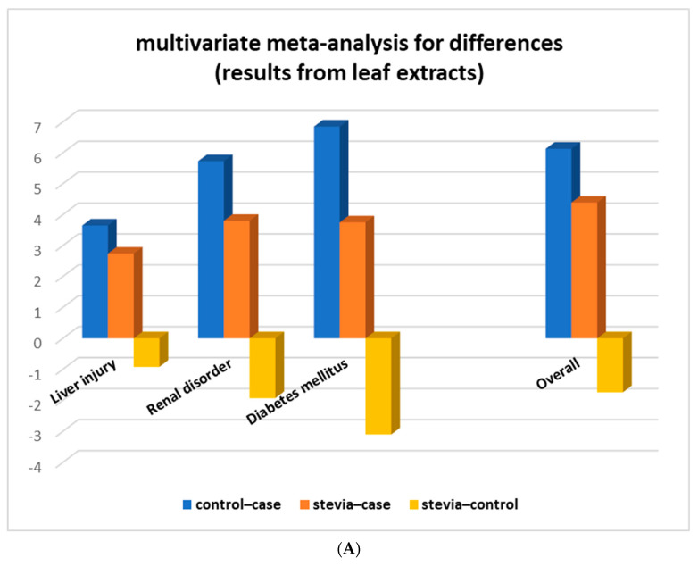
We next stratified our analysis according to the different types of disease that rats had suffered from (Table 4). The data were grouped into three main categories: liver injury (acute liver injury, hepatotoxic, liver cirrhosis, liver fibrosis), renal disorder (hyperuricemia, renal ischemia/reperfusion), and diabetes mellitus. The results of the multivariate meta-analysis demonstrated full restoration of antioxidant SOD, CAT, and GSH activity of diseased rats suffering from diabetes mellitus as the differences ‘stevia – control’ were not statistically significant (SOD: d = −0.14 with p-value = 0.94, CAT: d = −0.14 with p-value = 0.94, and GSH: d = −1.73 with p-value = 0.06). For diseased rats that suffered from liver injury or renal disorder, the multivariate meta-analysis results revealed a significant restoration, though not full, of the oxidative stress markers when they were administered stevia leaf extracts (Table 4 and Figure 5A).
Table 4.
Results of the multivariate meta-analysis for the assays superoxide dismutase (SOD), catalase activity (CAT), reduced glutathione (GSH), and total antioxidant capacity (TAC) together, and malondialdehyde (MDA) and glutathione peroxidase (GPx) separated according to disease, and stratification according to different types of disease. Listed information includes differences between groups with the 95% confidence intervals.
| SOD, CAT, GSH, TAC | SOD | CAT | GSH | TAC | ||||||||||||||||
|---|---|---|---|---|---|---|---|---|---|---|---|---|---|---|---|---|---|---|---|---|
| Difference | Coef. |
p- value |
95% CI | # Studies | Coef. |
p- value |
95% CI | # Studies | Coef. |
p- value |
95% CI | # Studies | Coef. |
p- value |
95% CI | # Studies | Coef. |
p- value |
95% CI | # Studies |
| Overall | ||||||||||||||||||||
| control–case | 6.13 | 0.00 | 4.42, 7.84 | 50 | 4.72 | 0.00 | 3.08, 6.37 | 15 | 7.67 | 0.00 | 3.01, 12.33 | 13 | 7.67 | 0.00 | 3.72, 11.61 | 20 | 1.24 | 0.00 | 0.46, 2.03 | 2 |
| stevia–case | 4.39 | 0.00 | 3.03, 5.74 | 50 | 2.77 | 0.00 | 1.39, 4.15 | 15 | 5.73 | 0.00 | 1.97, 9.5 | 13 | 5.89 | 0.00 | 2.79, 8.99 | 20 | 1.44 | 0.12 | −0.39, 3.27 | 2 |
| stevia–control | −1.74 | 0.00 | −2.46, −1.02 | 50 | −1.95 | 0.00 | −3.18, −0.72 | 15 | −1.93 | 0.07 a | −4.07, 0.19 | 13 | −1.77 | 0.00 | −2.92, −0.62 | 20 | 0.19 | 0.79 a | −1.24, 1.62 | 2 |
| Diabetes mellitus | ||||||||||||||||||||
| control–case | 6.85 | 0.00 | 4.92, 8.80 | 34 | 3.41 | 0.00 | 2.28, 4.54 | 8 | 6.35 | 0.00 | 2.28, 10.43 | 6 | 8.04 | 0.00 | 3.70, 12.37 | 10 | ||||
| stevia–case | 3.75 | 0.00 | 2.22, 5.29 | 34 | 1.67 | 0.07 | −0.16, 3.49 | 8 | 6.22 | 0.07 | −0.43, 12.86 | 6 | 6.31 | 0.00 | 2.69, 9.92 | 10 | ||||
| stevia–control | −3.1 | 0.00 | −4.73, −1.48 | 34 | −1.74 | 0.08 a | −3.67, 0.19 | 8 | −0.14 | 0.94 a | −3.46, 3.19 | 6 | −1.73 | 0.06 a | −3.51, 0.05 | 10 | ||||
| Liver injury | ||||||||||||||||||||
| control–case | 3.65 | 0.00 | 2.76, 4.55 | 17 | 3.31 | 0.00 | 1.25, 5.37 | 2 | 5.49 | 0.04 | 0.35, 10.63 | 3 | 2.77 | 0.00 | 2.02, 3.52 | 5 | ||||
| stevia–case | 2.74 | 0.00 | 1.98, 3.49 | 17 | 2.13 | 0.04 | 0.08, 4.17 | 2 | 3.82 | 0.08 | −0.44, 8.09 | 3 | 2.14 | 0.00 | 1.48, 2.79 | 5 | ||||
| stevia–control | −0.92 | 0.00 | −1.3, −0.54 | 17 | −1.18 | 0.00 | −1.88, −0.48 | 2 | −1.67 | 0.00 | −2.72, −0.62 | 3 | −0.63 | 0.01 | −1.09, −0.17 | 5 | ||||
| Renal disorder | ||||||||||||||||||||
| control–case | 5.73 | 0.00 | 4.43, 7.03 | 10 | 5.53 | 0.00 | 4.05, 7.02 | 4 | 6.96 | 0.00 | 5.31, 8.61 | 2 | 5.65 | 0.00 | 2.52, 8.77 | 4 | ||||
| stevia–case | 3.80 | 0.00 | 2.63, 4.96 | 10 | 3.97 | 0.00 | 2.21, 5.73 | 4 | 3.15 | 0.01 | 0.69, 5.61 | 2 | 4.06 | 0.00 | 1.72, 6.39 | 4 | ||||
| stevia–control | −1.93 | 0.00 | −2.66, −1.19 | 10 | −1.56 | 0.00 | −2.43, −0.70 | 4 | −3.82 | 0.02 | −7.07, −0.55 | 2 | −1.59 | 0.00 | −2.51, −0.66 | 4 | ||||
p-values with a represent non-statistically significant differences between ‘stevia’ and ‘control’ groups; #: number of.
Figure 5.
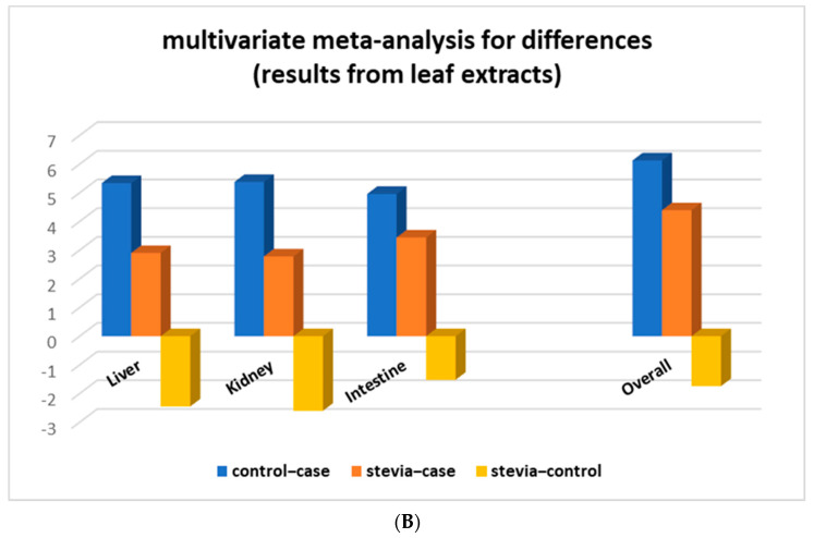
Stratification of the meta-analysis according to (A) type of disease and (B) type of tissue. Overall: results from combined SOD, CAT, GSH, and TAC assays.
We next wondered whether specific rat tissues or cell types could have been affected differently from stevia leaf extract administration to the experimental animal. Thus, we stratified our analysis on the combined (only leaf extract) datasets from the four assays (SOD, CAT, GSH, and TAC) according to different types of animal tissue (Table 5). The results revealed that complete restoration of the antioxidant enzyme SOD’s activity was seen in liver samples, since d = −0.32 with p-value = 0.67. However, it should be noted that in all types of tissues, the values of the differences ‘stevia – control’ suggested a significant restoration of the oxidative stress markers of the diseased animals when receiving stevia leaf extracts (Table 5 and Figure 5B). Taken together, the above results support that stevia leaf extracts do exert antioxidant activity on diseased animals and especially to those that suffer from diabetes mellitus.
Table 5.
Results of the multivariate meta-analysis for the assays superoxide dismutase (SOD), catalase activity (CAT), reduced glutathione (GSH), malondialdehyde (MDA), and total antioxidant capacity (TAC)) together and separated, malondialdehyde (MDA) and glutathione peroxidase (GPx), and stratification according to different types of tissues. Listed information includes differences between groups with the 95% confidence intervals.
| SOD, CAT, GSH, TAC | SOD | CAT | GSH | TAC | ||||||||||||||||
|---|---|---|---|---|---|---|---|---|---|---|---|---|---|---|---|---|---|---|---|---|
| Difference | Coef | p-value | 95% CI | # Studies | Coef | p-value | 95% CI | # Studies | Coef. | p-value | 95% CI | # Studies | Coef. | p-value | 95% CI | # Studies | Coef. | p-value | 95% CI | # Studies |
| Overall | ||||||||||||||||||||
| control–case | 6.13 | 0.00 | 4.42, 7.84 | 50 | 4.72 | 0.00 | 3.08, 6.37 | 15 | 7.67 | 0.00 | 3.01, 12.33 | 13 | 7.67 | 0.00 | 3.72, 11.61 | 20 | 1.24 | 0.00 | 0.46, 2.03 | 2 |
| stevia–case | 4.39 | 0.00 | 3.03, 5.74 | 50 | 2.77 | 0.00 | 1.39, 4.15 | 15 | 5.73 | 0.00 | 1.97, 9.5 | 13 | 5.89 | 0.00 | 2.79, 8.99 | 20 | 1.44 | 0.12 | −0.39, 3.27 | 2 |
| stevia–control | −1.74 | 0.00 | −2.46, −1.02 | 50 | −1.95 | 0.00 | −3.18, −0.72 | 15 | −1.93 | 0.07 a | −4.07, 0.19 | 13 | −1.77 | 0.00 | −2.92, −0.62 | 20 | 0.19 | 0.79 a | −1.24, 1.62 | 2 |
| Liver | ||||||||||||||||||||
| control–case | −5.34 | 0.00 | 3.73, 6.95 | 31 | 2.93 | 0.00 | 1.59, 4.28 | 6 | 4.73 | 0.00 | 1.85, 7.61 | 5 | 4.56 | 0.00 | 2.04, 7.07 | 9 | ||||
| stevia–case | 2.90 | 0.00 | 2.12, 3.67 | 31 | 2.62 | 0.00 | 0.79, 4.45 | 6 | 3.04 | 0.01 | 0.61, 5.47 | 5 | 3.49 | 0.01 | 0.97, 6.00 | 9 | ||||
| stevia–control | −2.45 | 0.00 | −3.99, −0.9 | 31 | −0.32 | 0.67 a | −1.79, 1.16 | 6 | −1.69 | 0.00 | −2.34, −1.04 | 5 | −1.07 | 0.04 | −2.09, −0.05 | 9 | ||||
| Kidney | ||||||||||||||||||||
| control–case | 5.38 | 0.00 | 2.96, 7.79 | 8 | 3.85 | 0.00 | 2.13, 5.57 | 2 | 4.85 | 0.01 | 1.36, 8.34 | 2 | 6.72 | 0.01 | 1.84, 11.60 | 4 | ||||
| stevia–case | 2.78 | 0.01 | 0.58, 4.99 | 8 | −0.12 | 0.94 | −3.28, 3.05 | 2 | 2.44 | 0.20 | −1.33, 6.21 | 2 | 4.53 | 0.01 | 1.00, 8.05 | 4 | ||||
| stevia–control | −2.60 | 0.00 | −3.46, −1.73 | 8 | −3.96 | 0.00 | −6.12, −1.81 | 2 | −2.41 | 0.00 | −3.35, −1.48 | 2 | −2.19 | 0.00 | −3.67, −0.71 | 4 | ||||
| Intestine (duodenum–Jejunum–ileum) | ||||||||||||||||||||
| control–case | 4.96 | 0.00 | 3.36, 6.56 | 6 | 4.28 | 0.00 | 1.96, 6.61 | 3 | ||||||||||||
| stevia–case | 3.44 | 0.00 | 1.96, 4.91 | 6 | 3.08 | 0.00 | 1.18, 4.98 | 3 | ||||||||||||
| stevia–control | −1.52 | 0.00 | −2.00, −1.05 | 6 | −1.20 | 0.00 | −1.92, −0.48 | 3 | ||||||||||||
p-values with a represent non-significant differences between ‘stevia’ and ‘control’ groups; #: number.
3.5. Meta-Analysis of Lipid Peroxidation (MDA) Assay Datasets
MDA is a lipid peroxidation product, and thus, the MDA assay is used as a marker for oxidative stress, the more the oxidative stress, the higher the MDA assay values. Thus, control experimental animal tissues show low MDA levels while diseased animals present higher MDA levels. Due to this particularity and because of the different biochemical principle that this assay is based on, MDA estimates could not be directly compared to the previously analyzed oxidation markers, and MDA results could not be analyzed together with the rest.
It is important to note here that whole leaf extracts reverted the oxidation status of diseased experimental animals while pure glycosides did not, as shown in Table 2 (difference ‘stevia–control’ leaf extract: 0.71, p-value 0.00; glycosides: 6.77, p-value 0.01). Consequently, a meta-analysis for stevia leaf extract datasets from 25 studies performed with the MDA assay showed significant restoration of the oxidation status of diseased rats when administered stevia leaf extracts of 87.07% (from −5.49 (p = 0.00) to −4.78 (p = 0.00)) (Table 6). It is worth noting that although the ‘stevia–control’ difference is statistically significant, the values of the differences between the ‘control’ and ‘case’ groups and the ‘control’ and ‘stevia’ groups decreased significantly.
Table 6.
Results of the multivariate meta-analysis for the assay of malondialdehyde (MDA) and stratification according to different types of extract, disease, and tissue. Listed information includes differences between groups with the 95% confidence intervals.
| Difference | Coef. | p-Value | 95% CI | # of Studies | |
|---|---|---|---|---|---|
| Leaf Extract | control–case | −5.49 | 0.00 | −7.15, 0.83 | 25 |
| stevia–case | −4.78 | 0.00 | −6.37, −3.19 | 25 | |
| stevia–control | 0.71 | 0.00 | 0.24, 1.17 | 25 | |
| Aqueous | control–case | −3.85 | 0.00 | −5.57, −2.14 | 8 |
| stevia–case | −3.3 | 0.00 | −4.94, −1.65 | 8 | |
| stevia–control | 0.56 | 0.01 | 0.15, 0.96 | 8 | |
| Organic | control–case | −5.68 | 0.00 | −8.97, −2.39 | 6 |
| stevia–case | −5.06 | 0.00 | −6.82, −3.30 | 6 | |
| stevia–control | 0.62 | 0.48 a | −1.10, 2.34 | 6 | |
| Hydroalcoholic | control–case | −8.33 | 0.00 | −14.08, −2.59 | 6 |
| stevia–case | −7.35 | 0.01 | −13.01, −1.69 | 6 | |
| stevia–control | 0.98 | 0.01 | 0.25, 1.71 | 6 | |
| Diabetes melittus | control–case | −7.33 | 0.00 | −12.25, 2.42 | 13 |
| stevia–case | −6.93 | 0.00 | −11.73, −2.12 | 13 | |
| stevia–control | 0.41 | 0.12 a | −0.10, 0.92 | 13 | |
| Liver injury | control–case | −6.33 | 0.00 | −10.04, −2.61 | 6 |
| stevia–case | −4.37 | 0.00 | −6.61, −2.13 | 6 | |
| stevia–control | 1.95 | 0.02 | 0.35, 3.55 | 6 | |
| Renal disorder | control–case | −4.53 | 0.00 | −5.77, −3.29 | 5 |
| stevia–case | −3.82 | 0.00 | −5.11, −2.53 | 5 | |
| stevia–control | 0.71 | 0.01 | 0.17, 1.25 | 5 | |
| Blood | control–case | −1.81 | 0.15 | −4.30, 0.67 | 2 |
| stevia–case | −1.32 | 0.19 | −3.29, 0.64 | 2 | |
| stevia–control | 0.49 | 0.26 a | −0.36, 1.35 | 2 | |
| Liver | control–case | −6.96 | 0.00 | −9.72, −4.19 | 11 |
| stevia–case | −5.91 | 0.00 | −8.66, −3.16 | 11 | |
| stevia–control | 1.05 | 0.04 | 0.04, 2.05 | 11 | |
| Kidney | control–case | −3.13 | 0.00 | −4.59, −1.66 | 4 |
| stevia–case | −2.45 | 0.00 | −3.91, −0.99 | 4 | |
| stevia–control | 0.68 | 0.13 a | −0.21, 1.57 | 4 | |
| Intestine (duodenum–jejunum–ileum) | control–case | −4.99 | 0.00 | −7.00, −2.98 | 3 |
| stevia–case | −4.64 | 0.00 | −6.54, −2.74 | 3 | |
| stevia–control | 0.35 | 0.23 a | −0.23, 0.93 | 3 | |
| Pancreas | control–case | −3.91 | 0.02 | −7.11, −0.71 | 2 |
| stevia–case | −3.89 | 0.01 | −6.64, −1.13 | 2 | |
| stevia–control | 0.02 | 0.95 a | −0.81, 0.85 | 2 |
p-values with a represent non-statistically significant differences in MDA assay results; #: number of.
Next, we stratified the multivariate meta-analysis of the MDA assay datasets according to type of stevia leaf extract, tissue, and disease and revealed that the restoration of the MDA content of samples of more than 70% was seen under every tested parameter. In particular, diseased rats administered organic extracts showed that the difference ‘stevia–control’ was not statistically significant (d = 0.62, p-value = 0.48), connotating a complete oxidative status restoration. This finding is in accordance with results from other assays (SOD, CAT, GSH, TAC) verifying that organic extracts possess high antioxidant potency.
Stratification of the meta-analysis according to the disease type (Table 6) highlighted that in diabetes, the low MDA levels of diseased animals are completely restored after leaf extract administration (‘stevia–control’ −0.41, p = 0.12). This observation supports our previous finding, that with all tested assays (SOD, CAT, GSH) whole stevia leaf extract administration completely restores all antioxidant markers levels, especially in diabetic experimental animals (Table 4).
Importantly, the ‘stevia–control’ differences were all found to not be statistically significant for all tissues tested, apart from liver (kidney: d = 0.68 (p-value = 0.13), duodenum–jejunum–ileum: d = 0.35 (p-value = 0.23), pancreas: d = 0.02 (p-value = 0.95)). Measurements in liver tissue showed a marginally statistically significant difference between the control and stevia groups (p = 0.04); however, restoration of the MDA measurements was up to 85% for the leaf-extract-treated diseased animals.
Taken together the above data show that even if complete restoration of antioxidant markers was not statistically significantly accomplished in diseased rats that were administered stevia, partial restoration did occur. As shown in Figure 3 this restoration rises from 65% to 85% for all antioxidant markers, reinforcing the notion that stevia leaf extracts do possess significant antioxidant activity when administered orally.
4. Discussion
Recent research towards sustainable solutions for global health is guided from the need to produce natural products with efficient health promoting properties that present a low incidence of side effects. Stevia is an aromatic plant known as a no-calories sweetener. By the identification of many bioactive compounds contained in its leaf extracts, stevia has emerged as a health beneficial aromatic herbal, characterizing it as high-added-value plant within the agrifood sector [69,74,75]. The antioxidant potency of stevia is currently being extensively studied with a variety of approaches (direct or indirect) entailing various (bio)chemical principles and cascades, using in vitro and in vivo systems. The present meta-analysis is the first effort to quantitatively synthesize all of the published evidence and assess the antioxidant effect of stevia phytoconstituents when administered to experimentally diseased animals.
Our findings indicate a statistically significant antioxidant restorative effect of oxidative status markers of experimentally diseased animals after they have been administered stevia leaf extracts. By combining data from 24 articles comprising 104 individual studies, we show this restorative activity is mainly exerted by stevia whole leaf extracts and to a minor extent by pure glycosides. Our findings are based on datasets coming from six different methods (SOD, CAT, GPx, GSH, TAC, and MDA) measuring either antioxidant enzyme activities (SOD, CAT, GPx) or products linked to the activities of antioxidant enzymes, or even oxidation products such as MDA, which measures the lipid peroxidation status of a cell. A stratification analysis of the stevia whole leaf extract datasets revealed a robust antioxidant effect of organic extracts of stevia leaves. Moreover, diabetes mellitus emerged as the disease with the highest restorative response to stevia leaf extract administration. Concerning the tissues on which the oxidative markers were tested, all showed significant restoration, from 65% to 85%, regardless of the type of assay employed.
The challenge that we faced in the present study was not only to summarize the evidence and draw conclusions for a positive relationship between stevia consumption and antioxidant activity, but to synthesize measurements coming from three different situations from six different assays on tissues from experimental rats.
Firstly, since we had to compare values from more than two animal groups (control, diseased, and stevia-treated) we employed multivariate meta-analysis [46,48,49]. This approach enabled us to perform multiple comparisons avoiding the need for the usual adjustments in order to avoid type I errors. Moreover, employing multivariate meta-analysis allowed us to obtain the estimated covariant matrix from which we could estimate the confidence intervals of the differences we measured and utilize the statistical significance of the differences [49,76].
The second challenge we faced was whether we could combine datasets from some or all assays in order to obtain results with more power. For this, multivariate meta-regression analysis was recruited to control for potential effect modifiers or any association between group differences and various assays [77,78]. The assay results were significantly dependent on the MDA assay (as expected), and on the GPx method as well. Interestingly, no dependence on the method was seen when meta-regression was employed with datasets from the SOD, CAT, GSH, and TAC assays. Given that the SOD, CAT, GPX, and GSH assays measure antioxidant enzyme activities and all act in the same pathway, i.e., one with being downstream from another, one would expect that their datasets could be combined. Employing multivariate meta-regression, we proved this assumption as correct; however, given the available data, only the SOD, CAT, and GSH assay datasets could be combined. In addition, we proved that the TAC assay datasets could also be pooled to obtain more robust estimates. Collectively, multivariate meta-regression meta-analysis enabled us to pool data from different sources, thus increasing the power of our analysis, a result that would not be possible to obtain under a unified/univariate meta-analysis [49].
Our initial hypothesis, that both stevia leaf extracts and pure stevia glycosides could possess antioxidant activity, though to different extents, appears to have a biological basis. Polyphenolic compounds from a plethora of plant resources have repeatedly been shown to display antioxidant potential [79,80,81] in direct assays [20,82] and exert effective antioxidant activity in vivo, including cell lines [20], experimental animals [83], or even humans [84]. Our findings suggest that stevia glycosides do possess a limited antioxidant activity. Glycosides are complex molecules, constituting a sugar molecule (glycone) bound to another functional group (aglycone), found in many living organisms. Although sugars have never been reported to convey any antioxidant activity, other functional groups may possess various functions. The aglycone in stevia glycosides is the diterpene steviol that is the major metabolite produced in the lower gastrointestinal tract of mammals [3]. Thus, the limited antioxidant activity seen with stevia glycosides could be attributed to the diterpene steviol. This is in accordance with previous findings showing that a combination of stevia glycosides could increase the SOD and CAT activities of rat cardiac fibroblasts when exposed to hydrogen peroxide [85]. The antioxidant properties of stevia glycosides were also observed with direct assays on food and fruit beverages [86,87]. Moreover, steviol glycosides were found to restore oxidative stress status in fish [88].
We acknowledge that the present meta-analysis possibly has some limitations that could have affected the integrity of our results. The studies recruited in the present meta-analysis covered a diverse range of stevia extracts or pure glycoside dosages and time settings. The variability in the interventions concerning the time that the intervention started (before or after the animals were diseased) and the time that intervention started after the induction of disease, together with limited information given in each study, made it impossible to stratify according to dosage. A meta-regression analysis to estimate the association of antioxidant markers with extract preparation, dosage, or time of intervention would be of high value; yet, such data were not available from all studies to perform the corresponding tests. However, our analysis could account for the type of extracts given as intervention, the type of tissue, and the disease by stratifying our analysis accordingly. We could not account for intrinsic differences within studies that constitute confounding factors that can potentially influence the quality of the parameters assessed. For example, sample handling (time and temperature of sample storage before being assayed, homogenization protocols) or the different genetic backgrounds of rats used could have affected the assays’ outcomes, and consequently, partially, the results of the present meta-analysis. Nonetheless, we performed the meta-analysis carefully, taking all these considerations into account. Finally, despite our systematic approach and effort to include all existing studies, gray literature bias cannot be completely ruled out.
Increased stevia consumption has emerged from global awareness of (a) the risks of sugar-related diseases (obesity, diabetes); (b) the fact that synthetic sweeteners may lead to long-term health problems; and (c) that stevia, being a natural product, has, most probably, no long-term side effects. The bottleneck in the industrial use of stevia is the cost related to steviol glycoside extraction and purification. Sustainability issues have also emerged concerning stevia’s waste disposal [89]. The present meta-analysis is the first effort to quantitatively estimate the effect of stevia leaf extract consumption, in addition to steviol glycosides, highlighting an additional—yet unexploited—health promoting property that can give stevia (and perhaps stevia waste) a second chance to rescue human health and global sustainability. Only if uniform settings are used can we obtain accurate comparisons of types of stevia extracts, dosage, and time interventions, and more in vivo experiments can be performed in order to obtain accurate insights into, to what extent can human health benefit from stevia consumption.
Author Contributions
Conceptualization, G.G.B.; methodology, M.P., P.I.K., G.G.B. and P.G.B.; software, M.P., P.I.K., P.G.B. and G.G.B.; validation, M.P., P.I.K., P.G.B. and G.G.B.; formal analysis, M.P., P.I.K. and G.G.B.; investigation, M.P. and G.G.B.; resources, M.P., P.I.K., P.G.B. and G.G.B.; data curation, M.P., and G.G.B.; writing—original draft preparation, M.P. and G.G.B.; writing—review and editing, M.P., P.I.K., P.G.B. and G.G.B.; visualization, M.P. and G.G.B.; supervision, G.G.B.; project administration, G.G.B.; funding acquisition, G.G.B. and P.G.B. All authors have read and agreed to the published version of the manuscript.
Institutional Review Board Statement
The study did not require ethical approval.
Informed Consent Statement
Not applicable.
Data Availability Statement
All data are within the manuscript.
Conflicts of Interest
The authors declare no conflict of interest.
Funding Statement
PB and GB acknowledge funding from “Molecular identification and exploitation of indigenous hop varieties for the production of high-quality beers with designation of origin-PAZL (Production Analysis of Hop Beers)”, Grant Number STER1-0026139, funded by the Operational Program “STEREA ELLADA” 2014–2020 (Strengthening of Research Projects, Technological Development and Innovation in the fields of RIS3). MP and PK acknowledge funding from “GENOMIC-OASIS: GENOMIC analysis of Organisms of Agricultural and live Stock Interest in Sterea”, Grant Number (MIS) 5045902, funded by the Operational Programme” Competitiveness, Entrepreneurship and Innovation” (NSRF 2014–2020) and co-financed by Greece and the European Union (European Regional Development Fund). The funders had no role in the study design, data collection and analysis, decision to publish, or preparation of the manuscript.
Footnotes
Disclaimer/Publisher’s Note: The statements, opinions and data contained in all publications are solely those of the individual author(s) and contributor(s) and not of MDPI and/or the editor(s). MDPI and/or the editor(s) disclaim responsibility for any injury to people or property resulting from any ideas, methods, instructions or products referred to in the content.
References
- 1.Rojas E., Bermúdez V., Motlaghzadeh Y., Mathew J., Fidilio E., Faria J., Rojas J., de Bravo M.C., Contreras J., Mantilla L.P., et al. Stevia rebaudiana Bertoni and its effects in human disease: Emphasizing its role in inflammation, atherosclerosis and metabolic syndrome. Curr. Nutr. Rep. 2018;7:161–170. doi: 10.1007/s13668-018-0228-z. [DOI] [PubMed] [Google Scholar]
- 2.Peteliuk V., Rybchuk L., Bayliak M., Storey K.B., Lushchak O. Natural sweetener Stevia rebaudiana: Functionalities, health benefits and potential risks. EXCLI J. 2021;20:1412. doi: 10.17179/excli2021-4211. [DOI] [PMC free article] [PubMed] [Google Scholar]
- 3.Orellana-Paucar A.M. Steviol Glycosides from Stevia rebaudiana: An Updated Overview of Their Sweetening Activity, Pharmacological Properties, and Safety Aspects. Molecules. 2023;28:1258. doi: 10.3390/molecules28031258. [DOI] [PMC free article] [PubMed] [Google Scholar]
- 4.Geuns J.M.C. Stevioside. Phytochemistry. 2003;64:913–921. doi: 10.1016/S0031-9422(03)00426-6. [DOI] [PubMed] [Google Scholar]
- 5.De Oliveira A.J.B., Gonçalves R.A.C., Chierrito T.P.C., Dos Santos M.M., De Souza L.M., Gorin P.A.J., Sassaki G.L., Iacomini M. Structure and degree of polymerisation of fructooligosaccharides present in roots and leaves of Stevia rebaudiana (Bert.) Bertoni. Food Chem. 2011;129:305–311. doi: 10.1016/j.foodchem.2011.04.057. [DOI] [PubMed] [Google Scholar]
- 6.Chaturvedula V.S.P., Zamora J. Reversed-Phase HPLC Analysis of Steviol Glycosides Isolated from Stevia rebaudiana Bertoni. Food Nutr. Sci. 2014;5:1711–1716. doi: 10.4236/fns.2014.517184. [DOI] [Google Scholar]
- 7.Tavarini S., Angelini L.G. Stevia rebaudiana Bertoni as a source of bioactive compounds: The effect of harvest time, experimental site and crop age on steviol glycoside content and antioxidant properties. J. Sci. Food Agric. 2013;93:2121–2129. doi: 10.1002/jsfa.6016. [DOI] [PubMed] [Google Scholar]
- 8.Wölwer-Rieck U. The leaves of Stevia rebaudiana (Bertoni), their constituents and the analyses thereof: A review. J. Agric. Food Chem. 2012;60:886–895. doi: 10.1021/jf2044907. [DOI] [PubMed] [Google Scholar]
- 9.Ruiz-Ruiz J.C., Moguel-Ordoñez Y.B., Segura-Campos M.R. Biological activity of Stevia rebaudiana Bertoni and their relationship to health. Crit. Rev. Food Sci. Nutr. 2017;57:2680–2690. doi: 10.1080/10408398.2015.1072083. [DOI] [PubMed] [Google Scholar]
- 10.Borgo J., Laurella L.C., Martini F., Catalán C.A.N., Sülsen V.P. Stevia genus: Phytochemistry and biological activities update. Molecules. 2021;26:2733. doi: 10.3390/molecules26092733. [DOI] [PMC free article] [PubMed] [Google Scholar]
- 11.Pacifico S., Piccolella S., Nocera P., Tranquillo E., Dal Poggetto F., Catauro M. New insights into phenol and polyphenol composition of Stevia rebaudiana leaves. J. Pharm. Biomed. Anal. 2019;163:45–57. doi: 10.1016/j.jpba.2018.09.046. [DOI] [PubMed] [Google Scholar]
- 12.Karaköse H., Jaiswal R., Kuhnert N. Characterization and quantification of hydroxycinnamate derivatives in Stevia rebaudiana leaves by LC-MS n. J. Agric. Food Chem. 2011;59:10143–10150. doi: 10.1021/jf202185m. [DOI] [PubMed] [Google Scholar]
- 13.Myint K.Z., Wu K., Xia Y., Fan Y., Shen J., Zhang P., Gu J. Polyphenols from Stevia rebaudiana (Bertoni) leaves and their functional properties. J. Food Sci. 2020;85:240–248. doi: 10.1111/1750-3841.15017. [DOI] [PubMed] [Google Scholar]
- 14.Gopalakrishnan B., Bawane A.A., Akki K.S., Hukkeri V.I. Free radical scavenging activity of flavonoid containing leaf extracts of Stevia rebaudiana Bert. Anc. Sci. Life. 2006;25:44–48. [PMC free article] [PubMed] [Google Scholar]
- 15.Karakose H., Muller A., Kuhnert N. Profiling and quantification of phenolics in Stevia rebaudiana leaves. J. Agric. Food Chem. 2015;63:9188–9198. doi: 10.1021/acs.jafc.5b01944. [DOI] [PubMed] [Google Scholar]
- 16.Panche A.N., Diwan A.D., Chandra S.R. Flavonoids: An overview. J. Nutr. Sci. 2016;5:e47. doi: 10.1017/jns.2016.41. [DOI] [PMC free article] [PubMed] [Google Scholar]
- 17.Forman H.J., Zhang H. Targeting oxidative stress in disease: Promise and limitations of antioxidant therapy. Nat. Rev. Drug Discov. 2021;20:689. doi: 10.1038/s41573-021-00233-1. [DOI] [PMC free article] [PubMed] [Google Scholar]
- 18.Demirci-Çekiç S., Özkan G., Avan A.N., Uzunboy S., Çapanoğlu E., Apak R. Biomarkers of Oxidative Stress and Antioxidant Defense. J. Pharm. Biomed. Anal. 2022;209:114477. doi: 10.1016/j.jpba.2021.114477. [DOI] [PubMed] [Google Scholar]
- 19.He L., He T., Farrar S., Ji L., Liu T., Ma X. Antioxidants Maintain Cellular Redox Homeostasis by Elimination of Reactive Oxygen Species. Cell. Physiol. Biochem. 2017;44:532–553. doi: 10.1159/000485089. [DOI] [PubMed] [Google Scholar]
- 20.Munteanu I.G., Apetrei C. Analytical methods used in determining antioxidant activity: A review. Int. J. Mol. Sci. 2021;22:3380. doi: 10.3390/ijms22073380. [DOI] [PMC free article] [PubMed] [Google Scholar]
- 21.Culotta V.C., Yang M., O’Halloran T V. Activation of superoxide dismutases: Putting the metal to the pedal. Biochim. Biophys. Acta (BBA)-Mol. Cell Res. 2006;1763:747–758. doi: 10.1016/j.bbamcr.2006.05.003. [DOI] [PMC free article] [PubMed] [Google Scholar]
- 22.Alfonso-Prieto M., Biarnes X., Vidossich P., Rovira C. The molecular mechanism of the catalase reaction. J. Am. Chem. Soc. 2009;131:11751–11761. doi: 10.1021/ja9018572. [DOI] [PubMed] [Google Scholar]
- 23.Adeoye O., Olawumi J., Opeyemi A., Christiania O. Review on the role of glutathione on oxidative stress and infertility. JBRA Assist. Reprod. 2018;22:61. doi: 10.5935/1518-0557.20180003. [DOI] [PMC free article] [PubMed] [Google Scholar]
- 24.Del Rio D., Stewart A.J., Pellegrini N. A review of recent studies on malondialdehyde as toxic molecule and biological marker of oxidative stress. Nutr. Metab. Cardiovasc. Dis. 2005;15:316–328. doi: 10.1016/j.numecd.2005.05.003. [DOI] [PubMed] [Google Scholar]
- 25.Tsikas D. Assessment of lipid peroxidation by measuring malondialdehyde (MDA) and relatives in biological samples: Analytical and biological challenges. Anal. Biochem. 2017;524:13–30. doi: 10.1016/j.ab.2016.10.021. [DOI] [PubMed] [Google Scholar]
- 26.Jové M., Mota-Martorell N., Pamplona R., Pradas I., Martín-Gari M., Ayala V. The advanced lipoxidation end-product malondialdehyde-lysine in aging and longevity. Antioxidants. 2020;9:1132. doi: 10.3390/antiox9111132. [DOI] [PMC free article] [PubMed] [Google Scholar]
- 27.Lourenço S.C., Moldão-Martins M., Alves V.D. Antioxidants of natural plant origins: From sources to food industry applications. Molecules. 2019;24:4132. doi: 10.3390/molecules24224132. [DOI] [PMC free article] [PubMed] [Google Scholar]
- 28.Cichoz-Lach H., Michalak A. Oxidative stress as a crucial factor in liver diseases. World J. Gastroenterol. 2014;20:8082–8091. doi: 10.3748/wjg.v20.i25.8082. [DOI] [PMC free article] [PubMed] [Google Scholar]
- 29.Poprac P., Jomova K., Simunkova M., Kollar V., Rhodes C.J., Valko M. Targeting Free Radicals in Oxidative Stress-Related Human Diseases. Trends Pharmacol. Sci. 2017;38:592–607. doi: 10.1016/j.tips.2017.04.005. [DOI] [PubMed] [Google Scholar]
- 30.Forero D.A., Lopez-Leon S., González-Giraldo Y., Bagos P.G. Ten simple rules for carrying out and writing meta-analyses. PLoS Comput. Biol. 2019;15:e1006922. doi: 10.1371/journal.pcbi.1006922. [DOI] [PMC free article] [PubMed] [Google Scholar]
- 31.Moher D., Liberati A., Tetzlaff J., Altman D.G., PRISMA Group Preferred reporting items for systematic reviews and meta-analyses: The PRISMA statement. PLoS Med. 2009;6:e1000097. doi: 10.1371/journal.pmed.1000097. [DOI] [PMC free article] [PubMed] [Google Scholar]
- 32.Hopewell S., McDonald S., Clarke M.J., Egger M. Grey literature in meta-analyses of randomized trials of health care interventions. Cochrane Database Syst. Rev. 2007;2010:MR000010. doi: 10.1002/14651858.MR000010.pub3. [DOI] [PMC free article] [PubMed] [Google Scholar]
- 33.Simpson T., Pase M., Stough C. Bacopa monnieri as an Antioxidant Therapy to Reduce Oxidative Stress in the Aging Brain. Evid.-Based Complement. Altern. Med. 2015;2015:615384. doi: 10.1155/2015/615384. [DOI] [PMC free article] [PubMed] [Google Scholar]
- 34.Somogyi A., Rosta K., Pusztai P., Tulassay Z., Nagy G. Antioxidant measurements. Physiol. Meas. 2007;28:R41–R55. doi: 10.1088/0967-3334/28/4/R01. [DOI] [PubMed] [Google Scholar]
- 35.Kakkar P.M., Das B., Viswanathan P.N. A Modified Spectrophotometric Assay of Superoxide Dismutase. Indian J. Biochem. Biophys. 1984;21:130–132. [PubMed] [Google Scholar]
- 36.Hadwan M.H. New method for assessment of serum catalase activity. Indian J. Sci. Technol. 2016;9:1–5. doi: 10.17485/ijst/2016/v9i4/80499. [DOI] [Google Scholar]
- 37.Singh S., Yadav D., Garg V. Antihyperglycemic and antioxidative ability of Stevia rebaudiana (Bertoni) leaves in diabetes induced mice Nano-encapsulation of various herbal remedies View project antihyperglycemic and antioxidative ability of Stevia rebaudiana (bertoni) leaves in diabetes induced mice. Int. J. Pharm. Pharm. Sci. 2013;5:297–302. [Google Scholar]
- 38.Lawrence R.A., Burk R.F. Glutathione peroxidase activity in selenium-deficient rat liver. Biochem Biophys Res Commun. 1976;71:952–958. doi: 10.1016/0006-291X(76)90747-6. [DOI] [PubMed] [Google Scholar]
- 39.Ellman G.L. Tissue Sulfhydryl Groups. Arch. Biochem. Biophys. 1959;82:70–77. doi: 10.1016/0003-9861(59)90090-6. [DOI] [PubMed] [Google Scholar]
- 40.Ranjbar T., Nekooeian A.A., Tanideh N., Koohi-Hosseinabadi O., Masoumi S.J., Amanat S., Azarpira N., Monabati A. A comparison of the effects of Stevia extract and metformin on metabolic syndrome indices in rats fed with a high-fat, high-sucrose diet. J. Food Biochem. 2020;44:e13242. doi: 10.1111/jfbc.13242. [DOI] [PubMed] [Google Scholar]
- 41.El Nashar E.M., Obydah W., Alghamdi M.A., Saad S., Yehia A., Maryoud A., Kiwan N.A., Alasmari W.A., Hussein A.M. Effects of Stevia rebaudiana Bertoni extracts in the rat model of epilepsy induced by pentylenetetrazol: Sirt-1, at the crossroads between inflammation and apoptosis. J. Integr. Neurosci. 2022;21:21. doi: 10.31083/j.jin2101021. [DOI] [PubMed] [Google Scholar]
- 42.Ramos-Tovar E., Flores-Beltrán R.E., Galindo-Gómez S., Vera-Aguilar E., Diaz-Ruiz A., Montes S., Camacho J., Tsutsumi V., Muriel P. Stevia rebaudiana tea prevents experimental cirrhosis via regulation of NF-κB, Nrf2, transforming growth factor beta, Smad7, and hepatic stellate cell activation. Phytother. Res. 2018;32:2568–2576. doi: 10.1002/ptr.6197. [DOI] [PubMed] [Google Scholar]
- 43.Ramos-Tovar E., Flores-Beltrán R.E., Galindo-Gómez S., Camacho J., Tsutsumi V., Muriel P. An aqueous extract of Stevia rebaudiana variety Morita II prevents liver damage in a rat model of cirrhosis that mimics the human disease. Ann. Hepatol. 2019;18:472–479. doi: 10.1016/j.aohep.2018.10.002. [DOI] [PubMed] [Google Scholar]
- 44.Assaei R., Mokarram P., Dastghaib S., Darbandi S., Darbandi M., Zal F., Akmali M., Omrani G.H.R. Hypoglycemic effect of aquatic extract of Stevia in pancreas of diabetic rats: PPARγ-dependent regulation or antioxidant potential. Avicenna J. Med. Biotechnol. 2016;8:65. [PMC free article] [PubMed] [Google Scholar]
- 45.El-Mesallamy A., Mahmoud S.A., Elazab K.M., Hussein S.A.M., Hussein A.M. Attenuation of metabolic dysfunctions in the skeletal muscles of type 1 diabetic rats by Stevia rebaudiana extracts, via AMPK upregulation and antioxidant activities. Acta Sci. Pol. Technol. Aliment. 2018;17:289–297. doi: 10.17306/J.AFS.0567. [DOI] [PubMed] [Google Scholar]
- 46.Jackson D., Riley R., White I.R. Multivariate meta-analysis: Potential and promise. Stat. Med. 2011;30:2481–2498. doi: 10.1002/sim.4172. [DOI] [PMC free article] [PubMed] [Google Scholar]
- 47.Mavridis D., Salanti G. A practical introduction to multivariate meta-analysis. Stat. Methods Med. Res. 2013;22:133–158. doi: 10.1177/0962280211432219. [DOI] [PubMed] [Google Scholar]
- 48.Van Houwelingen H.C., Arends L.R., Stijnen T. Advanced methods in meta-analysis: Multivariate approach and meta-regression. Stat. Med. 2002;21:589–624. doi: 10.1002/sim.1040. [DOI] [PubMed] [Google Scholar]
- 49.Vennou K.E., Piovani D., Kontou P.I., Bonovas S., Bagos P.G. Multiple outcome meta-analysis of gene-expression data in inflammatory bowel disease. Genomics. 2020;112:1761–1767. doi: 10.1016/j.ygeno.2019.09.019. [DOI] [PubMed] [Google Scholar]
- 50.Kapoula G.V., Kontou P.I., Bagos P.G. The impact of pneumatic tube system on routine laboratory parameters: A systematic review and meta-analysis. Clin. Chem. Lab. Med. 2017;55:1834–1844. doi: 10.1515/cclm-2017-0008. [DOI] [PubMed] [Google Scholar]
- 51.Nikolopoulos G.K., Bagos P.G., Tsangaris I., Tsiara C.G., Kopterides P., Vaiopoulos A., Kapsimali V., Bonovas S., Tsantes A.E. The association between plasminogen activator inhibitor type 1 (PAI-1) levels, PAI-1 4G/5G polymorphism, and myocardial infarction: A mendelian randomization meta-analysis. Clin. Chem. Lab. Med. 2014;52:937–950. doi: 10.1515/cclm-2013-1124. [DOI] [PubMed] [Google Scholar]
- 52.Harbord R.M., Higgins J.P.T. Meta-regression in Stata. Stata J. 2008;8:493–519. doi: 10.1177/1536867X0800800403. [DOI] [Google Scholar]
- 53.White I.R., Barrett J.K., Jackson D., Higgins J.P.T. Consistency and inconsistency in network meta-analysis: Model estimation using multivariate meta-regression. Res. Synth Methods. 2012;3:111–125. doi: 10.1002/jrsm.1045. [DOI] [PMC free article] [PubMed] [Google Scholar]
- 54.StataCorp LP . Stata Multilevel Mixed-Effects Reference Manual. Volume 9 StataCorp LP; College Station, TX, USA: 2013. [Google Scholar]
- 55.Abdallah S.H., Mostafa N.M., Mohamed M.A.E.H., Nada A.S., Singab A.N.B. UPLC-ESI-MS/MS profiling and hepatoprotective activities of Stevia leaves extract, butanol fraction and stevioside against radiation-induced toxicity in rats. Nat. Prod. Res. 2022;36:5619–5625. doi: 10.1080/14786419.2021.2015594. [DOI] [PubMed] [Google Scholar]
- 56.Abdel-Aal R.A., Abdel-Rahman M.S., Al Bayoumi S., Ali L.A. Effect of stevia aqueous extract on the antidiabetic activity of saxagliptin in diabetic rats. J. Ethnopharmacol. 2021;265:113188. doi: 10.1016/j.jep.2020.113188. [DOI] [PubMed] [Google Scholar]
- 57.Casas-Grajales S., Reyes-Gordillo K., Cerda-García-Rojas C.M., Tsutsumi V., Lakshman M.R., Muriel P. Rebaudioside A administration prevents experimental liver fibrosis: An in vivo and in vitro study of the mechanisms of action involved. J. Appl. Toxicol. 2019;39:1118–1131. doi: 10.1002/jat.3797. [DOI] [PubMed] [Google Scholar]
- 58.Casas-Grajales S., Ramos-Tovar E., Chávez-Estrada E., Alvarez-Suarez D., Hernández-Aquino E., Reyes-Gordillo K., Cerda-García-Rojas C.M., Camacho J., Tsutsumi V., Lakshman M.R., et al. Antioxidant and immunomodulatory activity induced by stevioside in liver damage: In vivo, in vitro and in silico assays. Life Sci. 2019;224:187–196. doi: 10.1016/j.lfs.2019.03.035. [DOI] [PMC free article] [PubMed] [Google Scholar]
- 59.Deenadayalan A., Subramanian V., Paramasivan V., Veeraraghavan V.P., Rengasamy G., Sadagopan J.C., Rajagopal P., Jayaraman S. Stevioside attenuates insulin resistance in skeletal muscle by facilitating IR/IRS-1/Akt/GLUT 4 signaling pathways: An in vivo and in silico approach. Molecules. 2021;26:7689. doi: 10.3390/molecules26247689. [DOI] [PMC free article] [PubMed] [Google Scholar]
- 60.El-Hadary A., Sitohy M. Safely effective hypoglycemic action of stevia and turmeric extracts on diabetic Albino rats. J. Food Biochem. 2021;45:e13549. doi: 10.1111/jfbc.13549. [DOI] [PubMed] [Google Scholar]
- 61.Elsaid F.H., Khalil A.A., Ibrahim E.M., Mansour A., Hussein A.M. Effects of exercise and stevia on renal ischemia/reperfusion injury in rats. Acta Sci. Pol. Technol. Aliment. 2019;18:317–332. doi: 10.17306/J.AFS.0652. [DOI] [PubMed] [Google Scholar]
- 62.Hussein A.M., Eid E.A., Bin-Jaliah I., Taha M., Lashin L.S. Exercise and Stevia Rebaudiana (R) Extracts Attenuate Diabetic Cardiomyopathy in Type 2 Diabetic Rats: Possible Underlying Mechanisms. Endocr. Metab. Immune Disord. Drug Targets. 2020;20:1117–1132. doi: 10.2174/1871530320666200420084444. [DOI] [PubMed] [Google Scholar]
- 63.Mehmood A., Zhao L., Wang C., Hossen I., Nadeem M. Stevia residue extract alone and combination with allopurinol attenuate hyperuricemia in fructose–PO-induced hyperuricemic mice. J. Food Biochem. 2020;44:e13087. doi: 10.1111/jfbc.13087. [DOI] [PubMed] [Google Scholar]
- 64.Mehmood A., Zhao L., Wang C., Hossen I., Raka R.N., Zhang H. Stevia residue extract increases intestinal uric acid excretion via interactions with intestinal urate transporters in hyperuricemic mice. Food Funct. 2019;10:7900–7912. doi: 10.1039/C9FO02032B. [DOI] [PubMed] [Google Scholar]
- 65.Morsi A.A., Mersal E.A., Farrag A.R.H., Abdelmoneim A.M., Abdelmenem A.M., Salim M.S. Histomorphological Changes in a Rat Model of Polycystic Ovary Syndrome and the Contribution of Stevia Leaf Extract in Modulating the Ovarian Fibrosis, VEGF and TGF-β Immunoexpressions: Comparison with Metformin. Acta Histochem. Cytochem. 2022;55:9–23. doi: 10.1267/ahc.21-00081. [DOI] [PMC free article] [PubMed] [Google Scholar]
- 66.Moselhy S.S., Ghoneim M.A., Khan J.A. Vivo evaluation of antimicrobial and antioxidant potential of stevia extract. Afr. J. Tradit. Complement Altern Med. 2016;6:18–21. doi: 10.21010/ajtcam.v13i6.4. [DOI] [PMC free article] [PubMed] [Google Scholar]
- 67.Perumal V., Manickam T., Bang K.S., Velmurugan P., Oh B.T. Antidiabetic potential of bioactive molecules coated chitosan nanoparticles in experimental rats. Int. J. Biol. Macromol. 2016;92:63–69. doi: 10.1016/j.ijbiomac.2016.07.006. [DOI] [PubMed] [Google Scholar]
- 68.Ramos-Tovar E., Hernández-Aquino E., Casas-Grajales S., Buendia-Montaño L.D., Galindo-Gómez S., Camacho J., Tsutsumi V., Muriel P. Stevia prevents acute and chronic liver injury induced by carbon tetrachloride by blocking oxidative stress through Nrf2 upregulation. Oxid Med. Cell Longev. 2018;2018:3823426. doi: 10.1155/2018/3823426. [DOI] [PMC free article] [PubMed] [Google Scholar]
- 69.Ranjan R., Jena J., Ranjan R., Jaiswal J. Stevia as a natural sweetener. IJRPC. 2011;1:1199–1202. [Google Scholar]
- 70.S L., Chaudhary S., R S R. Hydroalcoholic extract of Stevia rebaudiana bert. leaves and stevioside ameliorates lipopolysaccharide induced acute liver injury in rats. Biomed. Pharmacother. 2017;95:1040–1050. doi: 10.1016/j.biopha.2017.08.082. [DOI] [PubMed] [Google Scholar]
- 71.Shivanna N., Naika M., Khanum F., Kaul V.K. Antioxidant, anti-diabetic and renal protective properties of Stevia rebaudiana. J. Diabetes Complicat. 2013;27:103–113. doi: 10.1016/j.jdiacomp.2012.10.001. [DOI] [PubMed] [Google Scholar]
- 72.Mostafa A.F., Elalfy M.M., Shata A., Elhadidy M.G. Prophylactic effect of aquatic extract of stevia on acetic acid induced-ulcerative colitis in male rats: A possible role of Nrf2 and PPARγ. J. Basic Clin. Physiol. Pharmacol. 2020;32:1093–1104. doi: 10.1515/jbcpp-2020-0039. [DOI] [PubMed] [Google Scholar]
- 73.Myint K.Z., Chen J.M., Zhou Z.Y., Xia Y.M., Lin J., Zhang J. Structural dependence of antidiabetic effect of steviol glycosides and their metabolites on streptozotocin-induced diabetic mice. J. Sci. Food Agric. 2020;100:3841–3849. doi: 10.1002/jsfa.10421. [DOI] [PubMed] [Google Scholar]
- 74.Arumugam B., Subramaniam A., Alagaraj P. Stevia as a natural sweetener: A review. Cardiovasc. Hematol. Agents Med. Chem. 2020;18:94–103. doi: 10.2174/1871525718666200207105436. [DOI] [PubMed] [Google Scholar]
- 75.Raspe D.T., da Silva C., Cláudio da Costa S. Compounds from Stevia rebaudiana Bertoni leaves: An overview of non-conventional extraction methods and challenges. Food Biosci. 2022;46:101593. doi: 10.1016/j.fbio.2022.101593. [DOI] [Google Scholar]
- 76.Altman D.G., Bland M., Altman D.G. Statistics Notes Interaction revisited: The difference between two estimates. BMJ. 2003;326:219. doi: 10.1136/bmj.326.7382.219. [DOI] [PMC free article] [PubMed] [Google Scholar]
- 77.Borenstien M., Hedges L., Higgins J.P., Rothstein H. Introduction to Meta-Analysis. John Wiley & Sons; West Sussex, UK: 2009. [Google Scholar]
- 78.Schisterman E.F., Cole S.R., Platf R.W. Overadjustment bias and unnecessary adjustment in epidemiologic studies. Epidemiology. 2009;20:488–495. doi: 10.1097/EDE.0b013e3181a819a1. [DOI] [PMC free article] [PubMed] [Google Scholar]
- 79.Xu D.P., Li Y., Meng X., Zhou T., Zhou Y., Zheng J., Zhang J.J., Li H.B. Natural antioxidants in foods and medicinal plants: Extraction, assessment and resources. Int. J. Mol. Sci. 2017;18:96. doi: 10.3390/ijms18010096. [DOI] [PMC free article] [PubMed] [Google Scholar]
- 80.Ji M., Gong X., Li X., Wang C., Li M. Advanced research on the antioxidant activity and mechanism of polyphenols from Hippophae species—A review. Molecules. 2020;25:917. doi: 10.3390/molecules25040917. [DOI] [PMC free article] [PubMed] [Google Scholar]
- 81.Shahat A.A., Ibrahim A.Y., Elsaid M.S. Polyphenolic content and antioxidant activity of some wild Saudi Arabian asteraceae plants. Asian Pac. J. Trop. Med. 2014;7:545–551. doi: 10.1016/S1995-7645(14)60091-2. [DOI] [PubMed] [Google Scholar]
- 82.Moon J.K., Shibamoto T. Antioxidant assays for plant and food components. J. Agric. Food Chem. 2009;57:1655–1666. doi: 10.1021/jf803537k. [DOI] [PubMed] [Google Scholar]
- 83.Bayat E., Rahpeima Z., Dastghaib S., Gholizadeh F., Erfani M., Asadikaram G., Mokarram P. Stevia rebaudiana extract attenuate metabolic disorders in diabetic rats via modulation of glucose transport and antioxidant signaling pathways and aquaporin-2 expression in two extrahepatic tissues. J. Food Biochem. 2020;44:e13252. doi: 10.1111/jfbc.13252. [DOI] [PubMed] [Google Scholar]
- 84.Mejia E., Pearlman M. Natural alternative sweeteners and diabetes management. Curr. Diab. Rep. 2019;19:1–10. doi: 10.1007/s11892-019-1273-8. [DOI] [PubMed] [Google Scholar]
- 85.Prata C., Zambonin L., Rizzo B., Maraldi T., Angeloni C., Vieceli Dalla Sega F., Fiorentini D., Hrelia S. Glycosides from Stevia rebaudiana Bertoni possess insulin-mimetic and antioxidant activities in rat cardiac fibroblasts. Oxid. Med. Cell Longev. 2017;2017:3724545. doi: 10.1155/2017/3724545. [DOI] [PMC free article] [PubMed] [Google Scholar]
- 86.Wozniak Ł., Marszałek K., Skapska S. Influence of steviol glycosides on the stability of vitamin C and anthocyanins. J. Agric. Food Chem. 2014;62:11264–11269. doi: 10.1021/jf504001t. [DOI] [PubMed] [Google Scholar]
- 87.Šic Žlabur J., Dobričević N., Brnčić M., Barba F.J., Lorenzo J.M., Franco D., Atanasov A.G., Voća S., Brnčić S.R. Evaluation of the behavior of phenolic compounds and steviol glycosides of sonicated strawberry juice sweetened with stevia (Stevia rebaudiana Bertoni) Molecules. 2019;24:1202. doi: 10.3390/molecules24071202. [DOI] [PMC free article] [PubMed] [Google Scholar]
- 88.Sánchez-Aceves L.M., Dublán-García O., López-Martínez L.X., Novoa-Luna K.A., Islas-Flores H., Galar-Martínez M., García-Medina S., Hernández-Navarro M.D., Gómez-Oliván L.M. Reduction of the oxidative stress status using steviol glycosides in a fish model (Cyprinus carpio) Biomed. Res. Int. 2017;2017:2352594. doi: 10.1155/2017/2352594. [DOI] [PMC free article] [PubMed] [Google Scholar]
- 89.Suckling J., Morse S., Murphy R., Astley S., Halford J.C.G., Harrold J.A., Le-Bail A., Koukouna E., Musinovic H., Perret J., et al. Environmental life cycle assessment of production of the high intensity sweetener steviol glycosides from Stevia rebaudiana leaf grown in Europe: The SWEET project. Int. J. Life Cycle Assess. 2023;28:221–233. doi: 10.1007/s11367-022-02127-9. [DOI] [PMC free article] [PubMed] [Google Scholar]
Associated Data
This section collects any data citations, data availability statements, or supplementary materials included in this article.
Data Availability Statement
All data are within the manuscript.


