

Abstract
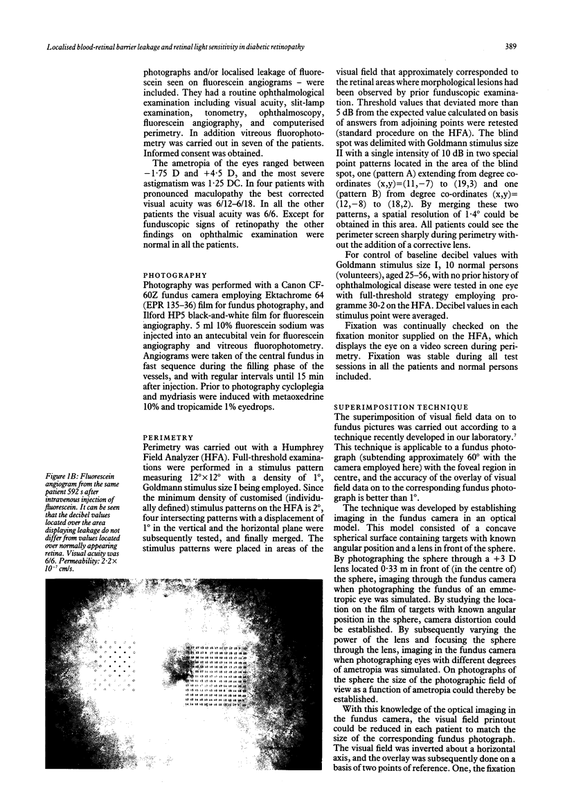
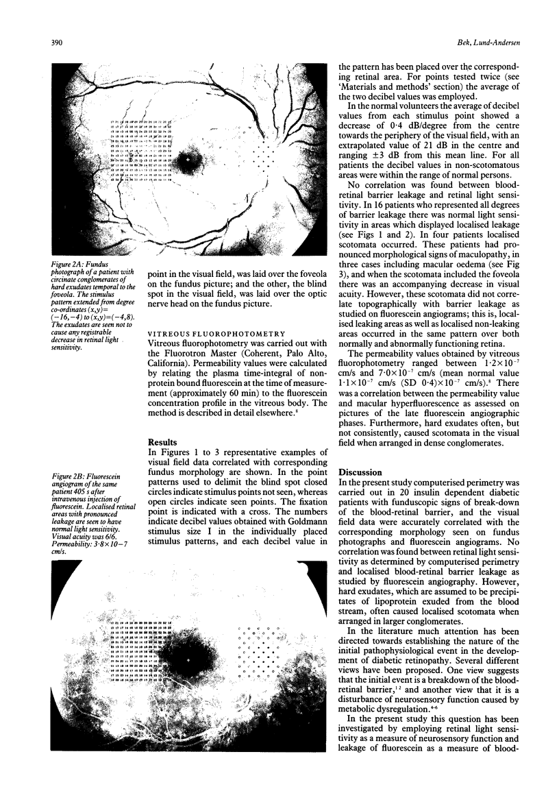
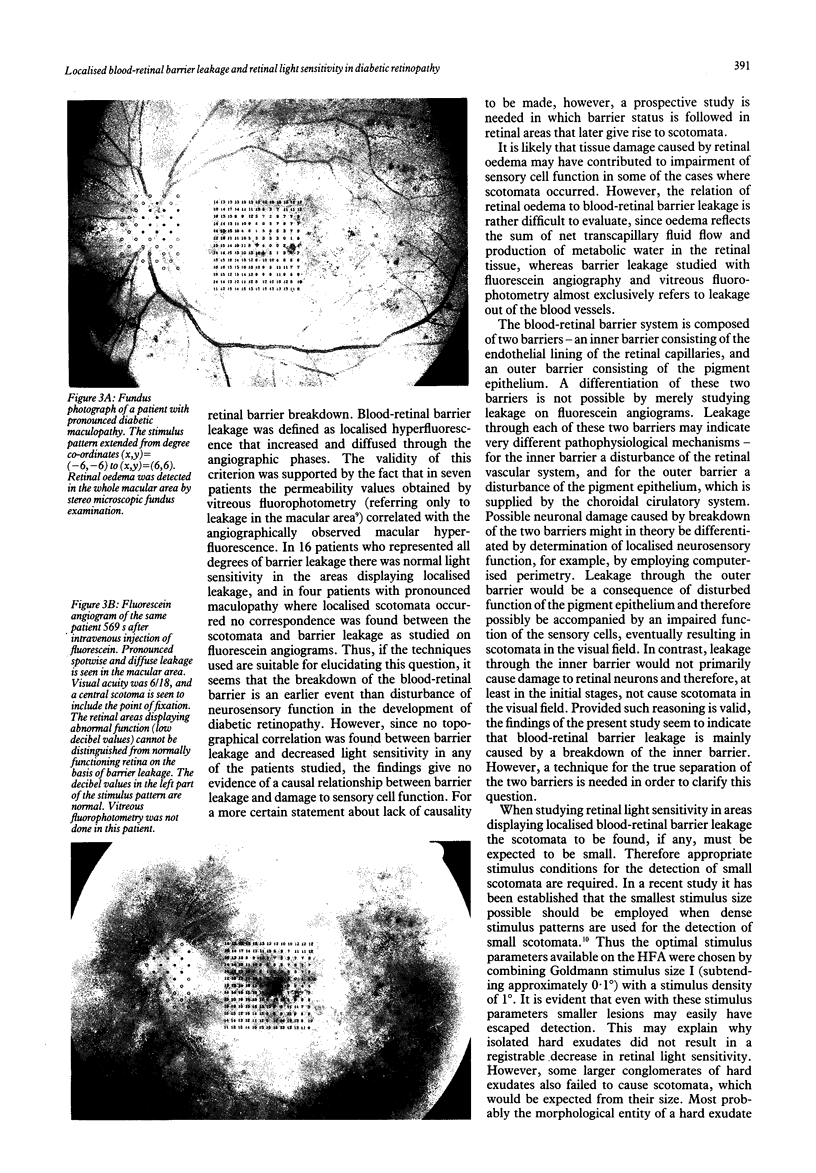
Twenty patients with insulin dependent diabetes mellitus were selected on the basis of morphological signs of blood-retinal barrier leakage--namely, hard exudates seen on fundus photographs and/or localised leakage of fluorescein seen on fluorescein angiograms. Computerised perimetry was carried out in visual field areas that corresponded to the morphological lesions, and the visual field data were accurately correlated with the morphology as seen on fundus photographs and fluorescein angiograms. In addition, in seven of the patients who represented the range of leakage among the patients studied, the blood-retinal barrier leakage was quantitated by vitreous fluorophotometry. In 16 cases normal light sensitivity was found in retinal areas showing localised leakage as studied on fluorescein angiograms. In four cases with pronounced maculopathy, where scotomata occurred, there was no topographical correlation between the scotomata and barrier leakage. Furthermore hard exudates often, but not consistently, caused localised scotomata when arranged in dense conglomerates. The permeability values correlated with angiographically observed hyperfluorescence in the macular area. On the basis of the techniques employed in the present study it seems that breakdown of the blood-retinal barrier is an earlier event than disturbance of neurosensory function in the development of diabetic retinopathy. However, the findings give no evidence of a causal relationship between barrier leakage and damage to sensory cell function.
Full text
PDF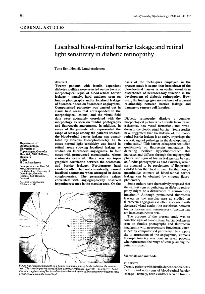
Images in this article


Selected References
These references are in PubMed. This may not be the complete list of references from this article.
- Bek T., Lund-Andersen H. Accurate superimposition of perimetry data onto fundus photographs. Acta Ophthalmol (Copenh) 1990 Feb;68(1):11–18. doi: 10.1111/j.1755-3768.1990.tb01642.x. [DOI] [PubMed] [Google Scholar]
- Bek T., Lund-Andersen H. The influence of stimulus size on perimetric detection of small scotomata. Graefes Arch Clin Exp Ophthalmol. 1989;227(6):531–534. doi: 10.1007/BF02169446. [DOI] [PubMed] [Google Scholar]
- Bresnick G. H. Diabetic retinopathy viewed as a neurosensory disorder. Arch Ophthalmol. 1986 Jul;104(7):989–990. doi: 10.1001/archopht.1986.01050190047037. [DOI] [PubMed] [Google Scholar]
- Cunha-Vaz J., Faria de Abreu J. R., Campos A. J. Early breakdown of the blood-retinal barrier in diabetes. Br J Ophthalmol. 1975 Nov;59(11):649–656. doi: 10.1136/bjo.59.11.649. [DOI] [PMC free article] [PubMed] [Google Scholar]
- Dalgaard P., Barker V. A., Lund-Andersen H. Vitreous fluorophotometry: mathematical analysis of the effect of peripheral leakage on axial scans. Invest Ophthalmol Vis Sci. 1989 Jul;30(7):1522–1526. [PubMed] [Google Scholar]
- Lund-Andersen H., Krogsaa B., la Cour M., Larsen J. Quantitative vitreous fluorophotometry applying a mathematical model of the eye. Invest Ophthalmol Vis Sci. 1985 May;26(5):698–710. [PubMed] [Google Scholar]