

Abstract
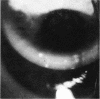
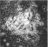
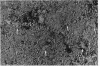
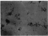
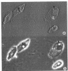
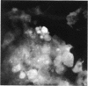
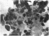
Corneal replicas were made from the severely affected eyes of 12 patients presenting with typical signs and symptoms of epidemic keratoconjunctivitis. Adenovirus was isolated from the eyes. Histopathological study of the replicas and cytology of diseased cells removed with the replica showed diffuse mild oedema of the epithelium, with scattered moderately swollen and deformed cells. The clinically observed punctate lesions histologically consisted of markedly swollen cells that had become oval or rounded. Fusion of these cells led to small syncytial formations, and the development of pseudopodia-like processes was occasionally observed. Loss of cell contents resulted in the formation of plicae on the surface of many cells. Two types of inclusion bodies were detected in the epithelial cells. The first type, intranuclear vacuolar inclusions, contained homogeneous material. The second type of inclusions were round, dense bodies, that developed in the homogeneous material of intranuclear vacuolar inclusions. The dense bodies contained the replicating and maturing virus particles. After cell degeneration the dense bodies become extracellular. Making a corneal replica had a beneficial effect on the clinical course of adenovirus keratitis.
Full text
PDF


Images in this article


Selected References
These references are in PubMed. This may not be the complete list of references from this article.
- Dawson C. R., Hanna L., Togni B. Adenovirus type 8 infections in the United States. IV. Observations on the pathogenesis of lesions in severe eye disease. Arch Ophthalmol. 1972 Mar;87(3):258–268. doi: 10.1001/archopht.1972.01000020260005. [DOI] [PubMed] [Google Scholar]
- Lund O. E., Stefani F. H. Corneal histology after epidemic keratoconjunctivitis. Arch Ophthalmol. 1978 Nov;96(11):2085–2088. doi: 10.1001/archopht.1978.03910060465016. [DOI] [PubMed] [Google Scholar]
- Maudgal P. C., Missotten L. Development of disseminating inclusion bodies in primary experimental herpes simplex keratitis. Bull Soc Belge Ophtalmol. 1977 Nov 27;179:25–36. [PubMed] [Google Scholar]
- Maudgal P. C., Missotten L. Histology and histochemistry of the normal superficial corneal epithelium of rabbit. Albrecht Von Graefes Arch Klin Exp Ophthalmol. 1978 Feb 22;205(3):167–174. doi: 10.1007/BF00414396. [DOI] [PubMed] [Google Scholar]
- Maudgal P. C., Missotten L. Histopathology and histochemistry of the superficial corneal epithelium in experimental herpes simplex keratitis. Albrecht Von Graefes Arch Klin Exp Ophthalmol. 1979 Feb 8;209(4):239–248. doi: 10.1007/BF00419058. [DOI] [PubMed] [Google Scholar]
- Maudgal P. C., Missotten L. Histopathology of human superficial herpes simplex keratitis. Br J Ophthalmol. 1978 Jan;62(1):46–52. doi: 10.1136/bjo.62.1.46. [DOI] [PMC free article] [PubMed] [Google Scholar]
- Maudgal P. C. The epithelial response in keratitis sicca and keratitis herpetica (an experimental and clinical study). Doc Ophthalmol. 1978 Aug 1;45(2):223–327. doi: 10.1007/BF00161670. [DOI] [PubMed] [Google Scholar]
- Maudgal P. C., Van Deuren H., Missotten L. Therapeutic effect of corneal replicas in herpetic keratitis. Bull Soc Belge Ophtalmol. 1979;185:39–45. [PubMed] [Google Scholar]
- Missotten L., Maudgal P. C. The replica technique used to study superficial corneal epithelium in vivo. Am J Ophthalmol. 1977 Jul;84(1):104–111. doi: 10.1016/0002-9394(77)90333-6. [DOI] [PubMed] [Google Scholar]
- Pereira H. G. Persistent infection by adenoviruses. J Clin Pathol Suppl (R Coll Pathol) 1972;6:39–42. [PMC free article] [PubMed] [Google Scholar]