

Abstract
Acute hemothorax is generally known to be a sequela of trauma to the chest, rupture of aortic aneurysm, or aortic dissection. Other causes of hemothorax have been attributed to iatrogenic, vascular, neoplastic, coagulopathy, or infectious processes. Although there has been a single report of spontaneous rupture of intercostal artery after coughing, there have been no reports of the same from sneezing. This rare presentation highlights the importance of a full evaluation for patients who present with viral symptoms such as sneezing or coughing along with a complaint of chest pain because massive hemothorax can be life‐threatening.
Keywords: ecchymosis, hemothorax, sneeze
1. INTRODUCTION
Sneezing is an upper airway reflex where air is expelled through the nose and mouth via forceful contractions of the intercostal muscles to remove irritants from the nasal cavity. It is a mundane phenomenon that many people experience multiple times per month and is usually regarded as benign. However, complications of sneezing have been described. 1 , 2 , 4
Hemothorax refers to a collection of blood within the pleural space. It is estimated that 300,000 cases of hemothorax occur annually in the United States. Of these, the overwhelming majority are related to trauma, with only 1.7% attributed to other causes. In nontraumatic hemothorax, other risk factors for bleeding into the chest are typically present, such as neurofibromatosis‐1, neoplasia, or use of anticoagulation. 5
2. CASE
A 65‐year‐old male with past medical history of type 2 diabetes and hypertension presented to the emergency department (ED) complaining of stabbing pain on the right side of the chest and right flank ecchymosis. His symptoms began after he sneezed approximately 10 days before presentation. At that time, the patient had been dealing with what he described as a “chest cold” with rhinorrhea, sneezing, and coughing.
After the sneeze, the patient sought medical attention at an outside hospital, where he was noted to be hypotensive and hypoxic with a right lower lobe opacity on chest x‐ray. He was admitted for sepsis secondary to community acquired pneumonia for 3 days. His course was complicated by an episode of atrial fibrillation with rapid ventricular response that spontaneously converted back to sinus rhythm. He was not started on anticoagulation for this.
He was discharged in improved condition but presented to our hospital shortly afterward for a second opinion because he felt that, although his pneumonia had been appropriately managed, the chest wall bruising and pain had not been addressed. He had noticed that the area of bruising to his flank was expanding and chest pain was worsening. He was concerned that he was “bleeding internally.”
On evaluation in our ED, we noted that the patient was hemodynamically stable with normal oxygen saturation. His examination was remarkable for decreased breath sounds on the right and a large area of ecchymosis to the right chest and flank, as pictured below (Figure 1).
FIGURE 1.
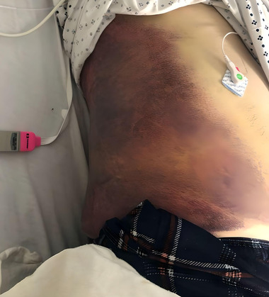
Left flank ecchymosis extending from the chest wall to the iliac crest.
Laboratory results showed normocytic anemia with a hemoglobin of 10.6, normal renal function, and normal coagulation studies.
The expanding ecchymosis, worsening pain, and normocytic anemia raised concern for uncontrolled or recurrent hemorrhage as a cause of the patient's symptoms. Parapneumonic effusion, empyema, and acute hemothorax were also considered in the differential because of the diminished right‐sided breath sounds. Therefore, a computed tomography (CT) scan of the chest and abdomen was obtained that demonstrated a hemothorax on the right. In addition, the intercostal space between the 7th and 8th ribs was abnormally widened, and the hemothorax appeared to be dissecting between those ribs causing the large flank ecchymosis (Figure 2).
FIGURE 2.
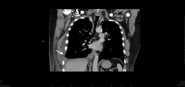
Coronal image of the patient's computed tomography scan. Notice the abnormal widening of the 7th intercostal space on the right compared to the left. There is also blunting of the costophrenic angle on the left side due to the hemothorax.
The patient was admitted for serial hemoglobin checks, hemodynamic monitoring, and thoracic surgery consultation. Plans were made for the placement of an ultrasound‐guided right pleural pigtail catheter that was attempted on day 2 of admission. By that time, however, most of the blood appeared to have clotted on sonographic assessment, so the procedure was aborted. He was monitored in the hospital for an additional 2 days during which his hemoglobin was stable and his pain gradually improved. He was discharged on admission day 4 in improved condition.
3. DISCUSSION
Although typically benign, serious complications of sneezing such as orbital blowout fracture, pneumomediastinum, and even aortic dissection have been described. In this case, we present another potentially life‐threatening complication of sneezing—hemothorax. This was diagnosed on a CT scan after the patient presented with pleuritic chest pain and chest wall ecchymosis triggered by a sneeze. The CT scan also showed abnormal widening of the intercostal space, with dissection of the hemothorax into the subcutaneous tissue. It is possible that the forceful contraction of the patient's intercostal muscles whereas sneezing caused a tear of the muscles and the intercostal artery, which led to bleeding both inward into the pleural space and outward into the skin of the chest wall. Although tears of the intercostal artery have not been previously described after sneezing, there is 1 case report of an intercostal artery tear after coughing. 3
Fortunately, this patient did not develop any hemodynamic instability or hypoxia during his stay in our hospital. However, one might speculate that this injury could have contributed to the hypotension and hypoxia he experienced at the outside hospital.
ED physicians frequently encounter patients like this one, who have chest pain associated with other viral symptoms. In these situations, it is important to maintain a broad differential to help guide further workup when clinically appropriate. This case demonstrates that acute hemothorax should be considered as a possibility—particularly when concerning examination findings such as flank ecchymosis, decreased breath sounds, or hemodynamic instability are present.
CONFLICT OF INTEREST STATEMENT
The authors report no conflicts of interest.
Neff A, Bradby C. Hemothorax after sneezing. JACEP Open. 2023;4:e13025. 10.1002/emp2.13025
Funding and support: By JACEP Open policy, all authors are required to disclose any and all commercial, financial, and other relationships in any way related to the subject of this article as per ICMJE conflict of interest guidelines (see www.icmje.org). The authors have stated that no such relationships exist.
Supervising Editor: Robert Levine, MD.
REFERENCES
- 1. Akyol PY, Ünlüer EE, Oyar O, Bilgin S. An unexpected complication of sneezing: blow‐out orbital fracture. J Emerg Trauma Shock. 2015;8(3):172‐173. doi: 10.4103/0974-2700.145409 [DOI] [PMC free article] [PubMed] [Google Scholar]
- 2. Faden D, Elackatuu A, Platt M. The “Closed‐airway sneeze”: an unusual cause of laryngeal fracture. Otolaryngol Head Neck Surg. 2011;145:515‐516. doi: 10.1177/0194599811402947 [DOI] [PubMed] [Google Scholar]
- 3. Jang JY, Lim YS, Woo JH, Jang JH. Spontaneous rupture of intercostal artery after severe cough. Am J Emerg Med. 2015;33(1):131.e1‐3. doi: 10.1016/j.ajem.2014.06.033. Epub 2014 Jul 1. PMID: 25085284. [DOI] [PubMed] [Google Scholar]
- 4. Setzen S, Platt M. The dangers of sneezing: a review of injuries. Am J Rhinol Allergy. 2019;33(3):331‐337. doi: 10.1177/1945892418823147. Epub 2019 Jan 8. PMID: 30616365. [DOI] [PubMed] [Google Scholar]
- 5. Zeiler J, Idell S, Norwood S, Cook A. Hemothorax: a review of the literature. Clin Pulm Med. 2020;27(1):1‐12. doi: 10.1097/CPM.0000000000000343. Epub 2020 Jan 10. PMID: 33437141; PMCID: PMC7799890. [DOI] [PMC free article] [PubMed] [Google Scholar]