Abstract
Introduction
Anatomical femoral tunnel placement is critical for anterior cruciate ligament reconstruction (ACLR). Tunnel placement may vary with different surgical techniques. The aim of this study was to compare the accuracy of femoral tunnel placement between the Anteromedial (AM) and Anterolateral (AL) visualisation portals on post-operative CT scans among a cohort of ACLR patients.
Materials and methods
This cross-sectional study was conducted from January 2018 to March 2020 after obtaining ethics clearance. Patients who went for arthroscopic ACLR in our institute were divided into an AM (group 1) and an AL (group 2) based on the visualisation portal for creating the femoral tunnel and a 3D CT scan was done. The femoral tunnel position was calculated in deep to shallow and high to low direction using the Bernard Hertel grid. Femoral tunnel angle was measured in the 2D coronal image. Statistical analysis was done with the data collected.
Results
Fifty patients with an average age of 26.36 (18-55) years ±7.216 SD were enrolled in the study. In this study, the AM technique was significantly more accurate (p<0.01) than the AL technique in terms of femoral tunnel angle. Furthermore, the deep to the shallow position was significantly (p= 0.018) closer to normative values, as determined by the chi-square test. The chances of error in tunnel angle in femoral condyle are 2.6 times greater in the AL technique (minimal clinical difference).
Conclusion
To conclude, in ACLR the anteromedial visualisation portal can facilitate accurate femoral tunnel placement compared to the anterolateral visualisation portal.
Keywords: tunnel placement, anatomic, anterior cruciate, portal
Introduction
Injury to the ACL alters the normal knee kinematics resulting in knee arthritis. The primary aim of ACLR is to restore the normal knee kinematics. The reconstruction should be as anatomic as possible to avoid failure and arthritis. The success of ACL reconstruction depends on several factors, among which femoral tunnel positioning plays a crucial role in the outcome.
There is a variation in femoral tunnel placement among surgeons for several reasons, including their expertise, training, literature support etc. The placement of the femoral tunnel in arthroscopic ACLR is also dependent on the technique chosen for drilling the femoral tunnel. While transtibial drilling places the femoral tunnel anteriorly and vertically in the distal femur, the transportal technique directed independent femoral tunnel placement. Recently, there has been an emphasis on more anatomical femoral tunnel placement in ACLR which is closer to the native anterior cruciate ligament (ACL) footprint. Studies have shown that anatomic femoral tunnel placement can be obtained better by transportal technique compared to a transtibial technique1-7.
Transportal femoral tunnel can be created either by visualisation from an anterolateral (AL) portal and making the tunnel from anteromedial portal or by visualisation from an anteromedial (AM) portal and making the tunnel from the accessory anteromedial portal. We hypothesise higher accuracy with AM portal-based femoral tunnel creation in ACLR. CT scan is the standard and validated imaging technique to evaluate the tunnel placement since plain radiographs are unreliable, challenging and particularly difficult to assess femoral tunnel position in the lateral view. Hence, this study aimed to compare the accuracy of femoral tunnel placement between the AM and AL visualisation portals on post-operative CT scan among a cohort of ACLR patients.
Materials and Methods
This cross-sectional study was conducted from January 2018 to March 2020 at a centre treating complex knee injuries. The institutional review board approved the study, and the study was completed in accordance with the Declaration of Helsinki. We searched the operation records to screen patients who underwent arthroscopic ACLR.
Patients with multi-ligamentous knee injuries, knee dislocations, and PCL injuries were excluded from the study. The cases were operated by two arthroscopic surgeons with more than 10 years of experience in sports medicine. We divided the cases into AM (group 1) and AL (group 2) based on the visualisation portal used for creating the femoral tunnel. In group 1 (AM), the femoral tunnel was drilled through the accessory anteromedial portal (AAM) by visualising from the anteromedial portal whereas in group 2 (AL), the femoral tunnel entry was made from the anteromedial portal by visualising through the anterolateral portal. The AL portal was placed high at the level of the inferior pole of patella adjacent to the lateral border of the patellar tendon. The AM portal was placed high at the same level as AL portal adjacent to the medial border of the patellar tendon. For the AAM portal, a 20G spinal needle was placed 1cm medial and lower to the AM portal and under direct vision by arthroscope from the AL portal it was confirmed to be above the anterior horn of medial meniscus as well as it had good access to the femoral tunnel entry site. In addition, care was also taken that the needle was not too close to the medial femoral condyle. Then, a portal was created with a 11-size blade.
For ACL reconstruction, a tourniquet was used for all the cases and the leg was in hanging position with a thigh holder. Different anatomical landmarks have been mentioned in the literature for anatomical femoral tunnel placement. In our institute, we routinely place the femoral tunnel slightly posterior to the centre of the native ACL footprint with the knee in 120° of flexion. In the absence of the native footprint, the femoral tunnel was usually placed inferior to the lateral intercondylar ridge and slightly posterior to the bifurcate ridge.
After obtaining informed consent, all these patients were contacted and included in this study. In the post-operative follow-up, a CT scan with 3D reconstruction was ordered for these patients.
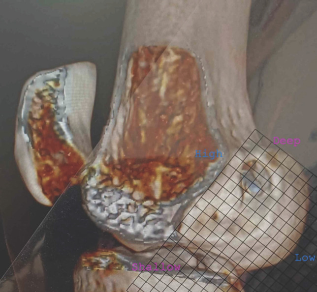
The three-dimensional sagittal image of the CT scan was formatted to subtract the medial condyle to assess the position of the femoral tunnel on a Bernard Hertel grid. The Bernard and Hertel’s grid were drawn on a plastic overhead transparent projector sheet of 10cm x 10cm dimension divided into multiple squares of 5mm x 5mm, and 1mm each of this grid was calibrated to 1%. The superior border of the grid was placed along the Blumensaat’s line. The Blumensaat’s line was drawn through the roof of the intercondylar notch placing the posterior border of the grid along the posterior femoral condyle. The femoral tunnel entry was recorded. The femoral tunnel position was calculated in deep to shallow and high to low direction (Fig. 1). Deep to shallow is defined as the femoral tunnel position, which is calculated parallel to the Blumensaat’s line as a percentage of the total sagittal diameter of the lateral femoral condyle and high to low is defined as femoral tunnel position calculated perpendicular to the Blumensaat’s line as a percentage to the maximum intercondylar notch height. A reference of 24-27% in the deep to shallow and 28-34% in the high to low direction was taken to estimate the femoral tunnel's position on the Bernard Hertel grid superimposed on the formatted sagittal image8-10. The values obtained from the operated cases were compared to the reference values described above.
Fig 1:

Figure showing the 3D reconstructed image of the medial side of the lateral femoral condyle with Bernard Hertel grid drawn in transparent sheet and placed over it to measure the femoral tunnel location in deep to shallow and high to low direction.
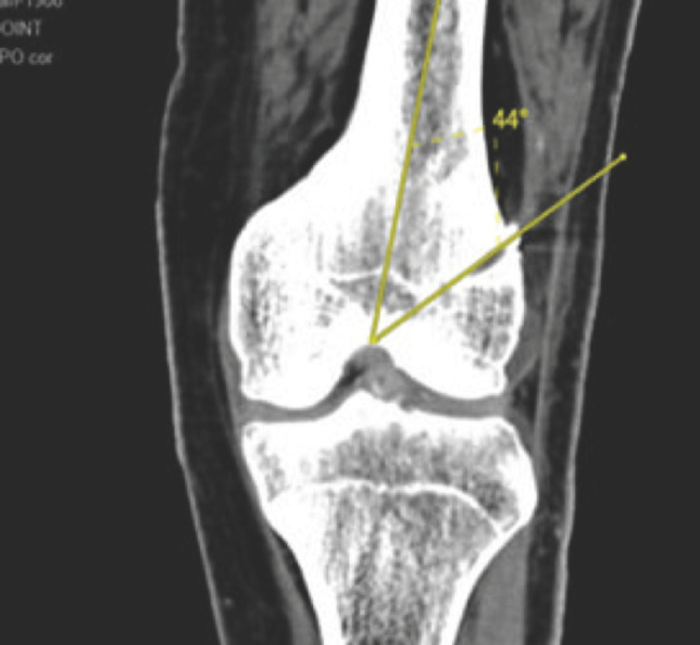
Femoral tunnel angle was measured in the 2D coronal image by a line drawn through the tunnel with the shaft of the femur (Fig. 2). Normal reference for femoral tunnel angle was taken as greater than 35° and compared to those obtained from cases. Statistical analysis was performed with the means of statistical software SPSS for Windows, version 19 [SPSS, Chicago, IL].
Fig 2:

Coronal cut of the CT scan of knee joint showing measurement of the femoral tunnel angle.
Results
Fifty patients with an average age of 26.36 (18-55) years ±7.216 SD were enrolled in the study. We had 46 (92%) males and 4 (8%) females among the enrolled patients. There were 21 (42%) left and 29 (58%) right ACLRs. 27 cases were in group 1, and 23 cases were in group 2.
The mean femoral tunnel position (deep-shallow, high-low, and in relation to Blumensaat’s line) and the femoral tunnel angle for both the techniques (Table I). Table II shows the percentage of near anatomic placement of the tunnel by both techniques.
Table I:
Shows the mean femoral tunnel position and angle between the two groups
| Femoral tunnel | Number | Group | Mean (in mm) | SD |
|---|---|---|---|---|
| Deep to shallow | 27 | 1 | 26.07 | 1.21 |
| 23 | 2 | 25.78 | 2.94 | |
| High to low | 27 | 1 | 29.96 | 2.49 |
| 23 | 2 | 30.65 | 1.87 | |
| Femoral tunnel angle | 27 | 1 | 40.37 | 5.98 |
| 23 | 2 | 35.00 | 11.27 |
Table II:
Shows near anatomic placement data between the two groups
| Femoral tunnel | Number | Group | Near anatomic placement | More than normal | Less than normal | Percentage |
|---|---|---|---|---|---|---|
| Deep to shallow | 27 | 1 | 25 | 2 | 0 | 92.6 |
| 23 | 2 | 16 | 1 | 6 | 69.5 | |
| High to low | 27 | 1 | 25 | 1 | 1 | 92.6 |
| 23 | 2 | 22 | 0 | 1 | 95.7 | |
| Femoral tunnel angle | 27 | 1 | 22 | 0 | 5 | 81.5 |
| 23 | 2 | 9 | 0 | 14 | 39.1 |
In this study, the AM technique was significantly more accurate (p<0.01) than the AL technique in terms of femoral tunnel angle. Furthermore, the deep to the shallow position was significantly (p=0.018) closer to normative values, as determined by the chi-square test. The chances of error in tunnel angle in femoral condyle are 2.6 times greater in the AL technique (minimal clinical difference).
Discussion
Our study shows that the deep to shallow position of the femoral tunnel placement using the AM visualisation portal (AM technique) was significantly closer to normative values than that of the AL visualisation portal (AL technique). In addition, the AM technique was found to be significantly more accurate than the AL technique in terms of femoral tunnel angle. Fourteen patients who underwent surgery by AL technique had femoral tunnel angles that were outside the normal range.
In ACLR, we found that AM visualisation-based AAM femoral tunnel creation was more accurate than AL-based AM femoral tunnel placement. The observed difference between the two techniques could be attributed to the poor visualisation of the ACL footprint through the 30° arthroscope from the AL portal. Viewing through the AL portal gives an inaccurate estimate of the depth of the posterior border of the lateral femoral condyle. The main advantage of the AM visualisation, on the other hand, is a direct view of the ACL footprint and the medial surface of the lateral femoral condyle. It prevents the femoral tunnel from being created non-anatomically. The incomplete visualisation of the ACL footprint from the AL portal also contributes to incorrect border estimation. For accurate tunnel placement, a 70° arthroscope should be used to view the medial surface of the lateral condyle. However, the 70° arthroscope's infrequent use and skewed visualisation makes it difficult to use on a regular basis.
Though the instrument crowding was blamed as one of the determinants for use against viewing and working portals in the AM technique, we placed the anteromedial portal at a higher level and the accessory anteromedial portal at a lower and medial level which made it convenient to drill the ACL femoral tunnel.
Anatomical femoral tunnel placement is critical for anterior cruciate ligament reconstruction (ACLR). Multicentre ACL Revision Study (MARS) reported that 80% of ACLR failure was at least partially secondary to a mal-positioned femoral tunnel11. Even experienced surgeons find it difficult to place femoral tunnels anatomically due to variations in surgical techniques, bone anatomy and patient morphology. Tunnel placement techniques have evolved over the years. Previous studies have compared transtibial technique with transportal techniques and reported better anatomic femoral tunnel placement with the latter1-7. In a study, Ahn et al2 found that the transportal technique allows for better anatomic femoral tunnel placement than the transtibial technique comparing the two techniques. Gadikota et al4 also found similar results in their study. Osti et al7 and Tompkins et al1 compared transtibial technique to AAM technique, and both found that AAM technique resulted in better placement. Shin et al12 discovered that the femoral tunnel was placed higher with the transtibial technique than with the low anteromedial technique in their study. They found no significant difference between deep and shallow positions.
Moon et al13 did a cadaveric study to show the influence of different anteromedial portals on the femoral tunnel orientation and reported that a mal-positioned AM portal results in abnormal tunnel orientation and significantly shorter tunnel length. They have also advised the use of AAM portal for better femoral tunnel placement. Our study also showed a statistically significant difference in deep to shallow position between the two techniques (AM technique superior to AL technique). Kim et al14 in their study showed the influence of knee flexion angles in creating the femoral tunnel. In our study, femoral tunnels were placed with knee in 120° flexion. Transportal technique has its own disadvantages particularly short tunnel length, medial femoral condyle cartilage damage, posterior blow out etc.
Recently, there has been emphasis on a horizontal angle of the femoral tunnel and a flat ACL graft for decreasing the risk of failure associated with a vertical graft. The use of a viewing anteromedial portal and drilling through an accessory anteromedial portal facilitated us to achieve this objective. Takeda et al15 reported that the AAM group's femoral tunnel angle is more horizontal than the TT group. Similarly, our study also revealed that the AM technique resulted in a more horizontal tunnel than the AL technique, which resulted in a more vertical tunnel, which was statistically significant.
Despite the study's main strength being CT-based measurement of femoral tunnel placements, there are a few limitations to this study. First and foremost, it is a retrospective study. Second, we have not examined the relationship between femoral tunnel malposition and clinical outcomes, highlighting the clinical significance. Third, we have not evaluated the complications associated with both techniques, such as medial femoral condyle damage, notch impingement, and so on, during tunnel placement.
Conclusion
While performing ACLR, the anteromedial visualisation portal can facilitate accurate femoral tunnel placement compared to anterolateral visualisation portal. However, the clinical implications and its long-term outcomes needs to be studied.
Conflict of Interest
The authors declare no potential conflict of interest.
References
- 1.Tompkins M, Milewski MD, Brockmeier SF, Gaskin CM, Hart JM, Miller MD. Anatomic femoral tunnel drilling in anterior cruciate ligament reconstruction: use of an accessory medial portal versus traditional transtibial drilling. Am J Sports Med. 2012;40(6):1313–21. doi: 10.1177/0363546512443047. doi: [DOI] [PubMed] [Google Scholar]
- 2.Ahn JH, Jeong HJ, Ko CS, Ko TS, Kim JH. Three-dimensional reconstruction computed tomography evaluation of tunnel location during single-bundle anterior cruciate ligament reconstruction: a comparison of transtibial and 2-incision tibial tunnel-independent techniques. Clin Orthop Surg. 2013;5(1):26–35. doi: 10.4055/cios.2013.5.1.26. doi: [DOI] [PMC free article] [PubMed] [Google Scholar]
- 3.Arno S, Bell CP, Alaia MJ, Singh BC, Jazrawi LM, Walker PS. Does Anteromedial Portal Drilling Improve Footprint Placement in Anterior Cruciate Ligament Reconstruction? Clin Orthop Relat Res. 2016;474(7):1679–89. doi: 10.1007/s11999-016-4847-7. et al. doi: [DOI] [PMC free article] [PubMed] [Google Scholar]
- 4.Gadikota HR, Sim JA, Hosseini A, Gill TJ, Li G. The relationship between femoral tunnels created by the transtibial, anteromedial portal, and outside-in techniques and the anterior cruciate ligament footprint. Am J Sports Med. 2012;40(4):882–8. doi: 10.1177/0363546511434276. doi: [DOI] [PMC free article] [PubMed] [Google Scholar]
- 5.Metso L, Nyrhinen KM, Bister V, Sandelin J, Harilainen A. Comparison of clinical results of anteromedial and transtibial femoral tunnel drilling in ACL reconstruction. BMC Musculoskelet Disord. 2020;21(1):341. doi: 10.1186/s12891-020-03351-w. doi: [DOI] [PMC free article] [PubMed] [Google Scholar]
- 6.Lee DH, Kim HJ, Ahn HS, Bin SI. Comparison of Femoral Tunnel Length and Obliquity Between Transtibial, Anteromedial Portal, and Outside-In Surgical Techniques in Single-Bundle Anterior Cruciate Ligament Reconstruction: A Meta-analysis. Arthroscopy. 2016;32(1):142–50. doi: 10.1016/j.arthro.2015.07.026. doi: [DOI] [PubMed] [Google Scholar]
- 7.Osti M, Krawinkel A, Ostermann M, Hoffelner T, Benedetto KP. Femoral and tibial graft tunnel parameters after transtibial, anteromedial portal, and outside-in single-bundle anterior cruciate ligament reconstruction. Am J Sports Med. 2015;43(9):2250–8. doi: 10.1177/0363546515590221. doi: [DOI] [PubMed] [Google Scholar]
- 8.Amis AA, Jakob RP. Anterior cruciate ligament graft positioning, tensioning and twisting. Knee Surg Sports Traumatol Arthrosc. 1998;6(Suppl 1):S2–12. doi: 10.1007/s001670050215. doi: [DOI] [PubMed] [Google Scholar]
- 9.Bird JH, Carmont MR, Dhillon M, Smith N, Brown C, Thompson P. Validation of a new technique to determine midbundle femoral tunnel position in anterior cruciate ligament reconstruction using 3-dimensional computed tomography analysis. Arthroscopy. 2011;27(9):1259–67. doi: 10.1016/j.arthro.2011.03.077. et al. doi: [DOI] [PubMed] [Google Scholar]
- 10.Parkar AP, Adriaensen ME, Strand T, Inderhaug E, Harlem T, Solheim E. How to read post-operative radiographs and CT scans after single-bundle anterior cruciate ligament reconstruction. Skeletal Radiol. 2013;42(11):1489–500. doi: 10.1007/s00256-013-1686-4. doi: [DOI] [PubMed] [Google Scholar]
- 11.MARS Group; Wright RW, Huston LJ, Spindler KP, Dunn WR, Haas AK. Descriptive epidemiology of the Multicenter ACL Revision Study (MARS) cohort. Am J Sports Med. 2010;38(10):1979–86. doi: 10.1177/0363546510378645. et al. doi: [DOI] [PMC free article] [PubMed] [Google Scholar]
- 12.Shin YS, Ro KH, Lee JH, Lee DH. Location of the femoral tunnel aperture in single-bundle anterior cruciate ligament reconstruction: comparison of the transtibial, anteromedial portal, and outside-in techniques. Am J Sports Med. 2013;41(11):2533–9. doi: 10.1177/0363546513500764. doi: [DOI] [PubMed] [Google Scholar]
- 13.Moon DK, Jo HS, Lee DY, Kang DG, Byun JH, Hwang SC. Influence of the different anteromedial portal on femoral tunnel orientation during anatomic ACL reconstruction. Acta Orthop Traumatol Turc. 2017;51:227–32. doi: 10.1016/j.aott.2017.02.004. doi: [DOI] [PMC free article] [PubMed] [Google Scholar]
- 14.Kim SH, Kim SJ, Choi CH, Kim D, Jung M. Optimal Condition to Create Femoral Tunnel Considering Combined Influence of Knee Flexion and Transverse Drill Angle in Anatomical Single-Bundle ACL Reconstruction Using Medial Portal Technique: 3D Simulation Study. Biomed Res Int. 2018;2018:2643247. doi: 10.1155/2018/2643247. doi: [DOI] [PMC free article] [PubMed] [Google Scholar]
- 15.Takeda Y, Iwame T, Takasago T, Kondo K, Goto T, Fujii K. Comparison of tunnel orientation between transtibial and anteromedial portal techniques for anatomic double-bundle anterior cruciate ligament reconstruction using 3-dimensional computed tomography. Arthroscopy. 2013;29(2):195–204. doi: 10.1016/j.arthro.2012.08.020. et al. doi: [DOI] [PubMed] [Google Scholar]


