. 2023 Aug 14;12:e82850. doi: 10.7554/eLife.82850

Figure 2. Position and extent of the prefrontal areas on the medial, lateral, and orbital views of the Yerkes19 surface.

The files with the parcellation scheme are available via EBRAINS platform of the Human Brain Project (https://search.kg.ebrains.eu/instances/Project/e39a0407-a98a-480e-9c63-4a2225ddfbe4) and the BALSA neuroimaging site (https://balsa.wustl.edu/study/7xGrm). Macroanatomical landmarks are marked in red letters, while black dashed lines mark fundus of sulci. arcs, spur of the arcuate sulcus; cgs, cingulate sulcus; cs, central sulcus; ias, inferior arcuate sulcus; lf, lateral fissure; ps, principal sulcus; sas, superior arcuate sulcus.

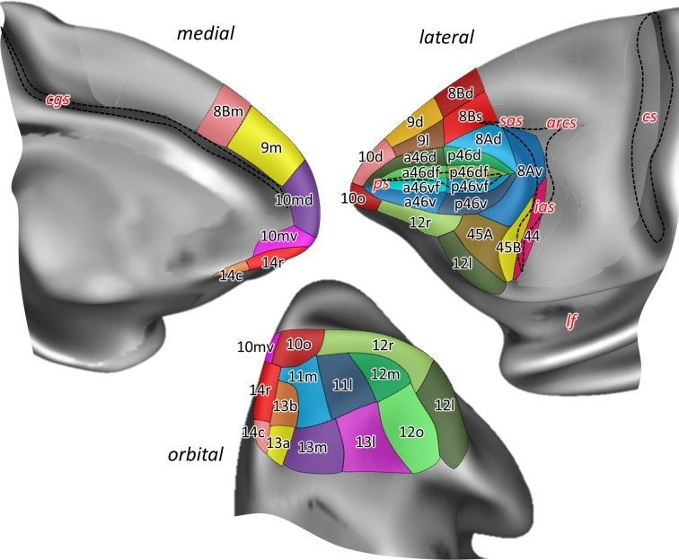
Figure 2.

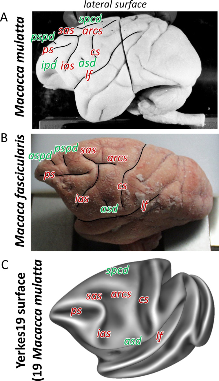
Figure 2—figure supplement 1. Macroanatomical landmarks (sulci labelled in red letters and dimples in green) shown on the lateral surface of the two related species of macaque monkey used in the present architectonic analyses.

Figure 2—figure supplement 1.

Photographs of two of the postmortem brains used in this study. Brain ID DP1. (A) Macaca mulatta, and brain ID 11530 (B) Macaca fascicularis. Average surface representations of the Yerkes19. (C) Macaca mulatta template brains. arcs, spur of the arcuate sulcus; asd, anterior supracentral dimple; aspd, anterior superior principal dimple; cs, central sulcus; ias, inferior arcuate sulcus; ps, principal sulcus; pspd, posterior superior principal dimple; sas, superior arcuate sulcus; spcd, superior precentral dimple.
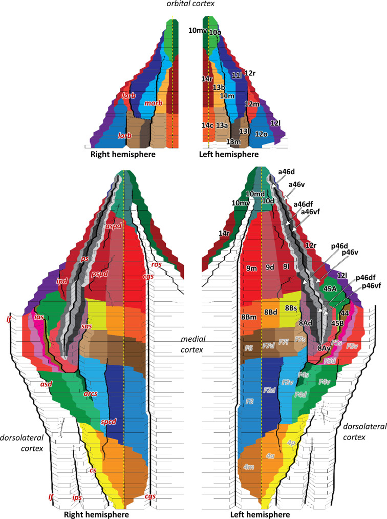
Figure 2—figure supplement 2. 2D flat map, based on the macroanatomical landmarks of every 40th section, displays orbital, medial, and dorsolateral hemispheric views with all defined areas within the macaque frontal lobe.

Figure 2—figure supplement 2.

Areas are labelled on the left hemisphere, that is, prefrontal areas in black and previously mapped (pre)motor areas (Rapan et al., 2021) in grey. Due to limited space on the map, we used white arrows to mark anterior and posterior subdivisions of 46. Dashed yellow line on the hemispheres represents the midline, which separates medial and dorsolateral cortex. Black full lines mark the fundus of sulci. Macroanatomical landmarks are marked on the right hemisphere; arcs, spur of the arcuate sulcus; asd, anterior supracentral dimple; aspd, anterior superior principal dimple; cgs, cingulate sulcus; cs, central sulcus; ias, inferior arcuate sulcus; ipd, inferior principal dimple; lf, lateral fissure; lorb, lateraral orbital sulcus; morb, medial orbital sulcus; ps, principal sulcus; pspd, posterior superior principal dimple; sas, superior arcuate sulcus; spcd, superior precentral dimple.