Abstract
In the Escherichia coli phage-plasmid P4, two partially overlapping replicons with bipartite ori sites coexist. The essential components of the oriI replicon are the α and cnr genes and the ori1 and crr sites; the oriII replicon is composed of the α gene, with the internal ori2 site, and the crr region. The P4 α protein has primase and helicase activities and specifically binds type I iterons, present in ori1 and crr. Using a complementation test for plasmid replication, we demonstrated that the two replicons depend on both the primase and helicase activities of the α protein. Moreover, neither replicon requires the host DnaA, DnaG, and Rep functions. The bipartite origins of the two replicons share the crr site and differ for ori1 and ori2, respectively. By deletion mapping, we defined the minimal ori1 and ori2 regions sufficient for replication. The ori1 site was limited to a 123-bp region, which contains six type I iterons spaced regularly close to the helical periodicity, and a 35-bp AT-rich region. Deletion of one or more type I iterons inactivated oriI. Moreover, insertion of 6 or 10 bp within the ori1 region also abolished replication ability, suggesting that the relative arrangement of the iterons is relevant. The ori2 site was limited to a 36-bp P4 region that does not contain type I iterons. In vitro, the α protein did not bind ori2. Thus, the α protein appears to act differently at the two origins of replication.
P4 is a natural phage-plasmid of Escherichia coli which can be propagated in different ways in the host cell. In the presence of a helper phage, such as P2, P4 can enter either the lytic cycle or the lysogenic state. P4 lacks morphogenetic genes and has developed specific mechanisms to exploit the helper phage functions for the construction of its capsid and tail and for lysis of the host cell. In the absence of the helper, P4 can either be maintained as a high-copy-number plasmid or integrate its genome in the host chromosome and establish the immune-lysogenic condition (for a review, see reference 28).
P4 DNA replication, which occurs both in the lytic cycle and in the plasmid condition, is independent of the helper P2 functions. The product of a single P4 gene, the α gene, is required for DNA replication. The α protein is multifunctional, with primase, helicase, and specific DNA binding activities (46). Thus, P4 DNA replication does not require the host functions, such as DnaA (initiator protein), DnaB (helicase), DnaC (complex with DnaB), and DnaG (primase), for initiation of DNA replication. Moreover, P4 is independent of both E. coli Rep helicase and RNA polymerase (2, 4, 27). In vitro, P4 DNA replication requires the α protein and several bacterial functions, including DNA polymerase III, SSB protein, gyrase, and topoisomerase I (14, 25).
The double-stranded P4 DNA molecule circularizes after infection, and replication proceeds bidirectionally in a θ-type manner from a single site, ori1 (26). With an in vivo test for complementation of plasmid replication, it was demonstrated that the P4 origin of replication is bipartite: in addition to ori1, a second cis-acting region essential for replication, crr, was identified about 4,500 bp from ori1 (15). Electron microscopic analysis of replication intermediate molecules, obtained both in vivo and in vitro, showed that replication initiates at ori1 (14, 26). No initiation at crr could be observed. These results were confirmed by in vitro P4 DNA replication experiments: evidence of replication initiation at the ori1 region, but not at the crr region, was found (26). In this same experiment, a second replication initiation site was detected within the α coding region (close to the 6273-to-6906 P4 fragment). This might represent the initiation point of the alternative P4 replicon oriII (see Discussion) (40).
Both the ori1 and crr regions are AT rich and present a decameric sequence, called the type I iteron, repeated several times in direct and inverted orientations. The α protein specifically binds to these repeats (46). In ori1, but not in crr, three consecutive direct repeats of a second decameric sequence, the type II iterons, have also been described (46). The crr site consists of two well-conserved (98 of 120 bp are identical) direct repeats of about 120 bp, separated by 60 bp. The two crr repeats are redundant, since Flensburg and Calendar (15) demonstrated that a single crr repeat is sufficient to drive P4 DNA replication.
P4 DNA replication is negatively regulated by the product of the cnr gene (39). The Cnr function is essential for P4 propagation in the plasmid state in order to control P4 copy number. In the absence of the Cnr protein, P4 overreplicates, and cell lethality ensues.
P4 mutants insensitive to Cnr control carry mutations in the α gene, suggesting that the α and Cnr proteins might interact (48). In vitro, the Cnr protein increases α binding affinity to ori1 and crr (48); since Cnr negatively regulates P4 DNA replication, it was hypothesized that the Cnr-α-DNA complex might be inactive for replication.
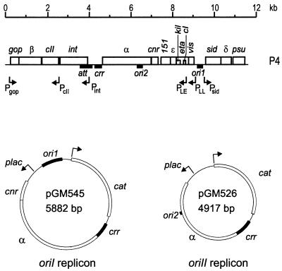
Using an in vivo test for complementation of plasmid replication, we have previously shown that two replicons coexist in phage-plasmid P4 (Fig. 1) (40): (i) the oriI replicon is made up of the α and cnr genes and the ori1 and crr sites, which constitute the bipartite oriI origin of replication; (ii) the oriII replicon is made up of the α gene and the bipartite oriII origin. This alternative origin of replication is composed of the crr and ori2 sites, the latter located within the 6186-to-6421 P4 region, internal to the α coding sequence. Where replication initiates in this second replicon has not been established.
FIG. 1.
Map of bacteriophage P4 and of the oriI and oriII replicons. The P4 map is drawn according to the P4 DNA sequence (17); the maps of pGM545 (replicon oriI) and pGM526 (replicon oriII) were described by Tocchetti et al. (40). Genes and open reading frames are indicated by open bars, sites are indicated by solid bars, and promoters are indicated by arrows. Expression of the cnr and/or α genes in pGM545 and pGM526 is from the lacp promoter.
The presence of the two replicons was confirmed by the construction of two plasmids, pGM545 (replicon oriI) and pGM526 (replicon oriII [Fig. 1]), in which the portions of the P4 genome constituting the oriI and oriII replicons, respectively, were ligated to the chloramphenicol resistance gene. In these plasmids, expression of the P4 cnr and/or α gene is under the control of lacp (40).
It was demonstrated (40) that both replicons depend on the α protein for replication, whereas the role of the Cnr protein differs. Replicon oriI requires the Cnr protein to avoid overreplication and cell killing. In fact, the construction of an oriI plasmid lacking the cnr gene failed, due to the absence of the negative regulation of replication (40). On the other hand, replicon oriII is inhibited by Cnr (40). This makes it rather unlikely that the oriII origin of replication is active in P4.
In this work we further characterize the two P4 replicons, demonstrating that both depend on α primase and helicase activities for replication, and we reduce the ori1 and ori2 regions, defining the minimal ori1 and ori2 sites.
MATERIALS AND METHODS
Bacterial strains, phages, and plasmids.
The bacterial strains that were derivatives of E. coli C were C-1a (prototrophic [34]), C-2107 (polA12; temperature-sensitive mutation in DNA polymerase I [31]), C-2428 [(recA-srl)Δ5 (40)], C-1414 (rep1 [8]), C-2307 (dnaA46 [4]), and C-5582 [dnaG3(Ts) rpoD::Tn10 by P1 transduction from PC3 (44) in C-1a]. The bacterial strains that were derivatives of E. coli K-12 were CM748 [polyauxotrophic; dnaA203(Ts) (5)] and DH5α (polyauxotrophic; recA [18]).
The bacteriophage strains used were P4 (36) and P4 vir1 (27). The P4 coordinates are from the complete P4 sequence (17) in the revised form (GenBank accession no. X51522).
The plasmids used are listed in Table 1.
TABLE 1.
Plasmids
| Plasmid | P4 region cloneda | Vector | Description and/or oligonucleotides used for PCR amplificationb (source or reference) |
|---|---|---|---|
| pACYC184 | Cmr (9) | ||
| pGB2ts | Spcr (12) | ||
| pGEX-4T1 | Apr (Pharmacia) | ||
| pGEX-α | 4595–6969 | pGEX-4T1 | Expresses the GST-α protein (40) |
| pGM526 | 4260–7041 | Replicon oriII (40) | |
| pGM545 | 4260–7631 and 9104–9463 | Replicon oriI (40) | |
| pGM614 | 8835–9465 and 9465–8835 | pUC19 | Two HindIII fragments of pRB3, containing the P4 8835–9465 region, were inserted in inverted orientation |
| pGM615 | 8835–9465 and 8835–9465 | pUC19 | Two HindIII fragments of pRB3, containing the P4 8835–9465 region, were inserted in direct orientation |
| pGM628 | 9167–9421 | pRB2 | 192PstI; 193HindIII |
| pGM629 | 9204–9421 | pRB2 | 207PstI; 193HindIII |
| pGM632 | 9298–9421 | pRB2 | 206PstI; 193HindIII |
| pGM633 | 4260–4595 | pGB2ts | Carries the P4 crr region (40) |
| pGM639 | 9176–9205 | pRB2c | 220PstI; 221PstI |
| pGM644 | 9176–9205 and 9298–9420 | pGM632c | 220PstI; 221PstI |
| pGM651 | 4260–4595 and 6186–6421 | pGB2ts | Carries the P4 crr and ori2 regions (40) |
| pGM660 | 9167–9350 | pRB2 | 192PstI; 279HindIII |
| pGM661 | 9167–9307 | pRB2 | 192PstI; 280HindIII |
| pGM669 | 4260–4595 and 8835–9465 | pGB2ts | Carries the P4 crr and ori1 regions (40) |
| pGM671 | 4260–4595 and 6186–6421 and 8835–9465 | pGB2ts | Carries the P4 crr, ori2, and ori1 regions (40) |
| pGM675 | 9212–9421 | pRB2 | 299PstI; 193HindIII |
| pGM682 | 9167–9467 | pRB2 | 192PstI; 300HindIII |
| pGM683 | 9245–9421 | pRB2 | 316PstI; 193HindIII |
| pGM686 | 4595–6969 carrying the αE214Q mutation | pGEX-4T1 | From pMS4Δ1E214Q (provided by R. Calendar [38]); expresses the GST-α primase-null proteind |
| pGM687 | 4595–6969 carrying the αK507T mutation | pGEX-4T1 | From pMS4Δ1K507T (provided by R. Calendar [47]); expresses the GST-α helicase-null proteind |
| pGM688 | 9338–9421 | pRB2 | 326PstI; 193HindIII |
| pGM689 | 9298–9397 | pRB2 | 206PstI; 327HindIII |
| pGM690 | 9298–9350 | pRB2 | 206PstI; 279HindIII |
| pGM693 | 6186–6329 | pGM633 | 249PstI; 344HindIII |
| pGM694 | 6269–6421 | pGM633 | 343PstI; 250HindIII |
| pGM695 | 6269–6329 | pGM633 | 343PstI; 344HindIII |
| pGM696 | 9351–9467e | pGM690 | 328HindIII; 300HindIII |
| pGM697 | 9361–9467e | pGM690 | 329HindIII; 300HindIII |
| pGM698 | 9351–9421e | pGM690 | 328HindIII; 193HindIII |
| pGM699 | 9361–9421e | pGM690 | 329HindIII; 193HindIII |
| pGM700 | 4260–4420 | pGM632f | 352BamHI; 353PstI |
| pGM704 | 9298–9467 | pRB2 | 206PstI; 300HindIII |
| pGM705 | 6186–6271 | pGM633 | 249PstI; 350HindIII |
| pGM706 | 6186–6254 | pGM633 | 249PstI; 351HindIII |
| pGM713 | 9351–9421d | pGM690 | 328HindIII; 193HindIII |
| pGM717 | 6208–6254 | pGM633 | 381PstI; 351HindIII |
| pGM735 | 6208–6242 | pGM633 | 381PstI; 425HindIII |
| pGM736 | 6219–6254 | pGM633 | 424PstI; 351HindIII |
| pGM737 | 6219–6242 | pGM633 | 424PstI; 425HindIII |
| pGM744 | 4260–7631 and 9167–9421 | pGM545 | By substitution of the 9104–9463 P4 region; 430XbaI; 193HindIII |
| pGM745 | 4260–7631 and 9297–9421 | pGM545 | By substitution of the 9104–9463 P4 region; 431XbaI; 193HindIII |
| pMK302 | 4260–10658 | pACYC184 | The cloned region derives from P4vir1 (26) |
| pRB2 | 4260–4595 | pUC19 | The P4 4260–4595 region, containing the crr site, is cloned in the polylinker region of pUC19 (11) |
| pRB3 | 8835–9465 | pUC19 | The P4 8835–9465 region, containing the ori1 site, is cloned in the polylinker region of pUC19 (11) |
| pRB4 | 4260–4595 and 8835–9465 | pUC19 | The P4 4260–4595 and 8835–9465 regions, containing the crr and ori1 sites, are cloned next to each other in the polylinker region of pUC19 (11) |
| pUC19 | Apr (43) |
The P4 fragments, obtained by PCR amplification with the pair of oligonucleotides indicated, were digested with the appropriate enzymes and cloned in the corresponding sites of the vector.
The oligonucleotides used are listed in Materials and Methods.
The P4 fragment was obtained by annealing of the oligonucleotides.
The amount of the GST-αE214Q and GST-αK507T mutant proteins expressed from pGM686 and pGM687, visualized by Coomassie blue staining, was comparable to the amount of the GST-α wild-type protein expressed from pGEX-α.
A 6- or 10-bp insertion was created between 9350 and 9351.
The P4 crr (4260-to-4595) fragment carried by pGM632 was replaced by the indicated fragment.
Oligonucleotides.
The oligonucleotides used in this work are listed below. The restriction site is in italic. The sequence complementary to P4 is underlined. 192PstI (GAGTCTGCAGTTCATCTCCACTTAAA); 193HindIII (CGGAAGCTTATTTTACTGTTCACCTCT); 206PstI (GACTCTGCAGCCCATCAACGG); 207PstI (GAGTCTGCAGCAATTTGTAATTTTTATAGTG); 220PstI (GCCACTTAAAGTCATTTAAAGCCACTTAAAGCTGCA); 221PstI (GCTTTAAGTGGCTTTAAATGACTTTAAGTGGCTGCA); 249PstI (ACTACTGCAGCACGGTCAGCGGCA); 250HindIII (GTCGAAGCTTCCGTAAGCGCACCCT); 279HindIII (CGCAAGCTTCGCAGTAATGACTGT); 280HindIII (CGGAAGCTTGATGGGCTTTTTG); 299PstI (AACGCTGCAGGTAATTTTTATAGTGAAATAC); 300HindIII (GTCAAGCTTCCAGGAAAAGGTCG); 316PstI (CTTCTGCAGCTTATTCATTCCCGG); 326PstI (AGTCTGCAGGTCATTACTGCGATTG); 327HindIII (CCCAAGCTTCCTTAATAAAAAAGATAAGTA); 328HindIII (CCGAAGCTTATTGTTCACCCTTTAAC); 329HindIII (CAGAAGCTTGCTACTTTAACTTACTGTATTACTTA); 343PstI (CCTACTGCAGAGCGCCACCATCACC); 344HindIII (CACCAAGCTTAGGGATACGCGACCG); 350HindIII (GGACAAGCTTGCTTTCTTCCGTGAACC); 351HindIII (GGACAAGCTTTCCTTTCTCTGGCCAGC); 352BamHI (CCGGGGATCCAAACCAGTGCAT); 353PstI (CATACTGCAGCGGCAGAATGCCGGAG); 381PstI (AGTACTGCAGCTGACAGGCGGGGTG); 424PstI (AGTACTGCAGGTGCTTCTGCCGGGCAA); 425HindIII (GAACAAGCTTCCAGCCTTGCCCGGC); 430XbaI (CTGAGTTCTAGATTCATCTCCACTTAAAG); 431XbaI (CTGAGATCTAGAAGCCCATCAACGG).
Integrative suppression of dnaA(Ts) and dnaG(Ts) mutants.
The C-2307 (dnaA46), CM748 (dnaA203), and C-5582 (dnaG3) strains were transformed by either pGM545 (oriI) or pGM526 (oriII), and chloramphenicol (30 μg/ml)-resistant transformants were selected at the permissive temperature (30°C) in the presence of IPTG (isopropyl-β-d-thiogalactopyranoside) (40 μg/ml). The transformant colonies obtained at 30°C were shifted to 42°C in the presence of chloramphenicol and IPTG. After 2 to 3 days, temperature-sensitive (Ts+) revertants appeared at a frequency of 10−7 to 10−8.
Segregation of chloramphenicol-sensitive clones was not observed when the Ts+ revertants were grown at 30°C in the absence of IPTG and chloramphenicol, thus suggesting that the pGM545 and pGM526 plasmids are integrated in the host chromosome.
Transformation.
Competent cells of strains C-2107 and C-2428 were obtained with CaCl2 treatment (39) from a culture grown at 30°C. After transformation with 0.1 μg of plasmid DNA, the cells were diluted 10 times in LD broth (16), divided into two subcultures, incubated at either 30 or 42°C for 1 h, plated on selective medium, and incubated at 30 and 42°C, respectively.
Affinity purification of GST-α fusion protein.
Glutathione S-transferase (GST)-α fusion protein was recovered from DH5α/pGEX-α as described by Smith and Johnson (37), modified as described in Polo et al. (33). Briefly, the expression of the fusion protein was induced with 1 mM IPTG for 90 min, and the fusion protein was purified with glutathione-Sepharose (Pharmacia). Analysis of the purified protein content was performed by sodium dodecyl sulfate 8% (wt/vol) polyacrylamide gel electrophoresis and Coomassie brilliant blue staining.
Fragment purification, end-labelling, and gel retardation.
The fragments used for the gel retardation experiments were obtained from pGM706 by digestion with either PstI and HindIII (ori2 fragment; 75 bp) or PstI and BglI (control fragment; 408 bp) and were obtained from pGM632 by digestion with PstI and BamHI (crr fragment; 340 bp). All of the above-mentioned fragments were purified with a QIAEXII kit and end labelled by the Klenow fill-in reaction in the presence of 10 μCi of [α-32P]dATP and 0.5 mM (each) dGTP, dCTP, and dTTP. After incubation at 30°C for 30 min, the reaction was terminated by heating the mixture at 75°C for 10 min, and the sample was phenol treated, precipitated with ethanol, and resuspended in TE (10 mM Tris-HCl [pH 7.5], 1 mM EDTA). The labelled fragments were run in polyacrylamide gels and purified by band excision and overnight elution in TE.
The gel retardation assay was performed as described in Polo et al. (33).
RESULTS
P4 oriI and oriII replicons do not depend on the bacterial genes dnaA, dnaG, and rep.
Replication of phage P4 does not depend on the bacterial DnaA initiation function or on the DnaG primase (4). We verified whether both P4 oriI and oriII replicons (pGM545 and pGM526, respectively) were independent of these host functions by testing their abilities to integratively suppress DnaA(Ts) and DnaG(Ts) host mutations. Two different DnaA(Ts) mutations, dnaA46 (C-2307) and dnaA203 (CM748), and the dnaG3 (C-5582) mutation were tested. Ts+ phenotypic revertants of the transformed host strains could be isolated with both plasmids (see Materials and Methods).
Growth of the Ts+ revertants at 42°C was IPTG dependent (replication of pGM545 and pGM526 is IPTG dependent, since the P4 cnr and/or α genes are expressed from lacp [Table 2]). Southern blot analysis of several independent Ts+ revertants demonstrated that in each strain an integrated copy of either pGM545 or pGM526 DNA was present in the bacterial chromosome; the integration sites were different in the independent isolates (data not shown). In most strains the presence of either free plasmid DNA or tandem integrated extra copies was also observed.
TABLE 2.
Integrative suppression of E. coli dnaA and dnaG mutants
| Straina | Viability at 42°Cb with:
|
||
|---|---|---|---|
| None | IPTG | IPTG + Cm | |
| C-2307 | − | − | − |
| C-2307(pGM545) | − | + | + |
| C-2307(pGM526) | − | + | + |
| CM748 | − | − | − |
| CM748(pGM545) | − | + | + |
| CM748(pGM526) | − | + | + |
| C-5582 | − | − | − |
| C-5582(pGM545) | − | + | + |
| C-5582(pGM526) | − | + | + |
Several independent Ts+ revertants of C-2307 (dnaA46), CM748 (dnaA203), and C-5582 (dnaG3) carrying either pGM545 or pGM526, isolated as described in the text, were tested.
Colonies were resuspended in a drop of LD broth and streaked on LD plates supplemented with IPTG (40 μg/ml) and chloramphenicol (Cm; 30 μg/ml) as indicated. −, no growth; +, growth.
The above results indicate that the DNA synthesis of the Ts+ strains at 42°C is driven by the integrated P4 replicons, and thus, the replication of neither the P4 oriI nor the oriII replicon requires the host initiation functions DnaA and DnaG.
Phage P4 is known to be independent of the bacterial Rep helicase (27). We transformed the C-1414 strain, which carries the rep1 mutation, with pGM545 and pGM526 and could obtain transformants containing freely replicating plasmid DNA, thus demonstrating that both replicons were able to replicate in a Rep− host.
Replicons oriI and oriII depend on the primase and helicase activities of the P4 α protein for replication.
Both replicons oriI and oriII depend on the product of the P4 α gene for replication. Since the α protein combines primase and helicase activities in the same molecule, we asked whether both functions were required for replication of the minireplicons. A complementation test of plasmid replication was performed (40). The primase-null (αE214Q) and the helicase-null (αK507T) α mutants (47) were each cloned in an expression vector (pGM686 and pGM687, respectively) and used to complement replication of plasmids carrying either the oriI or the oriII origin of replication. As shown in Table 3, neither mutant α protein could support replication of the plasmids. Thus, it appears that replication of both replicons depends on α primase and helicase activities.
TABLE 3.
Dependence on α helicase and primase activities
| Plasmida | Presence of P4 sitesb:
|
No. of transformantsc
|
|||||||||
|---|---|---|---|---|---|---|---|---|---|---|---|
| pGEX4T-1 (−)
|
pGEX-α (α)
|
pGM686 (αE214Q)
|
pGM687 (αK507T)
|
||||||||
| ori1 | ori2 | crr | 30°C | 42°C | 30°C | 42°C | 30°C | 42°C | 30°C | 42°C | |
| pGB2ts | − | − | − | 800 | 0 | 137 | 0 | 274 | 0 | 142 | 0 |
| pGM633 | − | − | + | 2,310 | 0 | 13 | 0 | 1 | 0 | 2,320 | 0 |
| pGM671 | + | + | + | 2,560 | 0 | 1,984 | 2,300 | 226 | 0 | 268 | 0 |
| pGM669 | + | − | + | 1,632 | 0 | 1,680 | 200 | 421 | 0 | 864 | 0 |
| pGM651 | − | + | + | 3,168 | 0 | 34 | 340 | 0 | 0 | 2,562 | 0 |
pGB2ts is thermosensitive for replication (12). The other plasmids are pGB2ts derivatives in which the indicated P4 sites are cloned.
Coordinates of the P4 sites are as follows: ori1, 8835 to 9465; ori2, 6186 to 6421; and crr, 4260 to 4595. The relative orientations of the fragments are the same as in the P4 genome. −, not present; +, present.
The transformed strains were C-2428 carrying the indicated plasmids. The P4 proteins, expressed as GST fusions from the plasmids, are indicated as follows: −, no protein; α, α wild type; αE214Q, α primase null; αK507T, α helicase null. Transformation was performed with 50 ng of DNA, and the transformants were selected at the indicated temperature in the presence of spectinomycin (100 μg/ml) and ampicillin (50 μg/ml).
Transformation at 30°C of a host strain carrying pGEX-α with either pGM633 or pGM651 gave rise to a low number of transformants, although transformation of the same strain with pGB2ts, pGM671, and pGM669 was efficient. This suggests that replication of pGB2ts derivatives carrying the crr site, but not the ori1 site, is inhibited at 30°C in the presence of the P4 α protein. A similar effect was observed when the α primase-null protein was expressed (pGM686), whereas the transformation efficiency was high in a strain expressing an α helicase-null mutant protein (pGM687). Thus, it appears that α helicase activity is responsible for this inhibition. Both the αE214Q and αK507T proteins retain DNA binding activity (47). Thus, it might be suggested that the α protein bound to the crr site causes DNA unwinding and that this might interfere with pGB2ts replication at 30°C. When both crr and ori1 are present in the same plasmid (pGM671 and pGM679), replication at 30°C was proficient, thus suggesting that in this condition either α does not interfere with pGB2ts replication or replication of the plasmids is driven by P4.
Definition of the minimal ori1 sequence is sufficient for replication of replicon oriI.
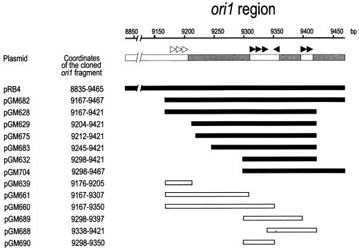
The ori1 site was previously located within the 8835-to-9465 P4 DNA region (26, 46). By sequence inspection, distinct features have been recognized (Fig. 2): three type II iterons (CAC/TTTAAAGT/C) at 9177 to 9206, followed by an AT-rich region of about 100 bp, the 9310-to-9415 region, containing six type I iterons (GGTGAACAGT/A [15, 46]), and the terminal 9416-to-9467 AT-rich region. The type I iterons within the 9310-to-9415 region are regularly spaced at multiples of 10 or 11 bp: three consecutive direct repeats followed by an inverted repeat 10 bp apart are separated from two consecutive direct repeats by 35 bp (see Fig. 4). This intervening 35-bp region is 83% AT-rich.
FIG. 2.
Identification of the minimal ori1 region. Diagrammatic representation of the ori1 region: the shaded bars indicate the AT-rich regions: 9207 to 9309, 70% AT; 9361 to 9395, 83% AT; and 9416 to 9467, 63.5% AT. The type I iterons are indicated by closed arrowheads, and the type II iterons are indicated by open arrowheads. The plasmids are derivatives of pRB2, which carries the P4 crr site (4260 to 4595); the bars indicate the cloned ori1 subfragment, whose coordinates are specified. The fragments able to support replication are solid.
FIG. 4.
Sequences of the minimal ori1, ori2, and crr sites. For ori1, the sequence of the 9298-to-9421 region is reported. For crr, the sequence of the 4260-to-4420 region is reported, corresponding to one of the two direct repeats in crr. The type I iterons are boxed. The arrows indicate the relative positions of the first base of each iteron, respective to the first iteron from the left. For ori2, the sequence of the 6219-to-6254 region is reported. The hairpin structure formed by this sequence is shown.
To define which elements within this region are essential for replication from ori1, we used an in vivo plasmid complementation test (40). We cloned in pUC19, which is unable to replicate in the polA(Ts) strain C-2107 at 42°C, both the crr site and different fragments of the previously identified ori1 region and tested whether such constructs could transform at 42°C C-2107 carrying pMK302, which provides the α and Cnr proteins in trans. The transformation abilities of the different plasmids are reported in Table 4 and summarized in Fig. 2. All the plasmids in which the ori1 fragment covers the 9298-to-9421 region were able to transform C-2107/pMK302 at 42°C. The transformation efficiency obtained with pGM632, which contains only the above-mentioned region, was comparable to that of the other plasmids carrying a more extended region, suggesting that this is the functional ori1 site.
TABLE 4.
Identification of the minimal ori1 sequence
| Plasmida | No. of transformantsb
|
Ratio (42°/30°C) | |
|---|---|---|---|
| 30°C | 42°C | ||
| pRB2 | 2,752 | 0 | <3 × 10⁻⁴ |
| pRB4 | 2,672 | 1,176 | 0.44 |
| pGM614 | 2,776 | 0 | <3 × 10⁻⁴ |
| pGM615 | 2,204 | 0 | <4 × 10⁻⁴ |
| pGM628 | 26,500 | 13,160 | 0.49 |
| pGM629 | 16,268 | 11,728 | 0.72 |
| pGM632 | 9,960 | 7,304 | 0.73 |
| pGM639 | 10,624 | 0 | <9 × 10⁻⁵ |
| pGM660 | 12,151 | 0 | <8 × 10⁻⁵ |
| pGM661 | 11,238 | 1 | 8 × 10⁻⁵ |
| pGM675 | 13,147 | 11,952 | 0.90 |
| pGM682 | 13,280 | 11,620 | 0.87 |
| pGM683 | 10,292 | 8,240 | 0.80 |
| pGM688 | 9,960 | 0 | <1 × 10⁻⁴ |
| pGM689 | 9,761 | 0 | <1 × 10⁻⁴ |
| pGM690 | 10,093 | 0 | <1 × 10⁻⁴ |
| pGM696 | 11,620 | 0 | <8 × 10⁻⁵ |
| pGM697 | 9,960 | 4,640 | 0.46 |
| pGM698 | 9,960 | 0 | <1 × 10⁻⁴ |
| pGM699 | 9,628 | 2 | 2 × 10⁻⁴ |
| pGM700 | 10,093 | 800 | 8 × 10⁻² |
| pGM704 | 15,936 | 13,088 | 0.82 |
| pGM713 | 8,632 | 0 | <1 × 10⁻⁴ |
The plasmids are derivatives of pUC19 carrying different P4 fragments, as indicated in Materials and Methods.
C-2107/pMK302 was transformed as described in Materials and Methods.
Further reduction of the 9298-to-9421 fragment by deletion of either the 9298-to-9337 (pGM688) or the 9398-to-9421 (pGM689) region impaired transformation ability. The minimal ori1 site contains all six type I iterons spaced by the 35-bp AT-rich region. Deletion of either the three type I repeats at the left end or the two repeats at the right end prevented replication ability.
These results indicate that the type II repeats and additional AT-rich regions are dispensable and might not be part of the functional ori1 site.
Construction of a minimal oriI replicon.
In order to confirm that the 9298-to-9421 ori1 region is sufficient for replication of replicon oriI, we cloned this fragment in pGM545, replacing the larger ori1 sequence (9104 to 9463). A second construct was obtained with the 9167-to-9421 fragment, which also includes the type II repeats. After transformation of strain C-1a, chloramphenicol-resistant transformants carrying the expected plasmids (pGM745 and pGM744) were isolated in the presence of IPTG. Replication of both pGM744 and pGM745 was IPTG dependent, and the plasmid copy number was similar to that of pGM545 (data not shown). This indicates that the P4 9298-to-9421 region contains the minimal ori1 sequence sufficient for replication and confirms that the type II iterons have no essential role in replication.
Characterization of the minimal ori1 sequence.
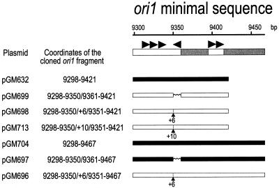
To investigate whether the single type I repeat in inverted orientation was essential for replication, we replaced it by a different 10-bp sequence with the same GC content (50%). pGM699, which carries the minimal ori1 fragment with the substitution, could not drive replication in the polA strain (Table 4 and Fig. 3), suggesting that the iteron is essential for replication. However, when the substitution was introduced in a larger ori1 fragment, which included the terminal AT-rich region (pGM697), replication ability was restored. Thus, the terminal AT-rich region may be part of a larger functional ori1 site.
FIG. 3.
Mutations in the ori1 minimal sequence. Diagrammatic representation of the minimal ori1 region (details are as for Fig. 2). The substitution of the type I iteron (9350 to 9360) is indicated by ∧∧∧; the insertion point at 9350 is marked by an arrow, and the number of the inserted nucleotide is indicated below. The fragments able to support replication are solid.
The type I iterons are arranged in ori1 with the first set of three adjacent direct repeats separated by approximately five helix turns from the last two repeats (Fig. 4). We changed the spacing between the iterons by inserting either 6 bp (pGM698) or 10 bp (pGM713) at 9350, after the first three iterons. Neither pGM698 nor pGM713 could transform at 42°C. In this case, the presence of the terminal AT-rich region (pGM696) did not restore replication. These results suggest that exact spacing of the iterons has an important role in ori1 architecture.
Role of the crr region in the replicon oriI.
The crr region consists of two directly repeated AT-rich sequences of 120 bp. It has been shown by Flensburg and Calendar (15) that a single 120-bp crr repeat, cloned beside the ori1 8835-to-9465 sequence, was able to drive replication. We tested whether this also occurred with the minimal ori1 sequence. pGM700, which carries the 4260-to-4420 crr sequence and the 9298-to-9421 ori1 sequence, was able to transform C-2107/pMK302 at 42°C, but the efficiency was reduced about 10-fold (Table 4).
We also tested whether the crr region could be replaced by the ori1 sequence. Plasmids pGM614 and pGM615 carry two ori1 sites (8835 to 9465) in direct or inverted orientation. Neither plasmid could replicate at 42°C in C-2107/pMK302 (Table 4). Thus, the crr and ori1 regions, although similar, carry out different roles in P4 DNA replication.
Identification of the ori2 minimal sequence.
The ori2 region was previously located within the 6186-to-6421 P4 region, internal to the α gene (40). Sequence comparison of the ori1 and ori2 regions did not reveal extended homology, except for the presence of several partially conserved type I-like repeats (Fig. 5). Moreover, a DnaA box (8 of 9 conserved bases; cTATCCACA at 6333 to 6341, with lowercase indicating the nonmatching base) was found.
FIG. 5.
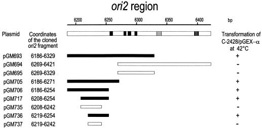
Identification of the minimal ori2 sequence. Diagrammatic representation of the ori2 site: the solid boxes indicate the type I-like sequences; the shaded box indicates the DnaA box sequence. All the plasmids carry the P4 crr site (4260 to 4595) and the indicated subfragment of the ori2 region, whose coordinates are reported. Strain C-2428/pGEX-α was transformed with the indicated plasmids, and its ability to promote replication at nonpermissive temperature was tested by transformation at 42°C and selection for spectinomycin resistance (100 μg/ml [40]). The fragments able to support replication are solid. +, able to transform; −, unable to transform.
In order to test whether these sequences were functionally important in replicon II, we used the complementation test, providing the α protein by the pGEX-α plasmid (40). Subfragments of the ori2 region were cloned in the thermosensitive plasmid pGM633, which carries the P4 crr region, and the transformation abilities of the hybrid plasmids at 42°C in C-2428/pGEX-α were tested (Fig. 5). pGM693, which lacks the DnaA box, and pGM706, in which all the type I-like repeats were deleted, still replicated at 42°C. Thus, neither the DnaA box nor the α protein binding sites are required for a functional ori2 site.
We further reduced the ori2 region: the shortest fragment able to transform at 42°C was 6219 to 6254, carried by pGM736. pGM735, which carries the 6208-to-6242 fragment, and pGM737, which carries the 6219-to-6242 fragment, did not replicate.
In order to confirm the replication ability of pGM736 at 42°C, transformants were selected at 30°C and their efficiency of plating at 42°C was tested. The ratio of efficiency at 42°C to efficiency at 30°C was about 0.5. Thus, the ori2 region was mapped within a 36-bp DNA fragment (Fig. 4).
The α protein does not bind to the ori2 site.
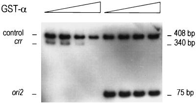
The P4 α protein binds to the type I iterons present in ori1 and crr (46). In the minimal ori2 region, no type I repeats are present. Thus, it might be supposed that α does not bind ori2. To test this, we performed band shift experiments with the purified GST-α fusion protein, expressed by pGEX-α (see Materials and Methods). As the complexes formed by the α protein and the bound DNA fragment are large aggregates which cannot enter the gel (46), the fraction of unbound DNA fragment at increasing α concentrations was estimated, in comparison to a control DNA. The DNA fragments used were (i) a 75-bp DNA fragment containing the 6186-to-6254 ori2 region, (ii) a 408-bp control DNA fragment, and (iii) a 240-bp fragment containing the 4260-to-4595 crr site. The results showed that the α protein does not bind ori2 (Fig. 6). Thus, in the oriII replicon, the α protein appears to bind only the crr site and not the ori2 site.
FIG. 6.
The ori2 site is not bound by the α protein. Gel retardation assays were performed as described in Polo et al. (33); the DNA fragments were prepared as described in Materials and Methods. ori2, 6286 to 6254 P4 region; crr, 4260 to 4595 P4 region; control, DNA fragment used for evaluating the binding specificity. Increasing amounts of the GST-α fusion protein (0, 25, 50, and 75 ng) were incubated with an equimolar mixture (4 fmol) of either the control and crr fragments or the control and ori2 fragments. After electrophoresis in a nondenaturing 6% polyacrylamide–10% glycerol gel at 12 V/cm at 4°C, the gel was dried and autoradiographed.
DISCUSSION
Common features and differences of the P4 oriI and oriII replicons.
We have previously shown that in phage-plasmid P4 two replicons coexist (40). In this work we have further characterized these replicons and we have found common features and differences.
Both the oriI and oriII replicons appear to be independent of some replication functions of the bacterial cell, such as the initiation protein DnaA, the primase DnaG, and the helicase Rep. It must be noted that our results do not definitely rule out a dependence on the DnaA protein, since it was demonstrated that the dnaA thermosensitive mutations might exhibit some leakiness (20, 23, 24).
On the other hand, we demonstrated that both replicons require the primase and helicase activities of the P4 α protein for replication.
In both replicons, the crr site is required in cis for replication. Since it is known that crr is bound by α, we can argue that both replicons also require the α DNA binding function.
Based on these results, we suggest that origin recognition and helicase and primase activities for initiation of DNA replication of both replicons oriI and oriII are provided not by the host but by the α protein. This is the most striking common feature of the two replicons.
A further apparent similarity is the presence of a bipartite origin of replication. The oriI origin is composed of the ori1 and crr sites, and oriII consists of ori2 and crr. The crr site, which is common to both origins and binds the α protein, might play the same role in the two replicons. No sequence similarities exist in ori1 and ori2: the ori2 site does not contain AT-rich regions, which are abundant in ori1, nor type I iterons, which are essential for the oriI origin. A detailed analysis of the ori1 and ori2 regions has been carried out in this work (see below). These results suggest that the ori1 and ori2 sites have different roles in replication and might support the hypothesis that the two P4 replicons replicate by different mechanisms. The identification of the replication initiation point and replication mode of the oriII replicon are necessary to verify this hypothesis.
Further differences in replication control of the two replicons have already been pointed out: the cnr gene is an essential component of the oriI replicon, since its product is necessary to prevent overreplication and cell killing; however, the Cnr protein has an inhibitory effect on the oriII replicon (40).
The oriI origin.
The previously identified ori1 site (8835 to 9465 [15, 26, 46]) presented several characteristic features: AT-rich stretches, which are usually found in replication origins; type I iterons, which are bound by the α protein; and type II direct repeats of unknown function. We have found that the minimal ori1 sequence sufficient for replication is located in the 9298-to-9421 P4 fragment. This region comprises all six type I iterons and a 35-bp AT-rich stretch. The surrounding AT-rich regions appear to be redundant.
We demonstrated that the substitution of the single type I iteron in inverted orientation impaired replication of the ori1 minimal origin; however, the inverted type I repeat appears not to be essential when the terminal AT-rich region is present. This suggests that the functional ori1 site may be larger than the “minimal origin” delimited by deletion analysis and that within this extended origin some architectural elements may be mutually redundant. An extended origin may thus tolerate mutations in one or more architectural elements, which may be an important factor in the diversification and evolution of replication origins.
No essential role could be found for the type II iterons. A replicon carrying the minimal ori1 sequence was able to replicate and be stably maintained in the bacterial cell; thus, the type II repeats are dispensable both for replication and for plasmid copy number control.
The type I iterons in ori1 are regularly spaced at multiples of 10 or 11 bp and thus are on the same side of the double helix of DNA. Deletions that further reduce the ori1 minimal fragment, eliminating either the first three or the last two direct repeats, as well as the substitution of the inverted repeat, abolish replication ability, suggesting that all sets of repeats are essential.
Insertions that change the distance between the first three and the last three iterons of either a half or whole helix turn abolish replication. This suggests that not only the helical periodicity of the type I iterons but also the distances among them are important for ori1 functionality.
Thus, we hypothesize that P4 replication initiation from ori1 occurs in a way similar to that of other iteron-containing origins, such as E. coli oriC or the phage P1 oriR (6, 7, 45), in which binding of the initiator protein causes the DNA to be bent and wrapped around a core of the protein. The adjacent AT-rich region responds to the distortion or to the binding of the initiator protein to the DNA by a specific strand-opening event. In P4, several α proteins, bound to the type I iterons, might have a regular disposition on the double helix of the DNA and constitute a nucleoprotein complex at the ori1 site, competent for replication. The central AT-rich region might be involved in specific unwinding and could be the initiation site of primer synthesis.
If this is a possible model for initiation of P4 replication, a relevant difference from the E. coli or P1 origins is given by the presence of the crr region, essential for replication.
In fact, the oriI origin is composed of the ori1 and crr sites, which lie about 4,500 bp apart in the P4 genome. By cloning these sites in a plasmid, it was demonstrated that the spacing between crr and ori1 can be reduced to less than 100 bp without affecting replication (15). However, the relative orientations of ori1 and crr were found to be essential for replication (11).
The crr site is formed by two 120-bp AT-rich repeats, each containing five type I iterons. Although the crr repeat is similar to the ori1 site, the type I iteron sequences are less conserved and their disposition does not follow the helical periodicity. As no initiation of replication has been observed from the crr site, it is likely that the α proteins bound to the type I iterons present in crr do not form a nucleoprotein complex competent for replication. A detailed analysis of the essential features of the crr region might elucidate the possible role of crr in replication.
We demonstrated that a single 120-bp crr repeat is sufficient to promote replication, even if the efficiency is reduced about 10-fold. Thus, one of the two 120-bp crr repeats is redundant, and we hypothesize that the presence of both repeats might be important to increase the efficiency of replication.
We also found that crr could not be replaced by an ori1 site: plasmids carrying two ori1 sites, in either direct or inverted orientation, could not replicate. This indicates that the ori1 and the crr sites have different roles in replication and are not interchangeable.
Proposal of a possible role for crr in P4 replication initiation should take into account the following observations: (i) the α protein binds to both ori1 and crr with approximately the same affinity (reference 46 and our unpublished results), (ii) the crr-ori1 relative orientation must be conserved, and (iii) the α protein causes looping of DNA molecules containing ori1 and crr (46).
We suggest that the α-crr and α-ori1 complexes may interact with each other, via α-α interactions (41), to form an ordered structure that is competent for replication initiation.
Several cases are known in which binding of a replication protein to specific sites causes DNA looping and/or intermolecular pairing of DNA molecules and controls DNA replication. Copy number control in phage P1 depends on binding of the RepA replication protein to two regions, ori and incA, which causes DNA looping and blocking of replication initiation (1, 10, 32). A similar “handcuffing” model, via intermolecular interactions, has been proposed for the copy number control of R6K and RK2 (22, 30, 35).
In P4 a different role should be hypothesized for crr, since this site is essential in cis for replication. In this case, DNA looping between ori1 and crr might activate replication.
The oriII origin.
The oriII origin is composed of the ori2 and crr sites. ori2 is located within the α gene coding sequence. In this work the ori2 site has been reduced to 36 bp at 6219 to 6254. Its location falls at the boundary between the primase and helicase domains of the α protein (46). This suggests that the multifunctional α gene might originate from the fusion of two ancestral genes, coding for the primase and helicase DNA binding functions, respectively, separated by the origin of replication.
Sequence analysis of the ori2 region did not reveal the presence of type I iterons or putative binding sites for other known P4 or E. coli factors, such as a DnaA box consensus sequence. Moreover, band shift experiments revealed that the α protein does not bind to the ori2 DNA region.
The orientation of the ori2 and crr sites in oriII, unlike ori1 and crr in oriI, is not relevant for replication (11).
The lack of structural and functional similarity between ori2 and ori1 suggests that their roles in replication are different.
The initiation site of replication in the oriII origin is still unknown. However, Krevolin et al. (26) detected a signal due to replication initiation in a region immediately to the right of ori2 in the P4 map (P4 HaeII 6273-to-6906 fragment), suggesting that replication starts at ori2. It might also be hypothesized that replication from ori2 proceeds unidirectionally, since no replication signals were detected to the left of ori2.
Secondary-structure predictions reveal the presence of two incomplete inverted repeats which could form a hairpin structure: a 10-bp imperfect long stem and a 6-bp long loop. Hairpin structures are normally found in the nic site (nick region) of double-stranded plasmids, which replicate by the rolling-circle mechanism (e.g., pT181, pC194, pLS1, and pVS1 [13, 21]). Based on these observations, we hypothesize that replication of the oriII replicon may occur via a rolling-circle mechanism. However, no significant homology was found by comparing the ori2 sequence with other known origins in GenBank; moreover, the primary sequence of the α protein does not contain any sequence motif common to proteins involved in initiation of rolling-circle replication, such as gpA of ΦX174, gene II protein of M13/fd, and the P2 A protein (3, 19, 29, 42). Analysis of replication intermediates of the oriII replicon might be useful in resolving this point.
The hypothesis that the two replicons replicate by different mechanisms, although both depend on the α protein, is suggestive.
ACKNOWLEDGMENTS
We are grateful to R. Calendar and E. Boye for kindly providing plasmids and strains used in this work. We thank E. Boye, E. Lanka, and P. Plevani for helpful discussions. We also thank I. Oliva for performing some experiments.
This work was supported by grant 97.04139.CT04 from Consiglio Nazionale delle Ricerche, Rome, Italy.
REFERENCES
- 1.Abeles A L, Reaves L D, Youngren-Grimes B, Austin S J. Control of P1 plasmid replication by iterons. Mol Microbiol. 1995;18:903–912. doi: 10.1111/j.1365-2958.1995.18050903.x. [DOI] [PubMed] [Google Scholar]
- 2.Barrett K J, Gibbs W, Calendar R. A transcribing activity induced by satellite phage P4. Proc Natl Acad Sci USA. 1972;69:2986–2990. doi: 10.1073/pnas.69.10.2986. [DOI] [PMC free article] [PubMed] [Google Scholar]
- 3.Beck E, Sommer R, Auerswald E A, Kurz C, Zink B, Osterburg G, Schaleer H, Sugimoto K, Sugisaki H, Okamoto T, Takanami M. Nucleotide sequence of bacteriophage fd DNA. Nucleic Acids Res. 1978;5:4495–4503. doi: 10.1093/nar/5.12.4495. [DOI] [PMC free article] [PubMed] [Google Scholar]
- 4.Bowden D W, Twersky R S, Calendar R. Escherichia coli deoxyribonucleic acid synthesis mutants: their effect upon bacteriophage P2 and satellite bacteriophage P4 deoxyribonucleic acid synthesis. J Bacteriol. 1975;124:167–175. doi: 10.1128/jb.124.1.167-175.1975. [DOI] [PMC free article] [PubMed] [Google Scholar]
- 5.Boye E, Stokke T, Kleckner N, Skarstad K. Coordinating DNA replication initiation with cell growth: differential roles for DnaA and SeqA proteins. Proc Natl Acad Sci USA. 1996;93:12206–12211. doi: 10.1073/pnas.93.22.12206. [DOI] [PMC free article] [PubMed] [Google Scholar]
- 6.Bramhill D, Kornberg A. A model for initiation at origins of DNA replication. Cell. 1988;54:915–918. doi: 10.1016/0092-8674(88)90102-x. [DOI] [PubMed] [Google Scholar]
- 7.Brendler T, Abeles A, Austin S. Critical sequences in the core of the P1 plasmid replication origin. J Bacteriol. 1991;173:3935–3942. doi: 10.1128/jb.173.13.3935-3942.1991. [DOI] [PMC free article] [PubMed] [Google Scholar]
- 8.Calendar R, Lindqvist B H, Sironi G, Clark A J. Characterization of rep-mutants and their interaction with P2 phage. Virology. 1970;40:72–83. doi: 10.1016/0042-6822(70)90380-6. [DOI] [PubMed] [Google Scholar]
- 9.Chang A C, Cohen S N. Construction and characterization of amplifiable multicopy DNA cloning vehicles derived from the P15A cryptic miniplasmid. J Bacteriol. 1978;134:1141–1156. doi: 10.1128/jb.134.3.1141-1156.1978. [DOI] [PMC free article] [PubMed] [Google Scholar]
- 10.Chattoraj D K, Mason R J, Wickner S H. Mini-P1 plasmid replication: the autoregulation-sequestration paradox. Cell. 1988;52:551–557. doi: 10.1016/0092-8674(88)90468-0. [DOI] [PubMed] [Google Scholar]
- 11.Christian R B. Studies on P4 bacteriophage DNA replication. Ph.D. thesis. Berkeley: University of California; 1990. [Google Scholar]
- 12.Churchward G, Belin D, Nagamine Y. A pSC101-derived plasmid which shows no sequence homology to other commonly used cloning vectors. Gene. 1984;31:165–171. doi: 10.1016/0378-1119(84)90207-5. [DOI] [PubMed] [Google Scholar]
- 13.del Solar G, Giraldo R, Ruiz-Echevarria M J, Espinosa M, Diaz-Orejas R. Replication and control of circular bacterial plasmids. Microbiol Mol Biol Rev. 1998;62:434–464. doi: 10.1128/mmbr.62.2.434-464.1998. [DOI] [PMC free article] [PubMed] [Google Scholar]
- 14.Dìaz-Orejas R, Ziegelin G, Lurz R, Lanka E. Phage P4 DNA replication in vitro. Nucleic Acids Res. 1994;22:2065–2070. doi: 10.1093/nar/22.11.2065. [DOI] [PMC free article] [PubMed] [Google Scholar]
- 15.Flensburg J, Calendar R. Bacteriophage P4 DNA replication. Nucleotide sequence of the P4 replication gene and the cis replication region. J Mol Biol. 1987;195:439–445. doi: 10.1016/0022-2836(87)90664-4. [DOI] [PubMed] [Google Scholar]
- 16.Ghisotti D, Chiaramonte R, Forti F, Zangrossi S, Sironi G, Dehò G. Genetic analysis of the immunity region of phage-plasmid P4. Mol Microbiol. 1992;6:3405–3413. doi: 10.1111/j.1365-2958.1992.tb02208.x. [DOI] [PubMed] [Google Scholar]
- 17.Halling C, Calendar R, Christie G E, Dale E C, Dehò G, Finkel S, Flensburg J, Ghisotti D, Kahn M L, Lane K B. DNA sequence of satellite bacteriophage P4. Nucleic Acids Res. 1990;18:1649. doi: 10.1093/nar/18.6.1649. [DOI] [PMC free article] [PubMed] [Google Scholar]
- 18.Hanahan D. Studies on transformation of Escherichia coli with plasmids. J Mol Biol. 1983;166:557–580. doi: 10.1016/s0022-2836(83)80284-8. [DOI] [PubMed] [Google Scholar]
- 19.Hanai R, Wang J C. The mechanism of sequence-specific DNA cleavage and strand transfer by ΦX174 gene A* protein. J Biol Chem. 1993;268:23830–23836. [PubMed] [Google Scholar]
- 20.Hansen E B, Yarmolinsky M B. Host participation in plasmid maintenance: dependence upon dnaA of replicons derived from P1 and F. Proc Natl Acad Sci USA. 1986;83:4423–4427. doi: 10.1073/pnas.83.12.4423. [DOI] [PMC free article] [PubMed] [Google Scholar]
- 21.Khan S A. Rolling-circle replication of bacterial plasmids. Microbiol Mol Biol Rev. 1997;61:442–455. doi: 10.1128/mmbr.61.4.442-455.1997. [DOI] [PMC free article] [PubMed] [Google Scholar]
- 22.Kittell B L, Helinski D R. Iteron inhibition of plasmid RK2 replication in vitro: evidence for intermolecular coupling of replication origins as a mechanism for RK2 replication control. Proc Natl Acad Sci USA. 1991;88:1389–1393. doi: 10.1073/pnas.88.4.1389. [DOI] [PMC free article] [PubMed] [Google Scholar]
- 23.Kline B C, Kogoma T, Tam J E, Shields M S. Requirement of the Escherichia coli dnaA gene product for plasmid F maintenance. J Bacteriol. 1986;168:440–443. doi: 10.1128/jb.168.1.440-443.1986. [DOI] [PMC free article] [PubMed] [Google Scholar]
- 24.Kogoma T, Kline B C. Integrative suppression of dnaA(Ts) mutations mediated by plasmid F in Escherichia coli is a DnaA-dependent process. Mol Gen Genet. 1987;210:262–269. doi: 10.1007/BF00325692. [DOI] [PubMed] [Google Scholar]
- 25.Krevolin M D, Calendar R. The replication of bacteriophage P4 DNA in vitro. Partial purification of the P4 α gene product. J Mol Biol. 1985;182:509–517. doi: 10.1016/0022-2836(85)90237-2. [DOI] [PubMed] [Google Scholar]
- 26.Krevolin M D, Inman R B, Roof D, Kahn M, Calendar R. Bacteriophage P4 DNA replication. Location of the P4 origin. J Mol Biol. 1985;182:519–527. doi: 10.1016/0022-2836(85)90238-4. [DOI] [PubMed] [Google Scholar]
- 27.Lindqvist B H, Six E W. Replication of bacteriophage P4 DNA in a nonlysogenic host. Virology. 1971;43:1–7. doi: 10.1016/0042-6822(71)90218-2. [DOI] [PubMed] [Google Scholar]
- 28.Lindqvist B H, Dehò G, Calendar R. Mechanisms of genome propagation and helper exploitation by satellite phage P4. Microbiol Rev. 1993;57:683–702. doi: 10.1128/mr.57.3.683-702.1993. [DOI] [PMC free article] [PubMed] [Google Scholar]
- 29.Liu Y, Haggard-Ljungquist E. Functional characterization of the P2 A initiator protein and its DNA cleavage site. Virology. 1996;216:158–164. doi: 10.1006/viro.1996.0042. [DOI] [PubMed] [Google Scholar]
- 30.McEachern M J, Bott M A, Tooker P A, Helinski D R. Negative control of plasmid R6K replication: possible role of intermolecular coupling of replication origins. Proc Natl Acad Sci USA. 1989;86:7942–7946. doi: 10.1073/pnas.86.20.7942. [DOI] [PMC free article] [PubMed] [Google Scholar]
- 31.Monk M, Kinross J. Conditional lethality of recA and recB derivatives of a strain of Escherichia coli K-12 with a temperature-sensitive deoxyribonucleic acid polymerase I. J Bacteriol. 1972;109:971–978. doi: 10.1128/jb.109.3.971-978.1972. [DOI] [PMC free article] [PubMed] [Google Scholar]
- 32.Pal S K, Chattoraj D K. P1 plasmid replication: initiator sequestration is inadequate to explain control by initiator-binding sites. J Bacteriol. 1988;170:3554–3560. doi: 10.1128/jb.170.8.3554-3560.1988. [DOI] [PMC free article] [PubMed] [Google Scholar]
- 33.Polo S, Sturniolo T, Dehò G, Ghisotti D. Identification of a phage-coded DNA-binding protein that regulates transcription from late promoters in bacteriophage P4. J Mol Biol. 1996;257:745–755. doi: 10.1006/jmbi.1996.0199. [DOI] [PubMed] [Google Scholar]
- 34.Sasaki I, Bertani G. Growth abnormalities in Hfr derivatives of Escherichia coli strain C. J Gen Microbiol. 1965;40:365–376. doi: 10.1099/00221287-40-3-365. [DOI] [PubMed] [Google Scholar]
- 35.Shah D S, Cross M A, Porter D, Thomas C M. Dissection of the core and auxiliary sequences in the vegetative replication origin of promiscuous plasmid RK2. J Mol Biol. 1995;254:608–622. doi: 10.1006/jmbi.1995.0642. [DOI] [PubMed] [Google Scholar]
- 36.Six E W, Klug C A C. Bacteriophage P4: a satellite virus depending on a helper such as prophage P2. Virology. 1973;51:327–344. doi: 10.1016/0042-6822(73)90432-7. [DOI] [PubMed] [Google Scholar]
- 37.Smith D B, Johnson K S. Single-step purification of polypeptides expressed in Escherichia coli as fusions with glutathione S-transferase. Gene. 1988;67:31–40. doi: 10.1016/0378-1119(88)90005-4. [DOI] [PubMed] [Google Scholar]
- 38.Strack B, Lessl M, Calendar R, Lanka E. A common sequence motif, -E-G-Y-A-T-A-, identified within the primase domains of plasmid-encoded I- and P-type DNA primases and the α protein of the Escherichia coli satellite phage P4. J Biol Chem. 1992;267:13062–13072. [PubMed] [Google Scholar]
- 39.Terzano S, Christian R, Espinoza F H, Calendar R, Dehò G, Ghisotti D. A new gene of bacteriophage P4 that controls DNA replication. J Bacteriol. 1994;176:6059–6065. doi: 10.1128/jb.176.19.6059-6065.1994. [DOI] [PMC free article] [PubMed] [Google Scholar]
- 40.Tocchetti A, Serina S, Terzano S, Deho G, Ghisotti D. Identification of two replicons in phage-plasmid P4. Virology. 1998;245:344–352. doi: 10.1006/viro.1998.9167. [DOI] [PubMed] [Google Scholar]
- 41.Tocchetti, A., S. Serina, G. Deho, and D. Ghisotti. Unpublished data.
- 42.van Wezenberg P M, Hulsebos T J, Schoenmakers J G. Nucleotide sequence of the filamentous bacteriophage M13 DNA genome: comparison with phage fd. Gene. 1980;11:129–148. doi: 10.1016/0378-1119(80)90093-1. [DOI] [PubMed] [Google Scholar]
- 43.Vieira J, Messing J. The pUC plasmids, an M13mp7-derived system for insertion mutagenesis and sequencing with synthetic universal primers. Gene. 1982;19:259–268. doi: 10.1016/0378-1119(82)90015-4. [DOI] [PubMed] [Google Scholar]
- 44.Wechsler J A, Gross J D. Escherichia coli mutants temperature-sensitive for DNA synthesis. Mol Gen Genet. 1971;113:273–284. doi: 10.1007/BF00339547. [DOI] [PubMed] [Google Scholar]
- 45.Woelker B, Messer W. The structure of the initiation complex at the replication origin, oriC, of Escherichia coli. Nucleic Acids Res. 1993;21:5025–5033. doi: 10.1093/nar/21.22.5025. [DOI] [PMC free article] [PubMed] [Google Scholar]
- 46.Ziegelin G, Scherzinger E, Lurz R, Lanka E. Phage P4 α protein is multifunctional with origin recognition, helicase and primase activities. EMBO J. 1993;12:3703–3708. doi: 10.1002/j.1460-2075.1993.tb06045.x. [DOI] [PMC free article] [PubMed] [Google Scholar]
- 47.Ziegelin G, Linderoth N A, Calendar R, Lanka E. Domain structure of phage P4 α protein deduced by mutational analysis. J Bacteriol. 1995;177:4333–4341. doi: 10.1128/jb.177.15.4333-4341.1995. [DOI] [PMC free article] [PubMed] [Google Scholar]
- 48.Ziegelin G, Calendar R, Ghisotti D, Terzano S, Lanka E. Cnr protein, the negative regulator of bacteriophage P4 replication, stimulates specific DNA binding of its initiator protein α. J Bacteriol. 1997;179:2817–2822. doi: 10.1128/jb.179.9.2817-2822.1997. [DOI] [PMC free article] [PubMed] [Google Scholar]


