SUMMARY
OBJECTIVE:
The aim of this study was to examine the isokinetic knee strength, H/Q ratio (%), and bone mineral density values between amputees (n=14; amputee soccer players) and healthy football players (n=14; non-amputee soccer players).
METHODS:
A total of 28 amputee soccer players and non-amputee soccer players participated in the study. An isokinetic dynamometer was used to determine the knee flexion/extension forces of the dominant legs of the athletes at 60, 180, and 240°/s. Bone mineral density scans were performed using dual-energy X-ray absorptiometry.
RESULTS:
H/Q ratio and 60º/s flexion and 180 and 240º/s flexion/extension strength (p<0.05) were found to be high (180º/s, p=0.03; 240º/s, p=0.048) in the non-amputee soccer player group. Accordingly, the bone mineral density values of the lumbar vertebra, femoral neck, proximal metaphysis of the femur (p<0.01), tibia/fibula proximal metaphysis, and tibia/fibula distal metaphysis (p<0.05) were found to be high. A correlation was observed between the 60º/s knee extension strength and tibia/fibula diaphyseal bone mineral density (p=0.025; r=0.594) and tibia/fibula distal metaphysis bone mineral density (p=0.017; r=0.623) values in the amputee soccer players group. The Z-scores of the amputee soccer players and non-amputee soccer players were in the expected range according to age (>-2).
CONCLUSION:
The bone mineral density, H/Q ratio, and all measured angular velocities of isokinetic strength were high in non-amputee soccer players. This finding made us think that lower extremity amputation may also be associated with losing strength. However, it was observed that the relationship between strength and bone mineral density in amputee athletes might vary according to different angular velocities. It is recommended that isokinetic strength measurement can be evaluated together with bone mineral density in athletes.
KEYWORDS: Amputee, Soccer, Bone mineral density, Muscle strength
INTRODUCTION
Although there are various types of para-sports events, the popularity of amputee football is spreading rapidly around the world and the awareness of the sport is increasing 1 . Amputee football players are required to run forward, move backward, turn around in their own axis, move laterally, show a high level of balance, and jump using single leg and forearm crutches while playing on the field 2 .
The low bone mineral density (BMD) experienced by amputees leads to an increased long-term risk of hip fragility fractures 3 . Therefore, physical exercise is extremely beneficial to increase BMD and bone mineral content as a potent protective factor to limit the occurrence of osteopenia, which leads to the development of osteoporosis 4 . Regarding the effects on muscle tissue, many studies investigating sports practice or resistance training during growth have shown a positive relationship between muscle mass and bone health 5–7 .
BMD is influenced by a variety of factors such as mechanical forces, hormonal changes, and exercise. The mechanical forces are, in part, influenced by muscle strength 8,9 .
When the literature is examined, different findings are found in BMD values in studies conducted with amputees and healthy individuals. However, it has been observed that the findings related to the subject of athletes are insufficient 10,11 . No study has been found in the literature investigating the relationship between isokinetic strength values and BMD (femur/tibia regions) of amputee soccer players (ASPs) and non-amputee soccer players (NASPs). In this study, the isokinetic muscle strength, BMD, and hamstring/quadriceps strength ratio of the dominant legs were evaluated together, and the differences between the groups were analyzed in ASPs and NASPs.
METHODS
Participants
Male amputee (n:14, age 29.21±5.87 years, height 172.50±8.04 cm, weight 76.71±16.26 kg, years in the sport 8.50±4.53 years, and amputation time 18.07±6.86 years) without any history of knee injury and non-amputee soccer (n:14, age 24.21±3.11 years, height 176.92±5, 92 cm, weight 73.85±8.42 kg, and years in the sport 10.35±0.53 years) voluntarily participated in the study. The lower extremity amputee levels of ASPs were determined as transtibial amputation, knee disarticulation, and transfemoral amputation.
Procedure
All athletes gave their informed consent according to the Declaration of Helsinki, and all experimental procedures were approved by the ethics committee of Ondokuz Mayıs University, Samsun, Turkey (No. 2017/164). All athletes voluntarily participated in the study and signed the voluntary consent form. Isokinetic strength measurements were performed in the sports sciences performance laboratory, and BMD measurements were made in the Nuclear Medicine Laboratory of the University Hospital (13:00 to 14:30). Athletes were asked not to participate in training and to be rested the day before and on the day of the test. Isokinetic knee extension/flexion strength and BMD measurements were performed on the intact leg for ASPs and the dominant leg for NASPs.
Assessment of muscle strength
A computer-controlled isokinetic dynamometer (Humac Norm Testing and Rehabilitation System, CSMI, USA) was used to measure the knee extension and flexion strengths of the athletes. Before the measurement, the athletes were asked to pedal for 5 min on the bicycle ergometer to warm up, and then stretching movements for the lower extremities were performed. The isokinetic strength measurement protocol in Cybex was evaluated as slow (60°/s) and fast (240°/s; 180°/s) 12 .
In the fixed protocol of the dynamometer, the knee extension and flexion measurements of the athletes were performed at angular velocities of 60°/s (15 s rest after four repeat trials and five repeats of the main test), 180°/s (four repeat trials, 15 s rest, and then five repeats of the main test), and 240°/s (four repeat trials followed by 15 s rest and 15 repeats of the main test), respectively. The rest intervals of 30 s were given to the athletes during the transitions between angular velocities 13 . The force values obtained in the tests were recorded in Newton meters.
Measurement of bone density
BMD measurements of the athletes were performed using the dual-energy X-ray absorptiometry (DEXA, Hologic QDR-2000, Discovery Series; Hologic, Inc., Waltham, MA, USA) device. For the lower extremity region, the femur and tibia/fibula regions of the dominant leg of the NASPs and the non-amputated leg of the ASPs were visualized, and BMD (g/cm2) was determined by drawing the relevant areas from each bone as proximal metaphysis, distal metaphysis, and diaphysis.
Data analysis
The SPSS 22.0 package program was used for the statistical analysis of the data obtained. An independent sample t-test was used for the group comparisons, whereas a Pearson correlation test was used to determine the relationships between the parameters. The alpha value was accepted as <0.05. When type I error (α) was 0.05 and type II error (β) was 0.20, at least 13 athletes were calculated by the power analysis with the NCSS-Pass v.2008 software.
RESULTS
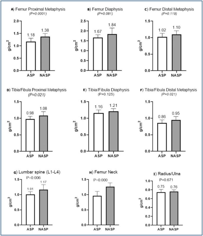
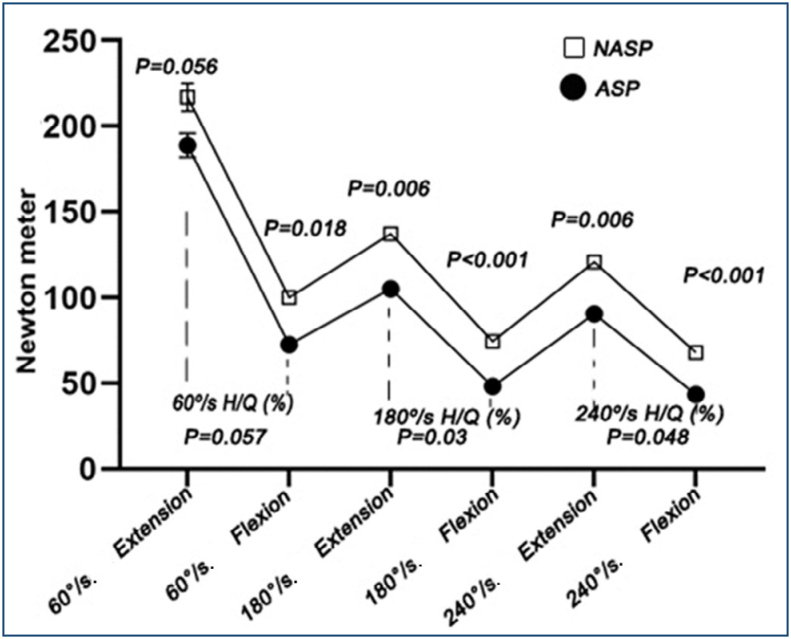
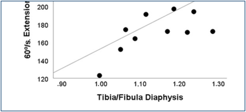
BMD values were found to be high in the femur proximal metaphysis (Figure 1A, p=0.0001), tibia/fibula proximal metaphysis (Figure 1D, p=0.021), tibia/fibula distal metaphysis (Figure 1F, p=0.021), lumbar vertebra (Figure 1G, p=0.006), and femoral neck (Figure 1H, p=0.000) in the NASP group. In the NASP group, 60º/s flexion (p=0.018), 180, and 240º/s flexion/extension strength was found to be high (p<0.01). No significant difference was found in the 60º/s extension strength (p=0.56). H/Q ratios of 180º/s (p=0.03) and 240º/s (p=0.048) were found to be significantly higher in the NASP group (Figure 2). In the NASP group, no correlation was observed between isokinetic knee strength parameters and BMD values in the leg regions. In the ASP group, a correlation was observed between the 60º/s knee extension strength and tibia/fibula diaphyseal BMD (p=0.025; r=0.594) and tibia/fibula distal metaphysis BMD (p=0.017; r=0.623) values (Figure 3).
Figure 1. Findings and significance levels of athletes’ bone mineral densities. A significant difference was found in the bone mineral density values of femur proximal metaphysis (1.38±0.03), tibia/fibula proximal metaphysis (1.08±0.03), and tibia/fibula distal metaphysis (0.95±0.03) (p<0.05). There was no significant difference in the bone mineral density values of femur diaphysis, femur distal metaphysis, and tibia/fibula diaphysis (p>0.05). A significant difference was found in the bone mineral density values of the lumbar vertebra and femoral neck (p<0.05). There was no significant difference in the radius/ulna bone mineral density values (p>0.05). (A) Femur proximal metaphysis. (B) Femur diaphysis. (C) Femur distal metaphysis. (D) Tibia/Fibula proximal metaphysis. (E) Tibia/Fibula diaphysis. (F) Tibia/Fibula distal metaphysis. (G) Lumbar spine (L1-L4). (H) Femur neck. (I) Radius/Ulna.
Figure 2. Findings of isokinetic knee strength and H/Q values of the dominant legs of the athletes. The non-amputee soccer players group had a higher flexion strength of 60 (100.07±8.26), 180 (74.57±4.22), and 240º/s (68.07±3.23) (p<0.05). Additionally, a significant difference was observed in the 180 (137.21±6.83) and 240º/s (120.64±6.50) extension strength (p<0.05). There was no significant difference in 60º/s extension strength (p>0.05). A significant difference was found in the 180º/s (54.64±1.89) and 240º/s (57.00±2.37) H/Q ratio (p<0.05). No significant difference was found at 60º/s (p>0.05).
Figure 3. Significant correlation findings between dominant leg bone mineral density values and peak torque in the amputee soccer players group. A correlation was observed between the 60º/s knee extension strength and tibia/fibula diaphysis bone mineral density (p=0.025; r=0.594) and tibia/fibula distal metaphysis bone mineral density (p=0.017; r=0.623) values in the amputee soccer players group.
There was no significant differences femur diaphysis (Figure 1B), femur distal metaphysis (Figure 1C), tibia/fibula diaphysis (Figure 1E), radius/ulna (Figure 1I) BMD between ASP and NASP (p>0.05).
DISCUSSION
In this study, the BMD values were examined separately by dividing the lower extremity into separate groups, and for the first time, their relationship with the isokinetic knee strength was evaluated.
Among the BMD parameters of the athletes, the lumbar vertebra and femoral neck were found to be significantly higher in the NASP group in our study. No significant difference was detected in the radius/ulna BMD value. BMD increases in athletes who continue high-impact loads in training and competition 14 . The occurrence of situations such as sprinting, jumping, sudden acceleration and deceleration, and change in direction in the soccer game also includes different loads on the muscles and bones and the ground reaction force. Thus, BMD develops most effectively with activities that produce greater loads on the bone, such as soccer 15 . From this point of view, we can predict that players who have been doing soccer training for many years and who are more exposed to loads may also show higher BMD values.
In our study, high BMD values were found in the NASP leg regions of the femur proximal metaphysis, tibia/fibula proximal metaphysis, and tibia/fibula distal metaphysis. No significant difference was found in the BMD values of the athletes’ femoral diaphysis, femur distal metaphysis, and tibia/fibula diaphysis.
It is plausible that there is a dynamic and dependent physiological link between muscle strength and BMD. However, it is known that multiple factors also affect BMD apart from the forces originating from muscle contraction. These factors include nutrition, age, hormones, impact, and genetics 16 . The reason for the high BMD values of NASPs may be that the leg area is exposed to more intense training compared to ASPs.
Some studies have suggested that the skeletal and muscular systems are structurally interdependent, and both adapt to mechanical loading. It is stated that, this way, muscle movement provides a stimulus for bone remodeling by pulling on the bone where the tendon attaches, and the skeleton adapts to the increasing magnitude of loading by accumulating bone 17 . Accordingly, it was thought that the findings of our study also affected the regional training of the athletes and the parts of the muscles at the attachment points.
In our study, 60º/s flexion and 180 and 240º/s flexion/extension strengths were high in the NASP group. H/Q ratios of 180 and 240º/s were significantly high in the NASP group. When the literature was examined, no study was found comparing the isokinetic knee strength values of ASPs and NASPs.
According to the literature, the high muscle strength values of soccer players can be explained by the fact that soccer includes various technical skills such as kicking, jumping, and landing, as well as the fact that strength exercises have an important place in training planning. Based on the findings of our study, it can be said that, due to the less active condition of ASPs, the muscle groups tend to be more atrophic and weaker.
Based on the H/Q ratios obtained in our study, it can be said that the risk of injury in ASPs is higher compared to NASPs as the H/Q ratios of both groups were found to be lower than the norm values indicated in the literature. The norm values of H/Q ratios are accepted as 50–70% for 60°/s18 and 70–90% for 180°/s 19,20 . The reason for the low H/Q ratios of both ASPs and NASPs can be shown as unilateral exercises in both groups and the neglect of the hamstring muscle group. H/Q ratios outside of the norm can pose a risk for joint and muscle injuries.
In our study, there was no correlation between the isokinetic knee strength parameters and leg region BMD values in the NASP group. In many studies, muscle strength and BMD values of athletes were found to be higher than non-athletic controls. The relationship between muscle strength and BMD was stronger in those with low-to-moderate physical training 21,22 . However, less or no relationship was observed between muscle strength and BMD in highly trained individuals. Such a relationship was not found in women athletes participating in sports that involve heavy body load such as soccer 21 . Such a relationship was not found in ice hockey athletes 17 . Based on these studies, it was concluded that high physical activity weakens this association. However, there are many studies reporting a relationship between muscle strength and BMD 23,24 . These conflicting results may be due to individual differences in athletes and training protocols.
In our study, a correlation was observed between 60º/s knee extension strength value and tibia/fibula diaphysis BMD and tibia/fibula distal metaphyseal BMD values in the ASP group. No correlation was found between the isokinetic knee strength parameters and leg region BMD values.
While few studies were found in the literature on isokinetic knee strength and BMD in amputees, these current studies do not focus on the relationship between isokinetic knee strength and BMD values of the leg regions.
Tugcu conducted a study with individuals with trans-tibial amputation and did not find a relationship between 30 and 1200/s knee muscle strength and femoral neck, total femur, and tibia BMD values 25 . The lack of strong correlations between many strength measurements and bone densities may be because strength is not only dependent on the size of the muscle attached to the bone but also on neural action to the muscle 14 . This can weaken the relationship between strength and bone.
CONCLUSION
The BMD, H/Q ratios, and all measured angular velocities of isokinetic strength were high in NASPs. This finding made us think that lower extremity amputation may also be associated with losing strength. However, it was observed that the relationship between strength and BMD in amputee athletes might vary according to different angular velocities. It is recommended that isokinetic strength measurement can be evaluated together with BMD in athletes. A limitation of this study is that the biochemical parameters that affect the BMD of the participants were not measured. Additionally, the small sample size and the sample consisting of only males can be seen as limitations of the study. Not being able to determine the effects of training protocols of participants on individual differences is also considered another limiting factor of the study.
ACKNOWLEDGMENTS
This article is produced by II from the PhD thesis entitled “The Investigation of the Isokinetic Muscle Strength and Bone Mineral Density of the Amputee And Non-Amputee Football Players” in Ondokuz Mayıs University, Graduate Education Institute, Coaching Education Department, Samsun, Turkey.
Footnotes
ETHICAL STATEMENT All athletes gave their informed consent according to the Declaration of Helsinki, and all experimental procedures were approved by the Ethics Committee of Ondokuz Mayis University Samsun, Turkey (No. 2017/164).
Funding: none.
REFERENCES
- 1.Mikami Y, Fukuhara K, Kawae T, Sakamitsu T, Kamijo Y, Tajima H, et al. Exercise loading for cardiopulmonary assessment and evaluation of endurance in amputee football players. J Phys Ther Sci. 2018;30(8):960–965. doi: 10.1589/jpts.30.960. [DOI] [PMC free article] [PubMed] [Google Scholar]
- 2.Wieczorek M, Wiliński W, Struzik A, Rokita A. Hand grip strength vs. sprint effectiveness in amputee soccer players. J Hum Kinet. 2015;48:133–139. doi: 10.1515/hukin-2015-0099. [DOI] [PMC free article] [PubMed] [Google Scholar]
- 3.Zaid MB, OʼDonnell RJ, Potter BK, Forsberg JA. Orthopaedic osseointegration: state of the art. J Am Acad Orthop Surg. 2019;27(22):e977–e985. doi: 10.5435/JAAOS-D-19-00016. [DOI] [PubMed] [Google Scholar]
- 4.Maïmoun L, Coste O, Mura T, Philibert P, Galtier F, Mariano-Goulart D, et al. Specific bone mass acquisition in elite female athletes. J Clin Endocrinol Metab. 2013;98(7):2844–2853. doi: 10.1210/jc.2013-1070. [DOI] [PubMed] [Google Scholar]
- 5.Saraiva BTC, Agostinete RR, Freitas IF, Júnior, Sousa DER, Gobbo LA, Tebar WR, et al. Association between handgrip strength and bone mineral density of Brazilian children and adolescents stratified by sex: a cross-sectional study. BMC Pediatr. 2021;21(1):207–207. doi: 10.1186/s12887-021-02669-1. [DOI] [PMC free article] [PubMed] [Google Scholar]
- 6.Bersotti FM, Mochizuki L, Brech GC, Soares ALS, Soares JM, Junior, Baracat EC, et al. The variability of isokinetic ankle strength is different in healthy older men and women. Clinics. 2022;77:100125–100125. doi: 10.1016/j.clinsp.2022.100125. [DOI] [PMC free article] [PubMed] [Google Scholar]
- 7.Felix ECR, Alonso AC, Brech GC, Fernandes TL, Almeida AM, Luna NMS, et al. Is 12 months enough to reach function after athletes’ ACL reconstruction: a prospective longitudinal study. Clinics. 2022;77:100092–100092. doi: 10.1016/j.clinsp.2022.100092. [DOI] [PMC free article] [PubMed] [Google Scholar]
- 8.Guimarães BR, Pimenta LD, Massini DA, Santos D, Siqueira LODC, Simionato AR, et al. Muscle strength and regional lean body mass influence on mineral bone health in young male adults. PLoS One. 2018;13(1):e0191769. doi: 10.1371/journal.pone.0191769. [DOI] [PMC free article] [PubMed] [Google Scholar]
- 9.Castillo GB, Brech GC, Luna NMS, Tarallo FB, Soares JM, Junior, Baracat EC, et al. Influence of invertor and evertor muscle fatigue on functional jump tests and postural control: a prospective cross-sectional study. Clinics. 2022;77:100011–100011. doi: 10.1016/j.clinsp.2022.100011. [DOI] [PMC free article] [PubMed] [Google Scholar]
- 10.Cavedon V, Sandri M, Peluso I, Zancanaro C, Milanese C. Body composition and bone mineral density in athletes with a physical impairment. PeerJ. 2021;9:e11296. doi: 10.7717/peerj.11296. [DOI] [PMC free article] [PubMed] [Google Scholar]
- 11.Finco MG, Kim S, Ngo W, Menegaz RA. A review of musculoskeletal adaptations in individuals following major lower-limb amputation. J Musculoskelet Neuronal Interact. 2022;22(2):269–283. [PMC free article] [PubMed] [Google Scholar]
- 12.Cress ME, Johnson J, Agre JC. Isokinetic strength testing in older women: a comparison of two systems. J Orthop Sports Phys Ther. 1991;13(4):199–202. doi: 10.2519/jospt.1991.13.4.199. [DOI] [PubMed] [Google Scholar]
- 13.Mameletzi D, Siatras T. Sex differences in isokinetic strength and power of knee muscles in 10-12 year old swimmers. Isokinetics Exerc Sci. 2003;11:231–237. doi: 10.3233/ies-2003-0152. [DOI] [Google Scholar]
- 14.Herbert AJ, Williams AG, Lockey SJ, Erskine RM, Sale C, Hennis PJ, et al. Bone mineral density in high-level endurance runners: part A-site-specific characteristics. Eur J Appl Physiol. 2021;121(12):3437–3445. doi: 10.1007/s00421-021-04793-3. [DOI] [PMC free article] [PubMed] [Google Scholar]
- 15.Tenforde AS, Fredericson M. Influence of sports participation on bone health in the young athlete: a review of the literature. Am Acad Phys Med Rehabil. 2011;3(9):861–867. doi: 10.1016/j.pmrj.2011.05.019. [DOI] [PubMed] [Google Scholar]
- 16.Sutter T, Toumi H, Valery A, El Hage R, Pinti A, Lespessailles E. Relationships between muscle mass, strength and regional bone mineral density in young men. PLoS One. 2019;14(3):e0213681. doi: 10.1371/journal.pone.0213681. [DOI] [PMC free article] [PubMed] [Google Scholar]
- 17.Pettersson U, Nordström P, Lorentzon R. A comparison of bone mineral density and muscle strength in young male adults with different exercise level. Calcif Tissue Int. 1999;64(6):490–498. doi: 10.1007/s002239900639. [DOI] [PubMed] [Google Scholar]
- 18.Dvir Z. Isocinética: avaliações musculares interpretações e aplicações clínicas. São Paulo: Manole; 2002. [Google Scholar]
- 19.Kellis E, Sahinis C, Baltzopoulos V. Is hamstrings-to-quadriceps torque ratio useful for predicting anterior cruciate ligament and hamstring injuries? A systematic and critical review. J Sport Health Sci. 2023;12(3):343–358. doi: 10.1016/j.jshs.2022.01.002. [DOI] [PMC free article] [PubMed] [Google Scholar]
- 20.Ruas CV, Pinto RS, Haff GG, Lima CD, Pinto MD, Brown LE. Alternative methods of determining hamstrings-to-quadriceps ratios: a comprehensive review. Sports Med Open. 2019;5(1):11–11. doi: 10.1186/s40798-019-0185-0. [DOI] [PMC free article] [PubMed] [Google Scholar]
- 21.Alfredson H, Nordström P, Lorentzon R. Total and regional bone mass in female soccer players. Calcif Tissue Int. 1996;59(6):438–442. doi: 10.1007/BF00369207. [DOI] [PubMed] [Google Scholar]
- 22.Kopiczko A, Adamczyk JG, Gryko K, Popowczak M. Bone mineral density in elite masters athletes: the effect of body composition and long-term exercise. Eur Rev Aging Phys Act. 2021;18(1):7–7. doi: 10.1186/s11556-021-00262-0. [DOI] [PMC free article] [PubMed] [Google Scholar]
- 23.Pelegrini A, Bim MA, Alves AD, Scarabelot KS, Claumann GS, Fernandes RA, et al. Relationship between muscle strength, body composition and bone mineral density in adolescents. J Clin Densitom. 2022;25(1):54–60. doi: 10.1016/j.jocd.2021.09.001. [DOI] [PubMed] [Google Scholar]
- 24.Montenegro Barreto J, Vidal-Espinoza R, Gomez Campos R, Arruda M, Urzua Alul L, Sulla-Torres J, et al. Relationship between muscular fitness and bone health in young baseball players. Eur J Transl Myol. 2021;31(1):9642–9642. doi: 10.4081/ejtm.2021.9642. [DOI] [PMC free article] [PubMed] [Google Scholar]
- 25.Tugcu I, Safaz I, Yilmaz B, Göktepe AS, Taskaynatan MA, Yazicioglu K. Muscle strength and bone mineral density in mine victims with transtibial amputation. Prosthet Orthot Int. 2009;33(4):299–306. doi: 10.3109/03093640903214075. [DOI] [PubMed] [Google Scholar]


