

Abstract
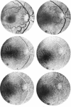
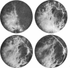
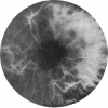
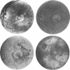
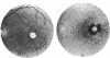
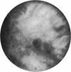
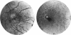
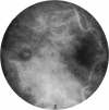
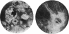
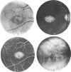
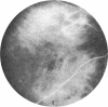
Indocyanine green fluorescence (ICG) angiography of the choroid gives better visualization of the choroidal vessels than does fluorescein angiography. We found that the detachment of the pigment epithelium seems bigger on ICG than on fluorescein angiograms, and pigmented lesions are more clearly delineated.
Full text
PDF


Images in this article


Selected References
These references are in PubMed. This may not be the complete list of references from this article.
- Craandijk A., Aan de Kerk A. L. Retinal photography using panchromatic and orthochromatic films. Br J Ophthalmol. 1969 Aug;53(8):568–573. doi: 10.1136/bjo.53.8.568. [DOI] [PMC free article] [PubMed] [Google Scholar]
- Hochheimer B. F. Angiography of the retina with indocyanine green. Arch Ophthalmol. 1971 Nov;86(5):564–565. doi: 10.1001/archopht.1971.01000010566014. [DOI] [PubMed] [Google Scholar]
- Kogure K., David N. J., Yamanouchi U., Choromokos E. Infrared absorption angiography of the fundus circulation. Arch Ophthalmol. 1970 Feb;83(2):209–214. doi: 10.1001/archopht.1970.00990030211015. [DOI] [PubMed] [Google Scholar]