Abstract
Targeted recombination within the S (spike) gene of transmissible gastroenteritis coronavirus (TGEV) was promoted by passage of helper respiratory virus isolates in cells transfected with a TGEV-derived defective minigenome carrying the S gene from an enteric isolate. The minigenome was efficiently replicated in trans and packaged by the helper virus, leading to the formation of true recombinant and pseudorecombinant viruses containing the S proteins of both enteric and respiratory TGEV strains in their envelopes. The recombinants acquired an enteric tropism, and their analysis showed that they were generated by homologous recombination that implied a double crossover in the S gene resulting in replacement of most of the respiratory, attenuated strain S gene (nucleotides 96 to 3700) by the S gene of the enteric, virulent isolate. The recombinant virus was virulent and rapidly evolved in swine testis cells by the introduction of point mutations and in-phase codon deletions in a domain of the S gene (nucleotides 217 to 665) previously implicated in the tropism of TGEV. The helper virus, with an original respiratory tropism, was also found in the enteric tract, probably because pseudorecombinant viruses carrying the spike proteins from the respiratory strain and the enteric virus in their envelopes were formed. These results demonstrated that a change in the tropism and virulence of TGEV can be engineered by sequence changes in the S gene.
Transmissible gastroenteritis virus (TGEV) is a member of the Coronaviridae family of the Nidovirales order (13, 18). TGEV replicates in both the villous epithelial cells of the small intestine and in the lung cells of newborn piglets, resulting in a mortality of nearly 100% (19, 51).
Coronaviruses attach to host cells through the spike (S) glycoprotein (25). TGEV entry into swine testis (ST) cells is also mediated by the S glycoprotein through interactions with porcine aminopeptidase N (pAPN), which is the cellular receptor (16). Aminopeptidase N also serves as the receptor for human, canine, and feline coronaviruses (3, 31, 61, 73). Interestingly, while porcine and human aminopeptidases show species specificity, the feline aminopeptidase seems to serve as a receptor for feline, canine, porcine, and human coronaviruses (3, 61).
TGEV enteric or respiratory tropism is conditioned by the primary structure of the spike gene (2). The S glycoprotein domain recognized by the cellular receptor on ST cells is located in the globular domain of the protein close to the antigenic sites A and D (58). A domain of the spike protein encoded by S gene nucleotides (nt) 1518 to 2184 is efficiently recognized by pAPN, and transfection of pAPN to nonpermissive cells makes them susceptible to TGEV (23). Nevertheless, this domain is present both in enteric and respiratory porcine coronaviruses, indicating that its presence in a virus is not in itself sufficient to allow for infections of the enteric tract. In fact, it has been demonstrated that a second factor mapping in the S gene around nt 655 drastically influences the enteric tropism of the PUR46 strain of TGEV (2).
Genetic alterations in either the virus or host cells can change the dynamics of virus-cell interaction. The regulatory elements that impact upon tissue-specific tropism and pathology may act at the levels of transcription, RNA processing, stability and transport, translation, and protein stability and processing (36). Nevertheless, very often the susceptibility to a virus is determined at the recognition and internalization level.
The development of RNA minigenomes has been very useful for studying the role of the different viral genes in the tropism and virulence of TGEV. These minigenomes are derived from defective TGEV viruses that are efficiently replicated by using a helper TGEV that provides the replicase and the structural proteins for minigenome packaging in trans (27, 42). This system in principle may be useful to complement a gene defect or to replace a gene by targeted recombination, as was found in the first studies on coronavirus genome modification performed on the nucleoprotein gene of the murine hepatitis virus (MHV) (30, 38, 43, 62) or as described in the more-recent report on the replacement of the S gene of this virus (35). In these studies, the coronavirus genome was modified by targeted recombination, involving a single crossover that replaced the 3′ end of the virus with a new one encoded in a coronavirus-derived minigenome. Since we were interested in studying the role of genes, such as the spike gene, located in an internal domain of the genome, we wondered whether targeted recombination involving two crossover events is a practical approach to modify the coronavirus genome to study viral function. The same system might also be useful to study the formation of pseudorecombinants that could provide the spike protein of a virus in trans to study its effect on virus tropism.
During the characterization of one of the oldest in vivo passages of the Purdue strain of TGEV (PUR46-SW11) (10, 24), we observed that this virulent strain was a mixture of at least two TGEV isolates, with remarkable differences in their in vivo and in vitro growth characteristics. One of them, isolate C11, replicated with high titers in the enteric tract and was virulent, while the other one (isolate C8) produced low virus titers in enteric tissues and was attenuated. The nucleotide differences within the one-third 3′ end sequences of these isolates have been determined, and most nucleotide substitutions are accumulated at the 5′ end of the S gene. Targeted recombination, implying two crossover events within the spike gene of an attenuated and respiratory TGEV, that replaced most of the endogenous spike gene by that of isolate C11 encoded by a TGEV-derived minigenome, provided enteric tropism and virulence to the attenuated virus. In addition, it has been shown that the recombinant virus rapidly evolved during its passage in ST cells by introduction of sequence modifications at the 5′ end of S gene, in a sequence domain previously associated with changes in viral tropism. The viral population evolved depending on the tissue used for virus replication. In the enteric tracts of swine, the recombinants carrying the S protein from the enteric virus (isolate C11) were selected, while in cultured ST cells the helper Purdue type virus (PTV) strain was favored.
MATERIALS AND METHODS
Cells and viruses.
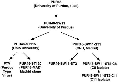
The origins of and relationship among the different TGEVs used in this study are summarized in Fig. 1. The PUR46-SW11 (SW11) strain was obtained from TGEV PUR46 by 11 passages in swine intestine; this virus was kindly provided as a 20% suspension of small intestine cells by M. Pensaert (Ghent, Belgium) (24, 34). The PUR46-SW11-ST2 (ST2) virus was obtained from SW11 after two passages in ST cells (40). The PUR46-SW11-ST2-C8 (C8) and PUR46-SW11-ST2-C11 (C11) isolates were plaque-purified clones derived (see Results) from the PUR46-SW11-ST1 (ST1). All viruses were grown in ST cells. The TGEV strain PUR46-MAD was derived from the Purdue strain by 120 passages on ST cells involving three cloning steps by plaque purification (54). Viruses were titrated in a plaque assay on ST cells as described previously (28).
FIG. 1.
Nomenclature and relationship among the TGEVs used in this work. All the TGEVs used were derived from an original isolate of the Purdue virus (24, 34) that has been named PUR46 in reference to the place and year that it was reported for the first time. The PUR46-SW11 (SW11) strain was obtained from PUR46 by 11 passages in swine intestine; this virus was kindly provided as a 20% suspension of small intestine cells by M. Pensaert (24, 34). The PUR46-SW11-ST2 (ST2) virus was obtained from SW11 after two passages in ST cells (40). The PUR46-SW11-ST2-C8 (C8) and PUR46-SW11-ST2-C11 (C11) isolates were plaque-purified isolates derived from the PUR46-SW11-ST1 (ST1) at CNB, Madrid, Spain. The PUR46-ST115 was independently derived from the PUR46-SW11 (or a similar passage number in swine) after 115 passages in ST cells, by L. Saif at the OARDC (Ohio State University) (50). From this strain we obtained the PUR46-MAD-ST120 by five additional passages on ST cells including three cloning steps by plaque purification (54). The PTV virus was derived from a Purdue type strain by sequential passage in gnotobiotic pigs by the pulmonary route, pig lung cell cultures, and diploid ST cell cultures. During this time the virus was exposed to an acidic (pH 3) environment and trypsin (66).
The PTV strain was previously named NEB72 (53). However, due to sequence similarity to the PUR46 strain (2), its name was changed to PTV (Purdue type virus).
TGEV growth kinetics in ST cells.
ST cell monolayers just at confluence, or 24 h after reaching confluence, were infected at multiplicities of infection (MOIs) of 1 and 10 with isolates C8 and C11. Aliquots of 200 μl were taken from the supernatant of each infected monolayer at different times postinfection, and virus titers were determined as described above.
TGEV growth kinetics in vivo.
Two- to three-day-old non-colostrum-deprived NIH miniswine (37, 49) were used to study isolates C8 and C11 in animal infections. Piglets were obtained from sows that were seronegative for TGEV tested by radioimmunoassay. Animals were oronasally (1.6 × 10⁸ PFU/pig) and intragastrically (3.4 × 10⁸ PFU/pig) inoculated with virus in final volumes of 0.7 and 1.5 ml, respectively, of phosphate-buffered saline (pH 7.2) (PBS) supplemented with 2% fetal calf serum. Groups of piglets were inoculated with C8, C11, a mixture of both isolates (70% C8 plus 30% C11), or, as a control, ST2, a virulent isolate. Piglets that had been inoculated with the same virus were grouped and housed in isolation chambers that were located in a P3-level containment facility at 18 to 20°C. Animals were fed three times per day with 30 ml of milk formula for newborns (Nidina1-Nestlé). At 1, 2, 3, and 4 days postinoculation (d p.i.) virus titers were determined in tissue extracts from the jejunum, ileum, lungs, mesenteric tissue, and mediastinal lymph nodes. Lung, jejunum, and ileum extracts were obtained by homogenizing the whole organs, in order to obtain representative samples, at 4°C by using a Pro-200 tissue homogenizer (Pro-Scientific). Infected animals were monitored daily to detect symptoms of disease (enteritis) and death.
RNA isolation.
Genomic RNA was extracted from partially purified virus as described previously (42). Briefly, ST cells from 10 roller bottles (500 cm2) were infected (MOI, 5). Medium was harvested at 22 hours postinfection (h p.i.). Virions were partially purified as described (28). The viral pellet was dissociated by resuspension in 500 μl of TNE buffer (0.04 M Tris-hydrochloride [pH 7.6], 0.24 M NaCl, 15 mM EDTA) containing 2% sodium dodecyl sulfate (SDS) and digested with 50 ng of proteinase K (Boehringer Mannheim) for 30 min at room temperature. RNA was extracted twice with phenol-chloroform and precipitated with ethanol.
Cytoplasmic RNA from infected ST cells was extracted as described previously (42). Briefly, ST cells grown in 8-cm2 wells were infected with TGEV at an MOI of 5. At 22 h p.i. cell extracts were prepared by washing 4 × 10⁶ cells in 1.5 ml of PBS. Cells were lysed in 200 μl of TSM buffer (0.15 M NaCl, 0.01 M Tris-hydrochloride [pH 7.6], 5 mM MgCl2) with 0.2% Nonidet P-40 in the presence of 10 mM vanadyl ribonucleoside complexes (New England BioLabs), on ice. Nuclei were pelleted by centrifugation at 13,000 × g for 30 s. The cell lysis was completed by resuspending the pellets in 100 μl of TSM buffer with 0.2% Nonidet P-40 in the presence of vanadyl ribonucleoside complexes and sedimenting the nuclei as described above. Supernatants were equilibrated at room temperature, and proteins were extracted with 300 μl of urea-SDS lysis buffer (1.5% SDS, 15 mM EDTA, 0.24 M NaCl, 0.04 M Tris-hydrochloride [pH 7.6], 8 M urea), vigorous vortexing, and phenol-chloroform extraction.
RNA from jejunum cells of swine infected with TGEV was extracted by using the procedure previously described (41) with minor modifications. Briefly, 50 μl of jejunum cell suspension (1 mg of tissue/ml) was diluted in PBS to a final volume of 400 μl, and the RNA was extracted in the manner that the TGEV-infected ST cells were extracted.
Cloning and sequencing.
Overlapping cDNA fragments to complete the sequence of the last 3′ end 8 kb of C8 and C11 viruses were synthesized by reverse transcription (RT)-PCR and were cloned into Bluescript SK M13− (Stratagene) or pGEM-T (Promega). Plasmid DNA was purified by using the FlexiPrep kit (Pharmacia) and sequenced by using an Applied Biosystems 373 DNA Sequencer. The sequence of the recombinant virus S gene was determined by using RT-PCR-amplified cDNAs. Sequence data were compiled by using the Wisconsin Package software, version 9.0, of the Genetics Computer Group (GCG) (Madison, Wis.). Sequences obtained were compared to those previously published for PUR46 virus strains (17, 29, 42, 44, 46, 53). Mutations were confirmed by sequencing three independently derived RT-PCR clones or by directly sequencing the viral RNA (20).
Construction of a cDNA encoding the spike gene of an enteric isolate (C11) within a TGEV-derived minigenome.
The S gene from the TGEV isolate C11 was cloned into TGEV-derived minigenome M54, producing the M54-Sc11 minigenome. This cDNA has been cloned after the T7 promoter and contains sequences derived from TGEV (44). M54 contains open reading frame (ORF) 1a nt 1 to 2144 and 12195 to 12368, ORF 1b nt 12338 to 13862 and 19275 to 20364, S gene nt 20365 to 20372, and gene 7 and 3′ untranslated region (UTR) sequences (nt 28093 to 28586, adding the six nucleotides of the S gene deletion, i.e., the 3′ end of the virus genome). The spike gene of the enteric isolate C11 was flanked at its 5′ end by the ORF 1b 3′ end 65 nt including the intergenic sequence CTAAAC and was cloned within the deletion that created the M54 minigenome from DI-C, i.e., between two NdeI sites at the ORF 1b positions 13858 and 19270. The S gene was flanked at the 3′ end by the sequence AATCACTAGTGCGGCCGCCTGCAGGTCGAC, containing the restriction endonuclease sites SpeI, NotI, PstI, and SalI, derived from the polylinker of plasmid pGEM-T. The minigenome is flanked at the 3′ end by a synthetic poly(A25) tract, the hepatitis delta virus ribozyme, and T7 terminator sequences (27).
Targeted recombination and formation of pseudorecombinant viruses.
To rescue the M54-Sc11 RNA in order to promote targeted recombination between the Sc11 gene and the S gene of the helper virus or to promote pseudorecombinant virus formation, in vitro transcription of linearized DNA templates was performed with T7 RNA polymerase according to the manufacturer’s instructions (Promega). The M54-Sc11 plasmid was linearized with SacII restriction endonuclease, downstream of the T7 terminator. The length of the in vitro-transcribed RNA was estimated in 1% agarose–Tris-borate-EDTA–0.1% SDS gels (52).
The in vitro-transcribed M54-Sc11 RNA was rescued as described previously (27). Briefly, ST cells were grown to confluence and infected with TGEV C8 or PTV strains (MOI, 10). At 4 to 6 h p.i., cells were trypsinized and resuspended in PBS. Cells were electroporated (200 V, 500 μF, single pulse) with in vitro-transcribed RNA (5 μg/106 cells) by using a Gene Pulser apparatus (Bio-Rad). Electroporated cells were resuspended in Dulbecco modified Eagle medium supplemented with 2% fetal calf serum and incubated at 37°C for 18 h. Supernatants from these cultures were passaged with fresh ST cells eight times in order to amplify the virions containing the minigenomes and to promote recombination. Within each passage, the virus was grown for 22 to 24 h. After the eighth passage the RNA was extracted as described above.
To select potential recombinant viruses and to see whether pseudorecombinant viruses based on the respiratory helper virus and the S protein from the enteric isolate Sc11 were formed, the virus pool from passage 8 was used to inoculate 2-day-old swine derived from a cross between Landrace and Large White swine, as described above for NIH minipigs. Virus growing in the gut was isolated from the jejunum, ileum, and intestinal content when the helper virus used was isolate C8. The presence of virus in the jejunum or ileum was evaluated when the PTV strain was used as a helper virus.
RNA analysis by Northern blotting.
Cytoplasmic RNA was extracted from helper virus-infected and RNA-transfected ST cells at passage 8, as described above. RNAs were separated in denaturing 1% agarose–2.2 M formaldehyde gels. Northern blot analysis was performed after blotting of the RNAs onto nylon membranes (Duralon-UV; Stratagene) by using an [α-32P]dATP-labeled 3′ UTR-specific single-stranded DNA probe complementary to sequences of gene 7 and the 3′ UTR (nt 28300 to 28544) of the TGEV PUR46-MAD strain genome (44), as described previously (27). RNA was quantified after Northern blot analysis by using a Molecular Imager FX System (Bio-Rad).
Screening of virus isolates.
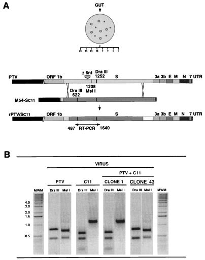
Two nucleotide substitutions in the sequence of strain PTV in relationship to that of strain C11 lead to differences in their restriction patterns. A nucleotide substitution (C to T) at position 622 of S gene (S622) implicated the loss of a DraIII restriction site in strain PTV, and the replacement of T with A at position S1208 generated a restriction site for MslI in this isolate. To screen for the presence of isolates with PTV- or C11-derived S genes after passage in tissue culture or in vivo, a cDNA fragment including S gene nt 487 to 1640 was synthesized by RT-PCR by using the RNA from infected cells. RT-PCR fragments derived from the different clones were digested with restriction endonucleases DraIII and MslI. cDNA fragments derived from the strain PTV were cut once by DraIII at nt 1252 and by MslI at nt 1208, while cDNAs derived from isolate C11 were cut twice by DraIII at positions 622 and 1252 and were not cut by MslI.
To confirm the sequence of the S gene, the isolates were plaque purified twice and amplified once in ST cell monolayers, the RNA was extracted as described above, and cDNAs were derived by RT-PCR and directly sequenced.
RESULTS
Isolation of two TGEV viruses with high and low replication levels in the enteric tract.
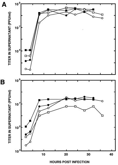
An uncloned stock of the original virulent TGEV Purdue strain that was passaged 11 times in swine (PUR46-SW11) (Fig. 1) was passaged once in ST cells, generating the isolate PUR46-SW11-ST1. This uncloned virus was plaqued on ST cells, and two types of isolates, with large (3-mm-diameter) and small (1-mm-diameter) plaques, were observed. Two isolates, PUR46-SW11-ST2-C8 (abbreviated C8; an isolate with small plaque) and PUR46-SW11-ST2-C11 (abbreviated C11; an isolate with large plaque), were plaque purified three times. The plaques maintained their morphology during the cloning. The growth kinetics of these isolates in ST cell monolayers at two cell densities (1 × 10⁵/cm2 and 2 × 10⁵ cell/cm2) at two MOIs (1 and 10) (Fig. 2) indicated that in all cases isolate C8 produced titers at least 10-fold higher than those produced by isolate C11.
FIG. 2.
Growth kinetics of isolates C8 and C11 in ST cells. The growth of isolates C8 (A) and C11 (B) in ST cells is shown. Virus replication in ST cells that just reached confluence (○, □) or were grown for 1 more day following confluence (●, ■) after infection at an MOI of 1 (○, ●) or 10 (□, ■) is shown. Aliquots (0.2 ml) were collected at the indicated times and titrated on ST cells. Results of a representative experiment among three that gave similar results are shown.
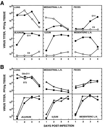
The in vivo growth of C8 and C11 isolates was determined by inoculating newborn NIH miniswine by the oronasal and intragastric routes. The piglets were sacrificed at 1, 2, 3, and 4 d p.i. Virus amounts present in the lungs, mediastinal and mesenteric lymph nodes, jejunum, ileum, and the intestinal content were determined. Isolates C8 and C11 had different growth patterns in vivo (Fig. 3A). Isolate C8 grew better in mediastinal lymph nodes than isolate C11. Isolate C8 grew to titers ranging between 102 and 103 PFU/g in the jejunum, ileum, and mesenteric lymph nodes, while isolate C11 grew with titers higher than 107 in these organs. Isolate C8 was not found in feces in significant amounts, while isolate C11 was shed after day 2 p.i. (Fig. 3A).
FIG. 3.
Growth kinetics of isolates C8 and C11 in swine. Two- to three-day-old non-colostrum-deprived NIH miniswine were used to study the growth kinetics of isolate C8 (○) or C11 (●) alone (A) or the growth kinetics of a mixture of isolates C8 and C11 (70% C8 plus 30% C11) (■) or a virulent TGEV isolate (ST2) (⧫) that includes both isolates (B). Groups of four minipigs were oronasally (2 × 10⁸ PFU/pig) and intragastrically (3 × 10⁸ PFU/pig) inoculated. Virus titers at the indicated number of days postinfection were determined in the indicated tissue extracts. L.N., lymph nodes. The whole organs were homogenized in order to obtain representative samples. Results of a representative experiment among three that gave similar results are shown in panels A and B.
Since in a mixture both isolates coexisted in vivo, the in vivo growth of an artificial mixture of isolates C8 and C11 (at the proportion of virus isolates found in the plaque assay, i.e., 70% isolate C8 and 30% isolate C11) and that of the uncloned PUR46-SW11-ST2 (abbreviated ST2) virus following two passages on ST cells were compared (Fig. 3B). Both virus stocks grew to the same extent in the respiratory and enteric tissues.
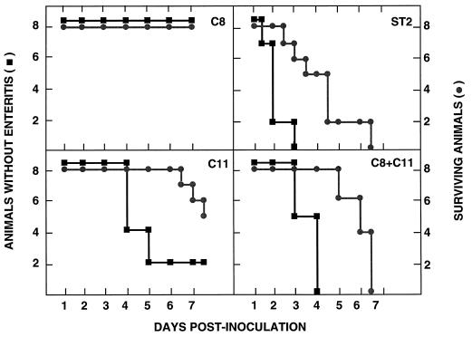
The onset of enteritis and mortality caused by isolates C8 and C11 (Fig. 4) were compared with those caused by the prototype TGEV strain used in our laboratory (PUR46-MAD), which has had a high passage number (120 times) in ST cells. Isolate C8 was highly attenuated (it produced very mild or no enteritis and no mortality), isolate C11 had an intermediate virulence (75% of animals had enteritis and mortality was 37% at 7 d p.i.), and the mixture of isolates C8 and C11 was highly virulent (100% of animals had enteritis at 4 d p.i. and mortality was 100% at 7 d p.i.). The uncloned PUR46-SW11-ST2 showed a slightly higher virulence than the mixture of isolates C8 and C11 (Fig. 4). The prototype strain (PUR46-MAD) behaved as did isolate C8, with very mild or no enteritis and no mortality in conventional (non-colostrum-deprived) piglets (data not shown).
FIG. 4.
Pathogenesis of isolates C8 and C11 in minipigs. Groups of eight minipigs were inoculated with the indicated TGEV isolates (isolates C8 and C11, a mixture of C8 and C11 [70% C8 plus 30% C11], or the virulent uncloned isolate ST2) as described in the legend for Fig. 3. The number of piglets without enteritis (■) or surviving (●) is shown at different days postinfection. Results of a representative experiment of two that gave similar results are shown.
Sequence differences between TGEV isolates C8 and C11.
The consensus RNA sequences of the 3′ 8,221 nt (from the beginning of S gene to the 3′ end of the genome) of isolates C8 and C11 were determined (Fig. 5). The sequence of isolate C8 was identical to that of PUR46-MAD (44). When the sequences of isolates C8 and C11 were compared, 15 nucleotide differences in the S gene, together with 7 nucleotide changes scattered from ORFs 3 to 6, were observed. No nucleotide difference was observed in ORF 7 or in the 3′ UTR. In addition, the 6-nt deletion of the S gene seen in all the Purdue strains described so far (47, 48, 53) was not present in the C11 isolate (Fig. 5). These nucleotide changes were responsible for 14 amino acid substitutions or deletions in the S gene and 1 amino acid substitution in each of the ORFs 3a, E, and M.
FIG. 5.
Sequence comparison of isolates C8 and C11. The nucleotide substitutions in isolate C8 in relationship to isolate C11 are shown for all genes except ORF 1. The top bar indicates the different viral genes, and the numbers above the second bar indicate the positions of the substituted nucleotides, with nt 1 of each gene considered to be the A of the initiation codon. Letters within the bars indicate the corresponding nucleotides at the indicated positions. Letters below the bars indicate the amino acid substitutions encoded by the nucleotides around the indicated position. Δ6 nt, deletion of six nucleotides. The arrow indicates the position of the S gene stop codon.
The frequency of nucleotide differences between isolates C8 and C11 within the 3′ 8,221 nt (Table 1) indicates that these changes are concentrated in the 5′ half of the S gene (with 5.8 nt substitutions/103 nt), as contrasted with substitution frequencies ranging from 1.1 to 2.3 per 103 in genes 3a to N. The second highest nucleotide substitution frequency (2.3/103 nt) was observed within genes 3a and 3b.
TABLE 1.
Nucleotide substitution frequency in isolate C8 relative to than in isolate C11
| Gene | No. of changes | Fraction of total no. of changes (%) | Frequencyb |
|---|---|---|---|
| S (complete)a | 17 | 70.8 | 3.8 |
| S 5′ 2,200 nta | 13 | 54.1 | 5.8 |
| S 3′ 2,147 nt | 4 | 16.7 | 1.8 |
| 3a-3b | 3 | 12.5 | 2.3 |
| E | 1 | 4.2 | 2.0 |
| M | 1 | 4.2 | 1.1 |
| N | 2 | 8.3 | 1.7 |
| 7 | 0 | 0 | 0 |
| 3′ UTR | 0 | 0 | 0 |
A two-codon deletion has been computed as two changes.
Number of substitutions per 103 nucleotides.
Generation of recombinant viruses by combination between TGEV strains displaying low and high replication levels in the enteric tract.
The different tropism and virulence properties shown by the C8 and C11 isolates could in principle be due to any of the sequence differences between isolates identified in the 8,221 nt of the 3′ end of the virus and also to the putative nucleotide substitutions within ORF 1. Based on our previous data demonstrating that two amino acid changes within the spike protein are responsible for the loss of enteric tropism of an attenuated strain of TGEV (2) and on the accumulation of most of the nucleotide differences between isolates C8 and C11 within gene S, we decided to study first whether the S gene differences were responsible for the observed differences in tropism and virulence between these isolates. To this end, the effect of the incorporation of the S gene from isolate C11 into isolate C8 by the formation of either true recombinant or pseudorecombinant viruses was studied (Fig. 6).
FIG. 6.
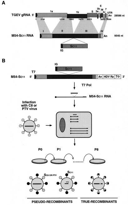
Diagram of the pseudorecombinant and true recombinant virus isolation protocol. (A) Structure of the M54 minigenome indicating the sequence position where the S gene from the enteric C11 isolate was cloned. Letters and numbers above the top bar indicate the TGEV ORFs. Numbers below this bar indicate the nucleotide sequences of the helper virus incorporated into the M54 minigenome. Numbers above the second bar indicate the four sequence domains that were linked during the generation of the minigenome. Numbers to the right of the bars indicate sizes of the genomes in nucleotides. gRNA, genomic RNA. IG, intergenic sequences preceding the S gene of the C11 isolate, which is identical to that of the PUR46-MAD strain of TGEV. An, poly(A). (B) Generation of recombinants. The S gene from an enteric TGEV (isolate C11) was cloned into minigenome M54, generating the minigenome M54-Sc11 with the structure diagrammatically shown in panel A. This minigenome has been cloned after the T7 promoter (T7) (black box) and preceding the hepatitis delta virus ribozyme (HDV Rz) sequences and the T7 terminator sequences (TΦ). ST cells were infected with C8 or PTV viruses. At 4 to 6 h p.i., cells were electroporated with in vitro-transcribed RNA and the supernatants from these cultures were passaged by using ST cells. The potential pseudorecombinants with the S protein from the respiratory helper viruses (light circles) or from the enteric C11 isolate (dark circles) containing either the genome of the helper virus (large bar) or the minigenome with the S gene of the C11 isolate are diagrammatically represented (bottom left). True recombinants with a chimeric S protein generated by two crossovers between the S gene from the helper virus and the S gene from isolate C11 are also indicated (bottom right).
The recombinant viruses were produced by the rescue of a minigenome with the isolate C11 S gene (M54-Sc11) by using either isolate C8 or PTV as the helper virus (Fig. 6). The minigenome M54 was derived from the PUR46-MAD strain of TGEV and has the sequences indicated in Fig. 6A (27). The S gene from isolate C11, preceded by the 5′ upstream 65 nt of the S gene containing the conserved intergenic sequence CUAAAC, was inserted in the unique NdeI restriction endonuclease site of minigenome M54 (27). In the first experiment, the minigenome RNA was in vitro transcribed with T7 polymerase and was transfected into ST cells that were infected with the C8 strain as helper virus (Fig. 6B). Supernatants from the infected cultures were passaged eight times to facilitate the recombination between the S genes present in the helper virus and the minigenome.
In principle, both pseudorecombinant and true recombinant viruses could be formed. Two types of pseudorecombinant viruses were expected (Fig. 6B). Both of them should contain the same envelope with spike proteins derived from the C8 and C11 isolates and should differ in their genomes, which should be derived either from the helper virus or from the minigenome M54-Sc11. In addition to the pseudorecombinant homotrimers of the spike protein derived from either the respiratory or the enteric virus, heterotrimers could also be formed.
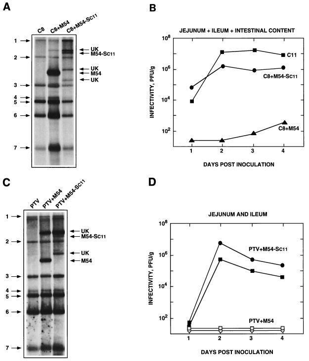
Northern blot analysis of the cytoplasmic RNAs of helper virus-infected ST cells, using a probe complementary to the 3′ end of the virus, showed the standard set of viral mRNAs (Fig. 7A). Analysis of the RNAs from the ST cells infected with the helper virus and transfected with the M54 or the M54-Sc11 minigenome showed, in addition to the standard RNA pattern of the helper virus, the RNAs corresponding to the minigenomes M54 and M54-Sc11, respectively. Other bands of unknown identity were also observed. The unknown band of about 12 kb present in the Northern blots shown in Fig. 7A and C did not appear when analyzed with a probe for the S gene. We do not know whether this band is derived from the helper virus, the minigenome, or a recombination between the two. Nevertheless, the 12-kb band is most likely derived from a recombination from the helper virus and the minigenome since this band appears only when the helper virus is passaged in the presence of the minigenome and it has a larger size than the minigenome.
FIG. 7.
Northern blot analysis and growth kinetics of the potential recombinants in the enteric tract. The characterization of the viruses obtained after minigenome M54-Sc11 rescue using as helper virus isolate C8 (A and B) or PTV (C and D) is shown. The Northern blot analysis of the viral RNAs obtained after infecting ST cells for eight passages with the indicated helper virus (either C8 or PTV) alone or with the helper virus plus the minigenome M54 or plus minigenome M54-Sc11 is shown (A and C). Numbers and arrows to the left of boxes A and C indicate the positions of the helper virus RNAs. Arrows and names to the right of these boxes indicate the expected positions for the minigenomes. UK, unknown RNA. Virus replication in the enteric tract was examined following oronasal and intragastrical inoculation of groups of four newborn piglets. Animals were sacrificed at the indicated days, and the virus content in representative samples of the whole tissue was determined by using the plaque assay on ST cells. Virus content was determined, and the arithmetic mean of the titers of virus recovered from the jejunum, ileum, and intestinal content is shown (panel B). ■, isolate C11; ●, isolate C8 plus minigenome M54-Sc11; ▴, isolate C8 plus minigenome M54. Virus replication using the strain PTV as the helper virus is also shown (panel D). Virus content was measured in the jejunum (●) or in the ileum (■) of piglets infected with strain PTV plus minigenome M54-Sc11; virus content was measured in the jejunum ( ) or the ileum (░⃞) of piglets infected with the strain PTV plus the minigenome M54.
The minigenome M54 was replicated and packaged with high efficiency by the helper virus, since the ratio of minigenome RNA to full-length RNA genome was higher than 50-fold and the minigenome RNA also was at least 5-fold more abundant than the viral mRNAs (Fig. 7A). The minigenome M54-Sc11 carrying the S gene was replicated with an efficiency similar to that of the virus genome as determined by Northern blot analysis with probes specific for the 3′ end of TGEV (Fig. 7A) and for the S gene (data not shown). The S gene mRNA encoded by the minigenome was not detected by Northern blot analysis using a probe complementary to the 3′ end of TGEV or a probe specific for the S gene; nevertheless, the presence of this mRNA was detected by RT-PCR analysis (data not shown).
To study the growth of the recombinant viruses in respiratory and enteric tissues, three groups of four piglets each were infected by the intranasal, oral, and intragastric routes with the TGEV strains C8 and C11 or with the potential recombinant viruses generated after eight passages in ST cells (Fig. 7B). The C8 and C11 strains were recovered from the intestinal content with low (<103 PFU/g of tissue) and high (>107 PFU/g of tissue) titers, respectively, as expected. Interestingly, the replication of isolate C8 in the enteric tract was increased more than 104-fold in the virus passaged in the presence of the minigenome M54-Sc11, and the virus production in the gut persisted for at least 4 days.
In the first experiment, the replication in enteric tissues of a virus (isolate C8) that was already enteric, although it replicated to a small extent in the gut, was studied. To prove that the tropism of a respiratory virus that did not replicate at all in the enteric tract could be extended to the gut, we performed a rescue experiment identical to the one described in Fig. 7A except that as helper virus the TGEV strain PTV was used (53). The results (Fig. 7C and D) indicated that PTV efficiently replicated the minigenome M54 alone or the minigenome M54 carrying the Sc11 gene, since clear bands with the expected sizes for the corresponding RNAs were observed (Fig. 7C). Newborn piglets were infected with the pool of virus generated after eight passages in ST cells. The helper virus PTV passaged in the presence of the minigenome M54 did not replicate at all in the jejunum or in the ileum (Fig. 7D). In contrast, the PTV passaged in the presence of the M54-Sc11 RNA replicated in the gut, with titers ranging between 8 × 10⁴ and 8 × 10⁶ PFU/g of tissue.
Characterization of the viral isolates produced in tissue culture and in vivo.
To identify the RNAs synthesized during minigenome rescue in tissue culture and in vivo, a collection of isolates was obtained by two steps of plaque selection after passage 8 in ST cells and after isolating the virus from the enteric tracts of swine. Virus was recovered from the gut of one of the infected swine at 2 d p.i. (Fig. 7D). Plaques of two sizes, large (diameter, 3.5 mm) and small (diameter, 1 mm), were isolated (Fig. 8A). Since plaque purification implied infection at an MOI lower than 1, only replication-competent virus, including the helper virus (small plaque) and potential recombinant virus (large plaque), should be recovered in principle, while the defective minigenomes should be lost. In fact, that was the case when the RNAs present were analyzed by Northern blotting and RT-PCR (Table 2).
FIG. 8.
Restriction endonuclease analysis of the potential recombinants. (A) A diagram of the procedure followed in the selection of true recombinant and pseudorecombinant viruses growing in the enteric tract is shown. Gut tissue (jejunum), collected from a single pig at 2 d p.i., was homogenized, and lysis plaques were isolated on ST cells. Plaques of two sizes (3- and 1-mm diameter) were observed and cloned twice. Viruses in these plaques were expected to have either the genome of the helper respiratory virus (PTV) or the genome of a true recombinant virus (rPTV/Sc11) formed by a two-crossover event within the S gene (bottom bar). The positions of restriction endonuclease (DraIII and MslI) sites in cDNA, derived by RT-PCR, between nt 487 and 1640 of the S gene are indicated. (B) Prototype restriction endonuclease patterns of cDNAs derived from S genes of the strains PTV and C11 and two isolates (clones 1 and 43) with a restriction endonuclease pattern identical to that of strain C11 or PTV are shown. MWM, molecular weight markers (1-kb DNA ladder; Gibco). Numbers on the left of the MWM lane indicate the sizes of the markers in kilobases.
TABLE 2.
Restriction endonuclease analysis of the viral isolates
| Virus passagea | Presence of minigenomeb | Phenotype of the
virus
|
||
|---|---|---|---|---|
| No. of clones with the genotype indicated in the next columnc/total no. of clones | Restriction endonuclease susceptibility at
positionc:
|
|||
| S 622 | S1208 | |||
| PTV + M54-Sc11 | + | 12/12 | PTV | PTV |
| PTV + M54-Sc11-SW1 | ND | ND | ND | ND |
| PTV + M54-Sc11-SW1-ST1 | − | 29/29 | C11 | C11 |
| PTV + M54-Sc11-SW1-ST2 | − | ND | ND | ND |
| PTV + M54-Sc11-SW1-ST3 | − | 12/20 | C11 | C11 |
| 8/20 | PTV | PTV | ||
The numbers after SW and ST indicate the passage numbers given in swine or ST cells, respectively.
The presence of the minigenome was determined by Northern blot and RT-PCR analyses. ND, not determined.
cDNAs derived from the S gene of TGEV strain PTV have MslI and DraIII restriction endonuclease sites at nt 1208 and 1252, respectively. cDNAs derived from clone C11 have two DraIII sites at nt 622 and 1252 and no restriction site susceptible to MslI. PTV and C11 indicate restriction endonuclease patterns identical to cDNA derived from the TGEV strains PTV or C11, respectively. ND, not determined.
RNAs from infected cells and tissues were analyzed by synthesizing cDNA fragments of 1,153 nt (from nt 487 to 1640 of S gene) by RT-PCR and studying their restriction endonuclease patterns by using two enzymes (Fig. 8). One of them (DraIII) cuts the isolate C11-derived cDNA at nt 622 and 1252 of S gene, while the other (MslI) exclusively cuts the cDNA derived from isolate PTV at position 1208, giving a differential restriction endonuclease pattern (Fig. 8B). The restriction endonuclease patterns given by the cDNAs derived from the parental viruses PTV and C11 and by two representative isolates (R1 and C43) recovered from the gut are shown (Fig. 8B). These two recombinants gave the typical patterns of the C11 isolate and the PTV isolate, respectively.
After eight passages of the helper virus PTV and the minigenome M54-Sc11 in ST cells, the presence of this minigenome was shown by both Northern blotting and RT-PCR analyses (Fig. 7C and Table 2). The restriction endonuclease analysis showed that all 12 isolates analyzed had the pattern expected for the PTV, indicating that most of the isolates had the genotype of the helper virus, without excluding the possible presence of recombinant viruses (Table 2).
The viruses that grew in the gut of the infected animal were plaque purified. The restriction endonuclease patterns of 29 isolates selected after the first passage in ST cells were determined. In all isolates the pattern expected for the S gene of isolate C11 was observed (Table 2). The gut virus, after being passaged three times in ST cells, contained mostly (12 of 20 isolates) isolates with the C11 genotype; the rest (8 of 20 isolates) of the isolates had the genotype of the PTV helper virus. These data indicated that the virus population quickly evolves during its passage in vivo or in vitro.
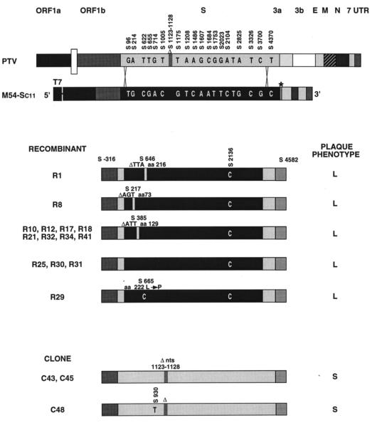
To further characterize the virus that replicated in the gut, the S genes of 17 isolates were sequenced. Fourteen of these isolates, selected after the first passage of the gut virus in ST cells, produced large plaques, like isolate C11 did. Isolates with the small-plaque phenotype, like the PTV strain, were not observed until the third virus passage in ST cells, indicating that the virus population was changing, and were then selected. RT-PCR sequence fragments starting 316 nt upstream of the S gene and covering the full length of this gene (a total of 4,898 nt) were sequenced. The 14 isolates with the large-plaque phenotype were recombinant viruses with two crossovers between S gene nucleotides −65 to 96 and 3700 to 4370 (Fig. 9), replacing most of the S gene of the helper virus by that of isolate C11. These isolates are true recombinants, since the described product cross-junctions do not exist in the helper virus or in the minigenome donor RNA. The origin of these sequences is the recombinant virus itself, since no defective minigenome with the S gene from isolate C11 was detected at this stage (Table 2).
FIG. 9.
Nucleotide sequences of TGEV recombinants isolated in enteric tissues. Virus was recovered from the enteric tract of a single pig at 2 d p.i. Recombinants with a large plaque size (no. 1, 8, 10, 12, 17, 18, 21, 25, 29, 30, 31, 32, 34, and 41) or a small plaque size (no. 43, 45, and 48) were plaque purified twice and expanded into ST cells, and their RNAs were copied into cDNA and sequenced. The nucleotide differences between the S genes of strains PTV and C11 are indicated (top bars). Numbers above bars indicate the positions of nucleotide substitutions or deletions. The approximate locations of the two crossovers in the S gene are indicated by two sets of crossed lines extending between the two top bars. The asterisk indicates the insertion sequences containing the restriction endonuclease sites SpeI, NotI, PstI, and SalI, derived from the polylinker of plasmid pGEM-T and located after the S gene. In isolates 1 to 41, a C was present at position 2136. This nucleotide (C) was different from that located at the equivalent positions in the S genes of the two parental viruses (T). The presence of codon deletions (▵) and nucleotide and amino acid (aa) substitutions is indicated above the bars. L and S indicate large- and small-plaque phenotypes.
All these isolates were probably derived from the same recombinant virus, since all of them had a C at position 2136 of the S gene (which did not cause an amino acid change), which was not present in the S genes of the helper virus or the minigenome M54-Sc11. Three of the recombinants (R25, R30, and R31) had S gene sequences (between nt 96 and 3700) identical to that of the C11 S gene. In recombinant R29, a single nucleotide substitution responsible for an amino acid change (replacement of leucine by proline at position 222) was observed in this sequence fragment. The other 10 recombinants showed one deletion of three nucleotides leading to removal of an amino acid at position 73 (R8), 129 (R10, R12, R17, R18, R21, R32, R34, and R41), or 216 (R1) (Fig. 9).
The growth in ST cells of all of the sequenced recombinants (Fig. 9) was determined in the first passage after their cloning, to reduce the diversification during their passage in ST cells. All the isolates that had introduced a codon deletion or a point mutation at the 5′ end of the S gene replicated in ST cells, producing virus titers 38- to 150-fold higher (the mean virus titer was 110-fold higher) than those of the recombinants with no change (Table 3). These recombinants also produced higher RNA levels as determined by semiquantitative RT-PCR (data not shown). These data suggested that the observed changes favored the growth in ST cells.
TABLE 3.
Recombinant virus replication in ST cells
| Recombinanta | (PFU/plaque)b |
|---|---|
| R1 | 7 × 10⁶ ± 3 × 10⁶ |
| R8 | 3 × 10⁶ ± 1 × 10⁶ |
| R10 | 8 × 10⁶ ± 2 × 10⁶ |
| R12 | 7 × 10⁶ ± 3 × 10⁶ |
| R17 | 2 × 10⁶ ± 2 × 10⁶ |
| R18 | 5 × 10⁶ ± 3 × 10⁶ |
| R21 | 7 × 10⁶ ± 3 × 10⁶ |
| R32 | 8 × 10⁶ ± 2 × 10⁶ |
| R34 | 7 × 10⁶ ± 1 × 10⁶ |
| R41 | 2 × 10⁶ ± 1 × 10⁶ |
| R25 | 1 × 10⁵ ± 1 × 10⁵ |
| R30 | 4 × 10⁴ ± 2 × 10⁴ |
| R31 | 2 × 10⁴ ± 1 × 10⁴ |
| R29 | 8 × 10⁶ ± 3 × 10⁶ |
| Clone 43 | 2 × 10⁶ ± 1 × 10⁶ |
| Clone 45 | 3 × 10⁶ ± 1 × 10⁶ |
| Clone 48 | 2 × 10⁶ ± 3 × 10⁶ |
The recombinants have been grouped according to the sequence of the S gene as indicated in Fig. 9.
Total virus in a lysis plaque grown on ST cells was determined in the first passage of the virus collected from the gut. Mean values of three independent determinations ± the standard errors of the means are shown.
The growth of a recombinant of each C11 type in the enteric tracts of newborn piglets was also evaluated by comparison with the virus isolates carrying the S gene sequence derived from the PTV virus. Some of these recombinants (e.g., R8, with a deletion of nt S217 to S219) infected the enteric tract, but others (e.g., R29, which has a nucleotide substitution at S665) did not (data not shown). Interestingly, the nucleotide change that affected the enterotropism maps very closely to the one (S655) that our group reported in a previous article (2) as being responsible for the loss of replication in the enteric tract. These results indicate that some nucleotide substitutions in the region from nt 217 to 665 abrogate the replication in the enteric tract. In contrast, isolate C43, with a sequence identical to the helper PTV, did not grow in the gut.
DISCUSSION
The isolation and characterization of two isolates of TGEV with different growth properties both in cell culture and in vivo are reported. These isolates replicate to a small or large extent in the enteric tracts of swine. Coinfection with both isolates increased the pathogenicity of each. These isolates presented differences in all structural and two nonstructural genes, although most of the differences were concentrated within the 5′ half of the S gene. Experiments designed for targeted recombination within the S genes from enteric and respiratory viruses led to the formation of true recombinant and pseudorecombinant viruses. Homologous recombination involving two crossover sites within the S gene has demonstrated that changes in the tropism and virulence of TGEV can be introduced by sequence changes in the S gene. The S gene from an enteric field strain rapidly evolved during its adaptation to grow in ST cells by changing its sequences at the 5′ end of the gene, between nt 217 and 665, a domain that was previously implicated in the control of the enteric tropism of TGEV.
TGEV isolates C8 and C11 were obtained from a historical sample of the Purdue strain of TGEV isolated by Haelterman’s group at Purdue University (West Lafayette, Ind.) (24, 34). The original virus (PUR46-SW11) (Fig. 1) was passaged exclusively in swine. This virus was adapted to grow in ST cells (9, 10), and after 115 passages on this cell line it was cloned and distributed to many scientists, including us. In our laboratory, the virus was recloned and named PUR46-MAD in reference to its place and year of isolation and the specific isolate name. A surprising observation was the high degree of conservation in the RNA sequence of this virus upon passage on ST cells, since almost one-third of its genome (8,221 nt), which encodes all the structural and three small nonstructural proteins, has complete sequence identity with C8, independently derived from the same original virus (PUR46-SW11) by only two passages on ST cells, which is the same cell line used in the high-passage isolate. This sequence identity may indicate that the selected virus has a sequence highly favored to grow in ST cells.
Isolate C8 has the 6-nt deletion in the S gene which has been considered a trademark of all TGEV Purdue virus strains (47, 48, 53). This deletion is not present in isolate C11 or in any TGEV isolates sequenced other than the Purdue strains (11, 14, 47, 53, 68). A comparison of the S gene sequences among 11 TGEV isolates showed that isolate C11 had the lowest computing distance (0.35) with isolate C8, while the computing distances with the Miller strain (MIL65) and the porcine respiratory coronaviruses (PRCoVs) were higher than 2.0 and 3.0, respectively (44). These data strongly suggest that isolate C8 is derived from C11 and not from other viruses circulating at the same time and in the same geographical area, such as the Miller strain, which was isolated in Fredericksburg, Ohio (8, 67). The relationship between the two isolates is in agreement with the results obtained by generating an evolutionary tree of TGEV (53). According to this epidemiological tree, the Purdue virus isolate C11 could be a recent ancestor of both PUR46-MAD (analogous to isolate C8) and the MIL65 strains of TGEV (53).
The availability of TGEV-derived minigenomes (27) allowed us to engineer a TGEV carrying S genes with the desired sequences by targeted recombination and to study the role of the S gene both in tropism and virulence.
First we have shown that the rescue of minigenomes carrying the S gene (Sc11) from an enteric virus, by a helper virus (isolate C8) that weakly replicated in the enteric tract, lead to an increase of 104-fold in its replication in the enteric tract. Then, a more stringent experiment was performed using a helper virus (PTV) which did not replicate at all in the enteric tract. The recombinant viruses were generated by the incorporation of most of the Sc11 gene, leading to the acquisition of the enteric tropism and virulence and demonstrating that the S gene alone modifies these virus activities.
The fact that the helper virus PTV was isolated from the enteric tract while this virus alone was never isolated from the gut strongly suggests that pseudorecombinant viruses with S proteins from the respiratory helper virus and from the enteric virus (Sc11) were formed, conferring enteric tropism to the pseudorecombinant. The enteric S protein could have been provided by either the minigenome or by true recombinant virus, since the minigenome was shown to be present and the recombinant viruses were probably originated during the eight passages in ST cells with minigenome M54-Sc11.
In the first passage in ST cells of the virus from the gut of the infected swine, PTV was in low proportion and it was not detected, but after the third passage a significant proportion (40%) of the analyzed isolates showed the PTV genotype. The increase in the proportion of PTV to isolate C11 was expected since PTV grows with higher titers than isolate C11 in ST cells (data not shown). These data indicate that the virus population quickly evolves during its passage in vivo and in ST cells. In vivo the isolates with an S gene derived from the C11 isolate were selected, while in ST cells the PTV strain was favored.
The formation of recombinants was studied by analyzing the restriction endonuclease pattern of cDNAs derived from the isolates and by sequencing their S genes. Viruses isolated from the infected swine gut were plaque purified, and all the isolates showed the genotype of the strain C11. Furthermore, all the plaques isolated were most likely derived from a unique recombination event, since all of them have a C at position 2136 which was present neither in the PTV nor in the Sc11 gene cloned in the minigenome. Sequence data demonstrated that a double crossover within the S gene had taken place, replacing most of the S gene of the helper virus by that of the enteric isolate. A double crossover within the S gene has been shown only in MHV by PCR, although in this case the recombinant viruses were not isolated (75). In contrast, viruses with a single crossover, in which the 3′ one-third of the genome has been replaced by that of another coronavirus, have recently been obtained. In this case, the role of the S gene in tissue tropism was also shown (35).
Three of the recombinants isolated from the enteric tract had a sequence identical to the Sc11 gene (with the exception of the marker mutation at position 2136). These isolates replicated in the enteric tract to the same extent as the parental virus isolate C11. Interestingly, the other four genotypes isolated showed either a codon deletion at nt 217, 385, or 646 or a nucleotide substitution at position 665 that caused an amino acid change (leucine to proline). The changes in the 5′ end of the S gene were most likely introduced during the passage of the recombinant viruses in ST cells, before they were used to infect the swine, and probably the nucleotide substitutions conferred the observed advantage of the selected viruses of growing in these cells. New virus variants of the enteric recombinant with the S gene of the C11 isolate were selected within a few passages of the virus on ST cells. The new recombinants had deletions or nucleotide substitutions within nt S217 to S665 (i.e., ΔS217 to S219, ΔS385 to S387, and ΔS646 to S648) that increased the replication of the virus in ST cells by a mean of 110-fold. Since the S gene is recognized by the cell receptor and no role has been associated with this gene in the macromolecular synthesis of the virus, most likely the changes on the S gene affected the recognition and internalization of the virus, suggesting that the S gene sequence between nt 217 and 665 plays a role in virus dissemination.
The binding of the TGEV S gene to aminopeptidase N, which is the receptor for group 1 coronaviruses including TGEV, human coronavirus 229E, and canine and feline coronaviruses (3, 16, 31, 61, 73), is mediated by an S protein domain encoded by nt 1518 to 2184 (23). We have described above that substitutions at nt S665 caused the loss of enteric tropism, in agreement with our previous data indicating that changes of nt S655 abrogate TGEV infection of the enteric tract. These data imply that the S protein domain recognized by aminopeptidase N and the domain of the S gene that has a decisive influence on TGEV tropism map in distal areas of the S gene. These results are also in agreement with the loss of enteric tropism and virulence in TGEV mutants such as PRCoV (12, 45, 54), a TGEV small-plaque mutant (69, 71), and hemadsorption-deficient mutants (6, 32) with alterations in a domain of the S gene different from the site recognized by the pAPN.
It is interesting that MHV tropism is also influenced by the binding of a domain located at the N terminus of murine coronavirus spike protein (33). In fact, the binding of MHV to its receptor is mediated by S protein amino acids encoded by nucleotides around positions 186, 612, 636, and 648 (59), which are located on S gene domains very similar to the ones that influence TGEV tropism. In MHV, it has also been shown that the expression of a functional viral receptor is not sufficient to establish MHV infection and that an additional factor is required for an early step of viral infection, possibly during virus entry, since transfection of the genomic RNA into nonsusceptible cells led to the production of infectious virus (74).
The involvement in virus entry of cellular factors additional to the cellular receptor has also been shown in coxsackievirus (4, 5, 55, 56), echoviruses (63, 64), aphtoviruses (21), herpes simplex virus (72), adenovirus (1, 26, 70), and human immunodeficiency virus (65). Unfortunately, the wide distribution of the receptors and accessory factors, which is broader than the observed tropism of the viruses mediated by these receptors and factors, suggests that host cell restriction is also mediated by additional limiting processes that probably take place after virus binding and internalization. In fact, in addition to the presence of a functional virus receptor and the required accessory molecule, a wide variety of mechanisms acting at different levels influence the host cell tropism. For instance, nonenvelope genes, such as human immunodeficiency virus Tat, Rev, and topoisomerase (15, 60), the UL5 gene encoding a component of the primase-helicase complex of herpes simplex virus (7), immediate-early gene products in human cytomegalovirus (57), and the polymerase gene in lymphocytic choriomeningitis virus (39) influence the tissue tropism of these viruses.
In conclusion, it has been shown that the enteric or respiratory tropism and virulence of TGEV can be engineered by changes exclusively within the S gene. This does not rule out the possibility that other genes also may influence the tropism and virulence of TGEV. In addition, data has been obtained reinforcing our previous conclusion (2) that the tissue specificity of TGEV is decided at the S gene level by a factor mapping at the 5′ end of this gene, located between nt 217 and 665, and not simply by the binding of this protein to aminopeptidase N, mediated by residues around spike protein site A (nt 1518 to 2184) (22, 23). We are interested in the genetic alterations that may help us to localize new signals regulating TGEV tropism and in identifying their functions. The possibility of TGEV genome modification by targeted recombination shown in this report should facilitate these studies.
ACKNOWLEDGMENTS
We are grateful to Victor Buckwold for critically reviewing the manuscript.
A.I. and S.A. received fellowships from the Department of Education, University and Research of the Gobierno Vasco, and I.S. and J.M.S. received fellowships from the Consejo Superior de Investigaciones Científicas of Spain and the Veterinary College of the Community of Madrid, respectively. This work has been supported by grants from the Comisión Interministerial de Ciencia y Tecnología (CICYT), La Consejería de Educación y Cultura de la Comunidad de Madrid, Fort-Dodge Veterinaria from Spain, and the European Union (Projects FAIR and Biotech).
REFERENCES
- 1.Bai M, Campisi L, Freimuth P. Vitronectin receptor antibodies inhibit infection of HeLa and A549 cells by adenovirus type 12 but not by adenovirus type 2. J Virol. 1994;68:5925–5932. doi: 10.1128/jvi.68.9.5925-5932.1994. [DOI] [PMC free article] [PubMed] [Google Scholar]
- 2.Ballesteros M L, Sanchez C M, Enjuanes L. Two amino acid changes at the N-terminus of transmissible gastroenteritis coronavirus spike protein result in the loss of enteric tropism. Virology. 1997;227:378–388. doi: 10.1006/viro.1996.8344. [DOI] [PMC free article] [PubMed] [Google Scholar]
- 3.Benbacer L, Kut E, Besnardeau L, Laude H, Delmas B. Interspecies aminopeptidase-Nchimeras reveal species-specific receptor recognition by canine coronavirus, feline infectious peritonitis virus, and transmissible gastroenteritis virus. J Virol. 1997;71:734–737. doi: 10.1128/jvi.71.1.734-737.1997. [DOI] [PMC free article] [PubMed] [Google Scholar]
- 4.Bergelson J M, Cunningham J A, Droguett G, Kurt-Jones E A, Krithivas A, Hong J S, Horwitz M S, Crowell R L, Finberg R W. Isolation of a common receptor for coxsackie B viruses and adenoviruses 2 and 5. Science. 1997;275:1320–1323. doi: 10.1126/science.275.5304.1320. [DOI] [PubMed] [Google Scholar]
- 5.Bergelson J M, Mohanty J G, Crowell R L, St. John N F, Lublin D M, Finberg R W. Coxsackievirus B3 adapted to growth in RD cells binds to decay-accelerating factor (CD55) J Virol. 1995;69:1903–1906. doi: 10.1128/jvi.69.3.1903-1906.1995. [DOI] [PMC free article] [PubMed] [Google Scholar]
- 6.Bernard S, Laude H. Site-specific alteration of transmissible gastroenteritis virus spike protein results in markedly reduced pathogenicity. J Gen Virol. 1995;76:2235–2241. doi: 10.1099/0022-1317-76-9-2235. [DOI] [PubMed] [Google Scholar]
- 7.Bloom D C, Stevens J G. Neuron-specific restriction of a herpes simplex virus recombinant maps to the UL5 gene. J Virol. 1994;68:3761–3772. doi: 10.1128/jvi.68.6.3761-3772.1994. [DOI] [PMC free article] [PubMed] [Google Scholar]
- 8.Bohl E H, Cross R F. Clinical and pathological differences in enteric infections in pigs caused by Escherichia coli. Ann N Y Acad Sci. 1971;176:150–161. [Google Scholar]
- 9.Bohl E H, Gupta P, Olquin F, Saif L J. Antibody responses in serum, colostrum, and milk of swine after infection or vaccination with transmissible gastroenteritis virus. Infect Immun. 1972;6:289–301. doi: 10.1128/iai.6.3.289-301.1972. [DOI] [PMC free article] [PubMed] [Google Scholar]
- 10.Bohl E H, Kumagai T. The use of cell cultures for the study of TGE virus of swine. Proc U S Livestock Sanitary Assoc. 1965;69:343–350. [Google Scholar]
- 11.Britton P, Mawditt K L, Page K W. The cloning and sequencing of the virion protein genes from a British isolate of porcine respiratory coronavirus: comparison with transmissible gastroenteritis virus genes. Virus Res. 1991;21:181–198. doi: 10.1016/0168-1702(91)90032-Q. [DOI] [PMC free article] [PubMed] [Google Scholar]
- 12.Callebaut P, Correa I, Pensaert M, Jiménez G, Enjuanes L. Antigenic differentiation between transmissible gastroenteritis virus of swine and a related porcine respiratory coronavirus. J Gen Virol. 1988;69:1725–1730. doi: 10.1099/0022-1317-69-7-1725. [DOI] [PubMed] [Google Scholar]
- 13.Cavanagh D, Brian D A, Britton P, Enjuanes L, Horzinek M C, Lai M M C, Laude H, Plagemann P G W, Siddell S, Spaan W, Talbot P J. Nidovirales: a new order comprising Coronaviridae and Arteriviridae. Arch Virol. 1997;142:629–635. [PubMed] [Google Scholar]
- 14.Chen C-M, Cavanagh D, Britton P. Cloning and sequencing of an 8.4-kb region from the 3′-end of a Taiwanese virulent isolate of the coronavirus transmissible gastroenteritis virus. Virus Res. 1995;38:83–89. doi: 10.1016/0168-1702(95)00046-S. [DOI] [PMC free article] [PubMed] [Google Scholar]
- 15.Dayton E T, Konings D A M, Lim S Y, Hsu R K S, Butini L, Pantaleo G, Dayton A I. The RRE of human immunodeficiency virus type 1 contributes to cell-type-specific viral tropism. J Virol. 1993;67:2871–2878. doi: 10.1128/jvi.67.5.2871-2878.1993. [DOI] [PMC free article] [PubMed] [Google Scholar]
- 16.Delmas B, Gelfi J, L’Haridon R, Vogel L K, Norén O, Laude H. Aminopeptidase N is a major receptor for the enteropathogenic coronavirus TGEV. Nature. 1992;357:417–420. doi: 10.1038/357417a0. [DOI] [PMC free article] [PubMed] [Google Scholar]
- 17.Eleouet J F, Rasschaert D, Lambert P, Levy L, Vende P, Laude H. Complete sequence (20 kilobases) of the polyprotein-encoding gene 1 of transmissible gastroenteritis virus. Virology. 1995;206:817–822. doi: 10.1006/viro.1995.1004. [DOI] [PMC free article] [PubMed] [Google Scholar]
- 18.Enjuanes, L., D. Brian, D. Cavanagh, K. Holmes, M. M. C. Lai, H. Laude, P. Masters, P. Rottier, S. G. Siddell, W. J. M. Spaan, F. Taguchi, and P. Talbot.Coronaviridae. In C. M. Fauquet, M. H. V. Van Regenmortel, D. H. L. Bishop and C. R. Pringle (ed.), Virus taxonomy, in press. Academic Press, New York, N.Y.
- 19.Enjuanes L, Van der Zeijst B A M. Molecular basis of transmissible gastroenteritis coronavirus epidemiology. In: Siddell S G, editor. The Coronaviridae. New York, N.Y: Plenum Press; 1995. pp. 337–376. [Google Scholar]
- 20.Fichot O, Girard M. An improved method for sequencing of RNA templates. Nucleic Acids Res. 1990;18:6162. doi: 10.1093/nar/18.20.6162. [DOI] [PMC free article] [PubMed] [Google Scholar]
- 21.Fox G, Parry N R, Barnett P V, McGinn B, Rowlands D J, Brown F. The cell attachment site on foot-and-mouth disease virus includes the amino acid sequence RGD (arginine-glycine-aspartic acid) J Gen Virol. 1989;70:625–637. doi: 10.1099/0022-1317-70-3-625. [DOI] [PubMed] [Google Scholar]
- 22.Gebauer F, Posthumus W A P, Correa I, Suñé C, Sánchez C M, Smerdou C, Lenstra J A, Meloen R, Enjuanes L. Residues involved in the formation of the antigenic sites of the S protein of transmissible gastroenteritis coronavirus. Virology. 1991;183:225–238. doi: 10.1016/0042-6822(91)90135-X. [DOI] [PMC free article] [PubMed] [Google Scholar]
- 23.Godet M, Grosclaude J, Delmas B, Laude H. Major receptor-binding and neutralization determinants are located within the same domain of the transmissible gastroenteritis virus (coronavirus) spike protein. J Virol. 1994;68:8008–8016. doi: 10.1128/jvi.68.12.8008-8016.1994. [DOI] [PMC free article] [PubMed] [Google Scholar]
- 24.Haelterman E O, Pensaert M B. Proceedings of the 18th World Veterinary Congress, Paris, France. 1967. Pathogenesis of transmissible gastroenteritis of swine; pp. 569–572. [Google Scholar]
- 25.Holmes K V, Lai M M C. Coronaviridae: the viruses and their replication. In: Fields B N, Knipe D M, Howley P M, editors. Fundamental virology. 3rd ed. Philadelphia, Pa: Lippincott-Raven; 1996. pp. 541–559. [Google Scholar]
- 26.Hong S S, Karayan L, Tournier J, Curiel D T, Boulanger P A. Adenovirus type 5 fiber knob binds to MHC class I α2 domain at the surface of human epithelial and B lymphoblastoid cells. EMBO J. 1997;16:2294–2306. doi: 10.1093/emboj/16.9.2294. [DOI] [PMC free article] [PubMed] [Google Scholar]
- 27.Izeta A, Smerdou C, Alonso S, Penzes Z, Mendez A, Plana-Duran J, Enjuanes L. Replication and packaging of transmissible gastroenteritis coronavirus-derived synthetic minigenomes. J Virol. 1999;73:1535–1545. doi: 10.1128/jvi.73.2.1535-1545.1999. [DOI] [PMC free article] [PubMed] [Google Scholar]
- 28.Jiménez G, Correa I, Melgosa M P, Bullido M J, Enjuanes L. Critical epitopes in transmissible gastroenteritis virus neutralization. J Virol. 1986;60:131–139. doi: 10.1128/jvi.60.1.131-139.1986. [DOI] [PMC free article] [PubMed] [Google Scholar]
- 29.Kapke P A, Brian D A. Sequence analysis of the porcine transmissible gastroenteritis coronavirus nucleocapsid protein gene. Virology. 1986;151:41–49. doi: 10.1016/0042-6822(86)90102-9. [DOI] [PMC free article] [PubMed] [Google Scholar]
- 30.Koetzner C A, Parker M M, Ricard C S, Sturman L S, Masters P S. Repair and mutagenesis of the genome of a deletion mutant of the coronavirus mouse hepatitis virus by targeted RNA recombination. J Virol. 1992;66:1841–1848. doi: 10.1128/jvi.66.4.1841-1848.1992. [DOI] [PMC free article] [PubMed] [Google Scholar]
- 31.Kolb A F, Hegui A, Siddell S G. Identification of residues critical for the human coronavirus 229E receptor function of human aminopeptidase N. J Gen Virol. 1997;78:2795–2802. doi: 10.1099/0022-1317-78-11-2795. [DOI] [PubMed] [Google Scholar]
- 32.Krempl C, Schultze B, Laude H, Herrler G. Point mutations in the S protein connect the sialic acid binding activity with the enteropathogenicity of transmissible gastroenteritis coronavirus. J Virol. 1997;71:3285–3287. doi: 10.1128/jvi.71.4.3285-3287.1997. [DOI] [PMC free article] [PubMed] [Google Scholar]
- 33.Kubo H, Yamada Y K, Taguchi F. Localization of neutralizing epitopes and the receptor-binding site within the amino-terminal 330 amino acids of the murine coronavirus spike protein. J Virol. 1994;68:5403–5410. doi: 10.1128/jvi.68.9.5403-5410.1994. [DOI] [PMC free article] [PubMed] [Google Scholar]
- 34.Lee K M, Moro M, Baker J A. Transmissible gastroenteritis in pigs. Am J Vet Res. 1954;15:364–372. [PubMed] [Google Scholar]
- 35.Leparc-Goffart I, Hingley S T, Chua M M, Phillips J, Lavi E, Weiss S R. Targeted recombination within the spike gene of murine coronavirus mouse hepatitis virus-A59: Q159 is a determinant of hepatotropism. J Virol. 1998;72:9628–9636. doi: 10.1128/jvi.72.12.9628-9636.1998. [DOI] [PMC free article] [PubMed] [Google Scholar]
- 36.Levine A J. Viruses and differentiation: the molecular basis of viral tissue tropism. In: Notkins A L, Oldstone M B S, editors. Concepts in viral pathogenesis. New York, N.Y: Springer-Verlag; 1984. pp. 130–134. [Google Scholar]
- 37.Lunney J K, Pescovitz M D, Sachs D H. The swine major histocompatibility complex: its structure and function. In: Tumbleson M E, editor. Swine in biomedical research. New York, N.Y: Plenum Press; 1986. pp. 1821–1836. [Google Scholar]
- 38.Masters P S, Koetzner C A, Kerr C A, Heo Y. Optimization of targeted RNA recombination and mapping of a novel nucleocapsid gene mutation in the coronavirus mouse hepatitis virus. J Virol. 1994;68:328–337. doi: 10.1128/jvi.68.1.328-337.1994. [DOI] [PMC free article] [PubMed] [Google Scholar]
- 39.Matloubian M, Kolhekar S R, Somasundaram T, Ahmed R. Molecular determinants of macrophage tropism and viral persistence: importance of single amino acid changes in the polymerase and glycoprotein of lymphocytic choriomeningitis virus. J Virol. 1993;67:7340–7349. doi: 10.1128/jvi.67.12.7340-7349.1993. [DOI] [PMC free article] [PubMed] [Google Scholar]
- 40.McClurkin A W, Norman J O. Studies on transmissible gastroenteritis of swine. II. Selected characteristics of a cytopathogenic virus common to five isolates from transmissible gastroenteritis. Can J Comp Med Vet Sci. 1966;30:190–198. [PMC free article] [PubMed] [Google Scholar]
- 41.McKittrick S P, Georgalis A M, Atchison B A. Efficient extraction of viral RNA for PCR amplification. BioTechniques. 1993;15:802–805. [PubMed] [Google Scholar]
- 42.Mendez A, Smerdou C, Izeta A, Gebauer F, Enjuanes L. Molecular characterization of transmissible gastroenteritis coronavirus defective interfering genomes: packaging and heterogeneity. Virology. 1996;217:495–507. doi: 10.1006/viro.1996.0144. [DOI] [PMC free article] [PubMed] [Google Scholar]
- 43.Peng D, Koetzner C A, McMahon T, Zhu Y, Masters P. Construction of murine coronavirus mutants containing interspecies chimeric nucleocapsid proteins. J Virol. 1995;69:5475–5484. doi: 10.1128/jvi.69.9.5475-5484.1995. [DOI] [PMC free article] [PubMed] [Google Scholar]
- 44.Penzes, Z., A. Izeta, C. Smerdou, A. Mendez, M. L. Ballesteros, and L. Enjuanes. Complete nucleotide sequence of transmissible gastroenteritis coronavirus strain PUR46-MAD. Submitted for publication.
- 45.Rasschaert D, Duarte M, Laude H. Porcine respiratory coronavirus differs from transmissible gastroenteritis virus by a few genomic deletions. J Gen Virol. 1990;71:2599–2607. doi: 10.1099/0022-1317-71-11-2599. [DOI] [PubMed] [Google Scholar]
- 46.Rasschaert D, Gelfi J, Laude H. Enteric coronavirus TGEV: partial sequence of the genomic RNA, its organization and expression. Biochimie. 1987;69:591–600. doi: 10.1016/0300-9084(87)90178-7. [DOI] [PMC free article] [PubMed] [Google Scholar]
- 47.Rasschaert D, Laude L. The predicted primary structure of the peplomer protein E2 of the porcine coronavirus transmissible gastroenteritis virus. J Gen Virol. 1987;68:1883–1890. doi: 10.1099/0022-1317-68-7-1883. [DOI] [PubMed] [Google Scholar]
- 48.Register K B, Wesley R D. Molecular characterization of attenuated vaccine strains of transmissible gastroenteritis virus. J Vet Diagn Investig. 1994;6:16–22. doi: 10.1177/104063879400600104. [DOI] [PubMed] [Google Scholar]
- 49.Sachs D, Leight G, Cone J, Schwarz S, Stuart L, Rosemberg S. Transplantation in miniature swine. I. Fixation of the major histocompatibility complex. Transplantation. 1976;22:559–567. doi: 10.1097/00007890-197612000-00004. [DOI] [PubMed] [Google Scholar]
- 50.Saif L J, Bohl E H. Passive immunity in transmissible gastroenteritis of swine: immunoglobulin classes of milk antibodies after oral-intranasal inoculation of sows with a live low cell culture-passaged virus. Am J Vet Res. 1979;40:115–117. [PubMed] [Google Scholar]
- 51.Saif L J, Wesley R D. Transmissible gastroenteritis. In: Leman A D, Straw B E, Mengeling W L, D’Allaire S, Taylor D J, editors. Diseases of swine. 7th ed. Ames, Iowa: Wolfe Publishing Ltd.; 1992. pp. 362–386. [Google Scholar]
- 52.Sambrook J, Fritsch E F, Maniatis T. Molecular cloning: a laboratory manual. 2nd ed. Cold Spring Harbor, N.Y: Cold Spring Harbor Laboratory; 1989. [Google Scholar]
- 53.Sanchez C M, Gebauer F, Suñé C, Mendez A, Dopazo J, Enjuanes L. Genetic evolution and tropism of transmissible gastroenteritis coronaviruses. Virology. 1992;190:92–105. doi: 10.1016/0042-6822(92)91195-Z. [DOI] [PMC free article] [PubMed] [Google Scholar]
- 54.Sanchez C M, Jiménez G, Laviada M D, Correa I, Suñé C, Bullido M J, Gebauer F, Smerdou C, Callebaut P, Escribano J M, Enjuanes L. Antigenic homology among coronaviruses related to transmissible gastroenteritis virus. Virology. 1990;174:410–417. doi: 10.1016/0042-6822(90)90094-8. [DOI] [PMC free article] [PubMed] [Google Scholar]
- 55.Shafren D R, Bates R C, Agrez M V, Herd R L, Furns G F, Barry R D. Coxsackieviruses B1, B3, and B5 use decay accelerating factor as a receptor for cell attachment. J Virol. 1995;69:3873–3877. doi: 10.1128/jvi.69.6.3873-3877.1995. [DOI] [PMC free article] [PubMed] [Google Scholar]
- 56.Shafren D R, Dorahy D J, Ingham R A, Burns G F, Barry R D. Coxsackievirus A21 binds to decay-accelerating factor but requires intercellular adhesion molecule 1 for cell entry. J Virol. 1997;71:4736–4743. doi: 10.1128/jvi.71.6.4736-4743.1997. [DOI] [PMC free article] [PubMed] [Google Scholar]
- 57.Sinzger C, Jahn G. Human cytomegalovirus cell tropism and pathogenesis. Intervirology. 1996;39:302–319. doi: 10.1159/000150502. [DOI] [PubMed] [Google Scholar]
- 58.Suñe C, Jiménez G, Correa I, Bullido M J, Gebauer F, Smerdou C, Enjuanes L. Mechanisms of transmissible gastroenteritis coronavirus neutralization. Virology. 1990;177:559–569. doi: 10.1016/0042-6822(90)90521-R. [DOI] [PMC free article] [PubMed] [Google Scholar]
- 59.Suzuki H, Taguchi F. Analysis of the receptor binding site of murine coronavirus spike protein. J Virol. 1996;70:2632–2636. doi: 10.1128/jvi.70.4.2632-2636.1996. [DOI] [PMC free article] [PubMed] [Google Scholar]
- 60.Takahashi H, Tatsumi M, Matsuda M, Nagashima K, Kurata T, Hall W W. The role of topoisomerase I in HIV-1 replication. Leukemia (Baltimore) 1997;11:26–28. [PubMed] [Google Scholar]
- 61.Tresnan D B, Levis R, Holmes K V. Feline aminopeptidase N serves as a receptor for feline, canine, porcine, and human coronaviruses in serogroup I. J Virol. 1996;70:8669–8674. doi: 10.1128/jvi.70.12.8669-8674.1996. [DOI] [PMC free article] [PubMed] [Google Scholar]
- 62.van der Most R G, Heijnen L, Spaan W J M, Degroot R J. Homologous RNA recombination allows efficient introduction of site-specific mutations into the genome of coronavirus MHV-A59 via synthetic co-replicating RNAs. Nucleic Acids Res. 1992;20:3375–3381. doi: 10.1093/nar/20.13.3375. [DOI] [PMC free article] [PubMed] [Google Scholar]
- 63.Ward T, Pipkin P A. Decay-accelerating factor CD55 is identified as the receptor for echovirus 7 using CELICS, a rapid immuno-focal cloning method. EMBO J. 1994;13:5070–5074. doi: 10.1002/j.1460-2075.1994.tb06836.x. [DOI] [PMC free article] [PubMed] [Google Scholar]
- 64.Ward T, Powell R M, Pipkin P A, Evans D J, Minor P D, Almond J W. Role for β2-microglobulin in echovirus infection of rhabdomyosarcoma cells. J Virol. 1998;72:5360–5365. doi: 10.1128/jvi.72.7.5360-5365.1998. [DOI] [PMC free article] [PubMed] [Google Scholar]
- 65.Weiss R A, Clapham P R. Hot fusion of HIV. Nature. 1996;381:647–648. doi: 10.1038/381647a0. [DOI] [PubMed] [Google Scholar]
- 66.Welter C J. Strategies for a successful coronavirus (TGE) vaccine for swine. In: Lai M M C, Stohlman S A, editors. Coronaviruses. New York, N.Y: Plenum Press; 1987. pp. 551–557. [DOI] [PubMed] [Google Scholar]
- 67.Wesley, R. D. 1999. Personal communication.
- 68.Wesley R D, Woods R D, Cheung A K. Genetic analysis of porcine respiratory coronavirus, an attenuated variant of transmissible gastroenteritis virus. J Virol. 1991;65:3369–3373. doi: 10.1128/jvi.65.6.3369-3373.1991. [DOI] [PMC free article] [PubMed] [Google Scholar]
- 69.Wesley R D, Woods R D, Cheung A K. Genetic basis for the pathogenesis of transmissible gastroenteritis virus. J Virol. 1990;64:4761–4766. doi: 10.1128/jvi.64.10.4761-4766.1990. [DOI] [PMC free article] [PubMed] [Google Scholar]
- 70.Wickham T J, Mathias P, Cheresh D A, Nemorow G R. Integrins αvβ3 and αvβ5promote adenovirus internalization but not virus attachment. Cell. 1993;73:309–319. doi: 10.1016/0092-8674(93)90231-e. [DOI] [PubMed] [Google Scholar]
- 71.Woods R D. Small plaque variant of transmissible gastroenteritis virus. J Am Vet Med Assoc. 1978;173:643–647. [PubMed] [Google Scholar]
- 72.WuDunn D, Spear P G. Initial interaction of herpes simplex virus with cells is binding to heparan sulfate. J Virol. 1989;63:52–58. doi: 10.1128/jvi.63.1.52-58.1989. [DOI] [PMC free article] [PubMed] [Google Scholar]
- 73.Yeager C L, Ashmun R A, Williams R K, Cardellichio C B, Shapiro L H, Look A T, Holmes K V. Human aminopeptidase N is a receptor for human coronavirus 229E. Nature. 1992;357:420–422. doi: 10.1038/357420a0. [DOI] [PMC free article] [PubMed] [Google Scholar]
- 74.Yokomori K, Asanaka M, Stohlman S A, Lai M M C. A spike protein-dependent cellular factor other than the viral receptor is required for mouse hepatitis virus entry. Virology. 1993;196:45–56. doi: 10.1006/viro.1993.1453. [DOI] [PubMed] [Google Scholar]
- 75.Zhang L, Homberger F, Spaan W, Luytjes W. Recombinant genomic RNA of coronavirus MHV-A59 after coreplication with a DI RNA containing the MHV-RI spike gene. Virology. 1997;230:93–102. doi: 10.1006/viro.1997.8460. [DOI] [PMC free article] [PubMed] [Google Scholar]


