

Abstract
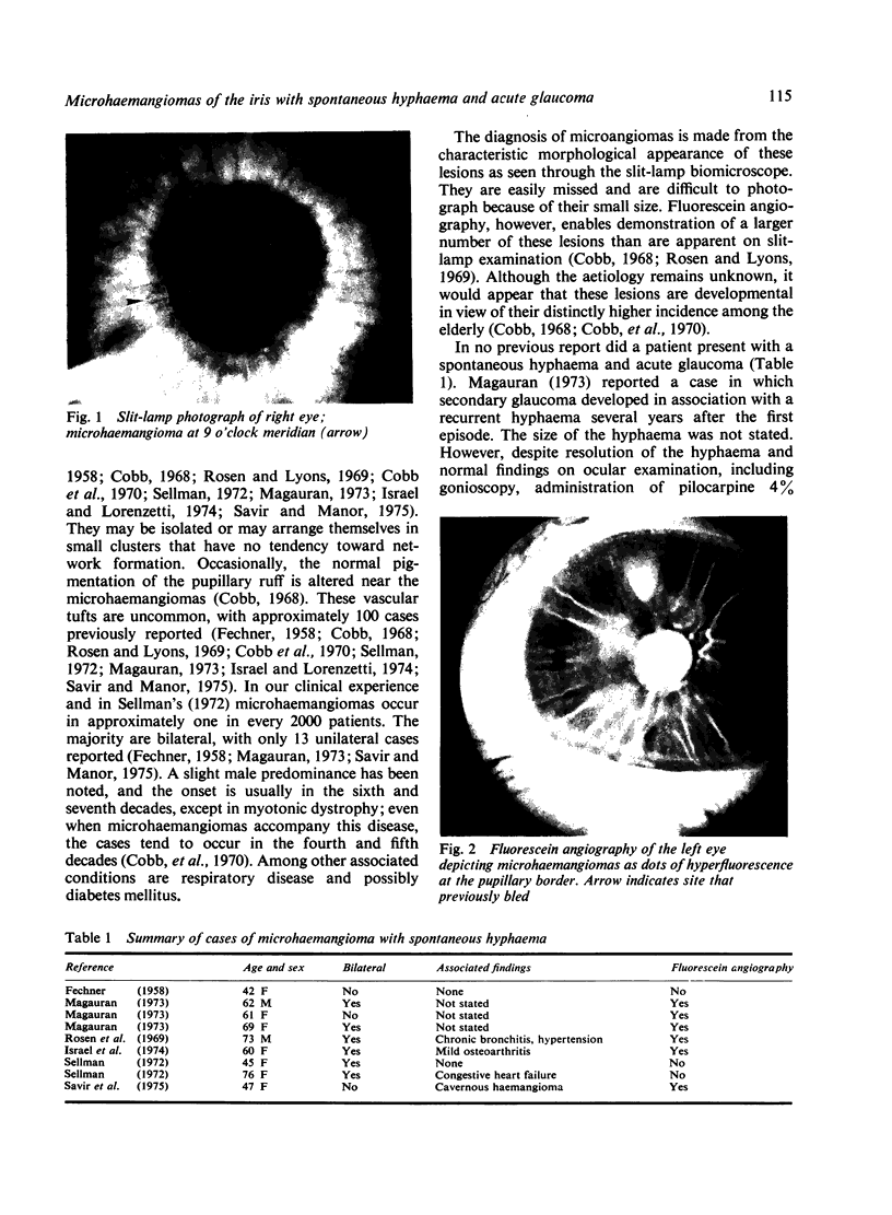
A case of bilateral microhaemangiomas of the pupillary borders in a 69-year-old woman is described. When first seen she presented with acute angle closure glaucoma and spontaneous hyphaema. Photographs and iris fluorescein angiography are presented and microhaemangiomas are discussed.
Full text
PDF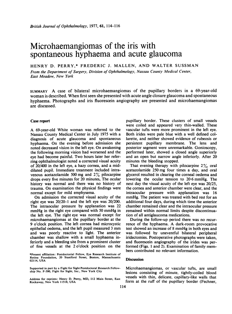
Images in this article
Selected References
These references are in PubMed. This may not be the complete list of references from this article.
- Cobb B., Shilling J. S., Chisholm I. H. Vascular tufts at the pupillary margin in myotonic dystrophy. Am J Ophthalmol. 1970 Apr;69(4):573–582. doi: 10.1016/0002-9394(70)91622-3. [DOI] [PubMed] [Google Scholar]
- Cobb B. Vascular tufts at the pupillary margin: a preliminary report on 44 patients. Trans Ophthalmol Soc U K. 1969;88:211–221. [PubMed] [Google Scholar]
- FECHNER P. U. Spontaneous hyphaema with abnormal iris vessels. Br J Ophthalmol. 1958 May;42(5):311–313. doi: 10.1136/bjo.42.5.311. [DOI] [PMC free article] [PubMed] [Google Scholar]
- Israel M. P., Lorenzetti D. W. Bilateral microhemangiomas of the pupillary border with later hyphema. Can J Ophthalmol. 1974 Jan;9(1):138–140. [PubMed] [Google Scholar]
- Magauran D. M. Unilateral spontaneous hyphaema. Br J Ophthalmol. 1973 Dec;57(12):945–947. doi: 10.1136/bjo.57.12.945. [DOI] [PMC free article] [PubMed] [Google Scholar]
- Rosen E., Lyons D. Microhemangiomas at the pupillary border demonstrated by fluorescein photography. Am J Ophthalmol. 1969 Jun;67(6):846–853. doi: 10.1016/0002-9394(69)90077-4. [DOI] [PubMed] [Google Scholar]
- Savir H., Manor R. S. Spontaneous hyphema and vessel anomaly. Arch Ophthalmol. 1975 Oct;93(10):1056–1058. doi: 10.1001/archopht.1975.01010020830018. [DOI] [PubMed] [Google Scholar]
- Sellman A. Hyphaema from microhaemangiomas. Acta Ophthalmol (Copenh) 1972;50(1):58–61. doi: 10.1111/j.1755-3768.1972.tb05641.x. [DOI] [PubMed] [Google Scholar]