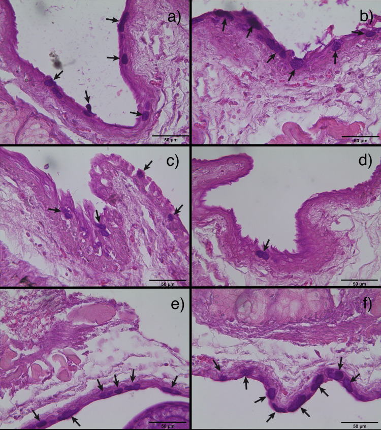
Figure 2. Merged images of PAS staining on conjunctival tissues.
Goblet cells, which exhibited a PAS-positive result (marked in black arrows), seen using 50x magnification. a) and b): control group right and left; c) and d): untreated group right and left; e) and f): lutein-treated group right and left.

