Abstract
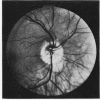
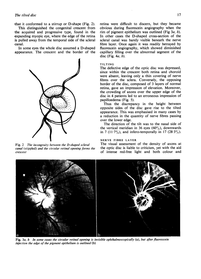
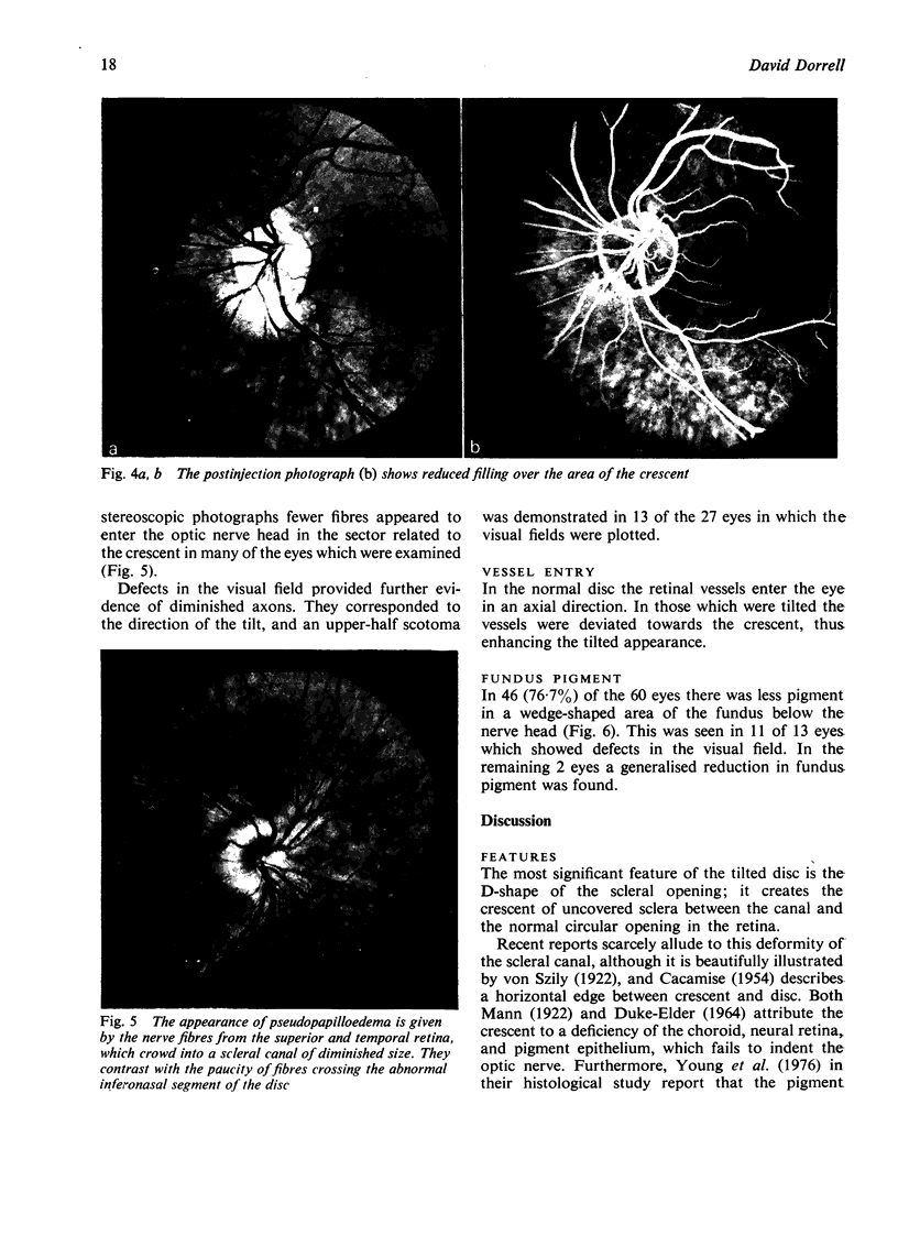
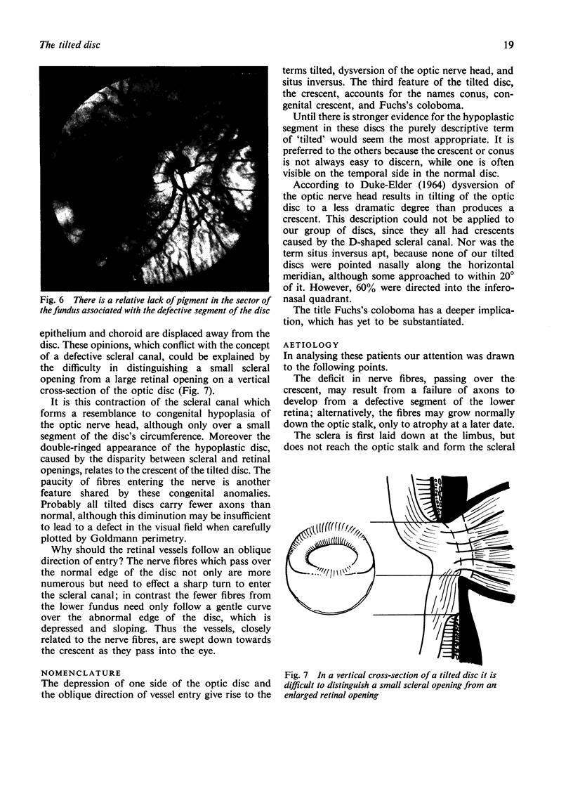
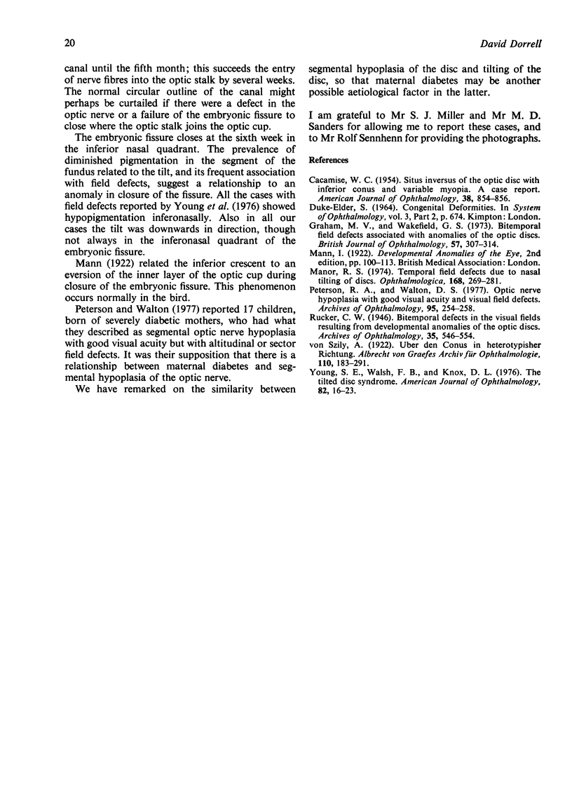
Sixty tilted discs were studied by colour photography, including some by fluorescein angiography. Attention was drawn to the contracted, D-shaped outline of the scleral canal, and it was suggested that fewer fibres than normal enter the defective side of the disc. This was supported by examination of the nerve fibre layer and the discovery of field defects in 13 of the 27 eyes in which the visual fields were examined. The similarity of these features with congenital hypoplasia of the optic nerve head was noted.
Full text
PDF
Images in this article
Selected References
These references are in PubMed. This may not be the complete list of references from this article.
- CACCAMISE W. C. Situs inversus of the optic disc with inferior conus and variable myopia: a case report. Am J Ophthalmol. 1954 Dec;38(6):854–856. doi: 10.1016/0002-9394(54)90418-3. [DOI] [PubMed] [Google Scholar]
- Graham M. V., Wakefield G. J. Bitemporal visual field defects associated with anomalies of the optic discs. Br J Ophthalmol. 1973 May;57(5):307–314. doi: 10.1136/bjo.57.5.307. [DOI] [PMC free article] [PubMed] [Google Scholar]
- Manor R. S. Temporal field defects due to nasal tilting of discs. Ophthalmologica. 1974;168(4):269–281. doi: 10.1159/000307049. [DOI] [PubMed] [Google Scholar]
- Petersen R. A., Walton D. S. Optic nerve hypoplasia with good visual acuity and visual field defects: a study of children of diabetic mothers. Arch Ophthalmol. 1977 Feb;95(2):254–258. doi: 10.1001/archopht.1977.04450020055011. [DOI] [PubMed] [Google Scholar]
- Young S. E., Walsh F. B., Knox D. L. The tilted disk syndrome. Am J Ophthalmol. 1976 Jul;82(1):16–23. doi: 10.1016/0002-9394(76)90658-9. [DOI] [PubMed] [Google Scholar]


