

Abstract
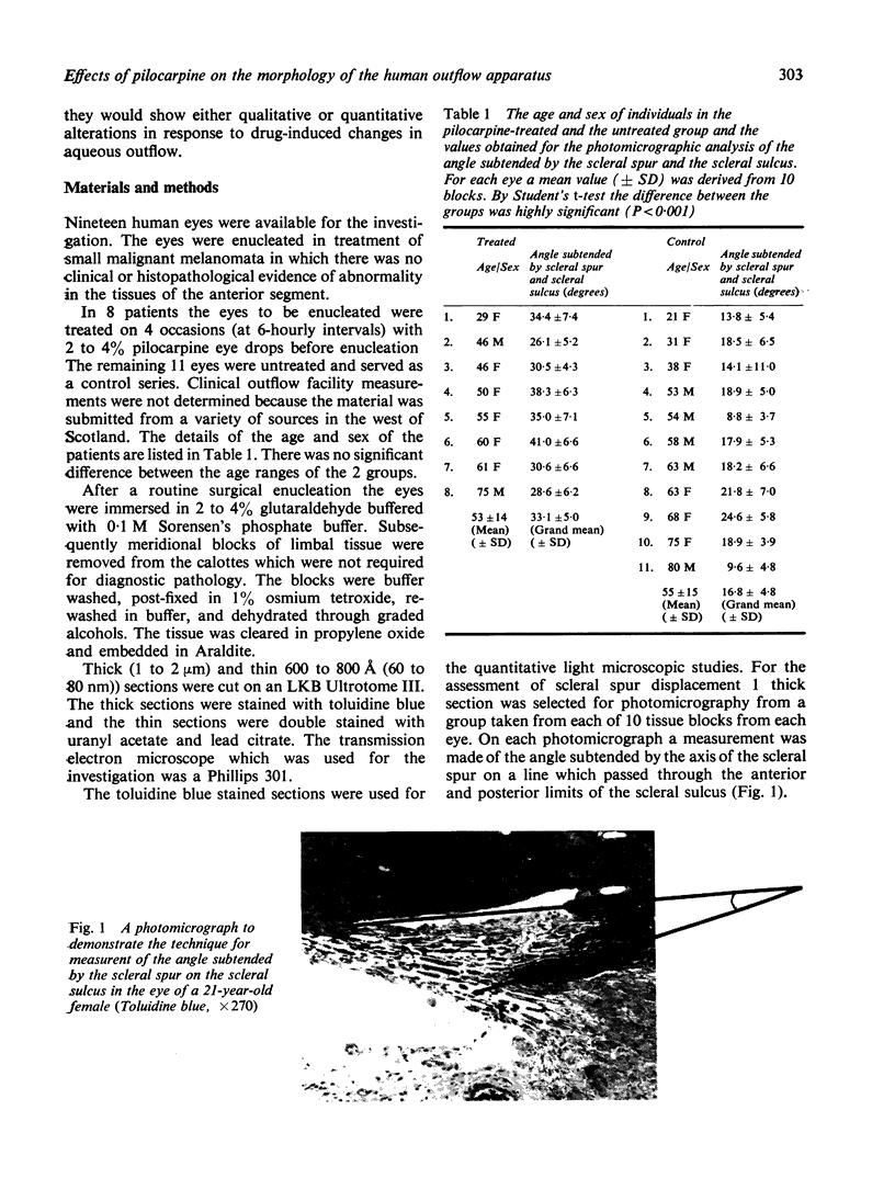
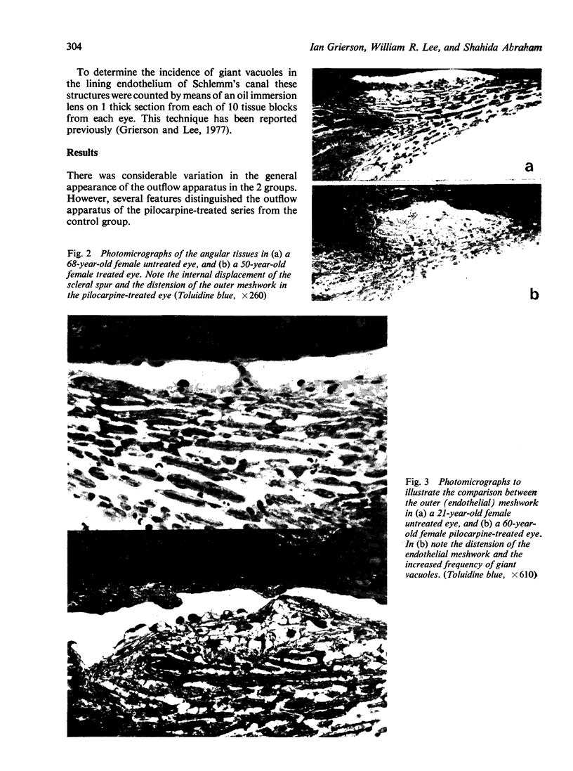
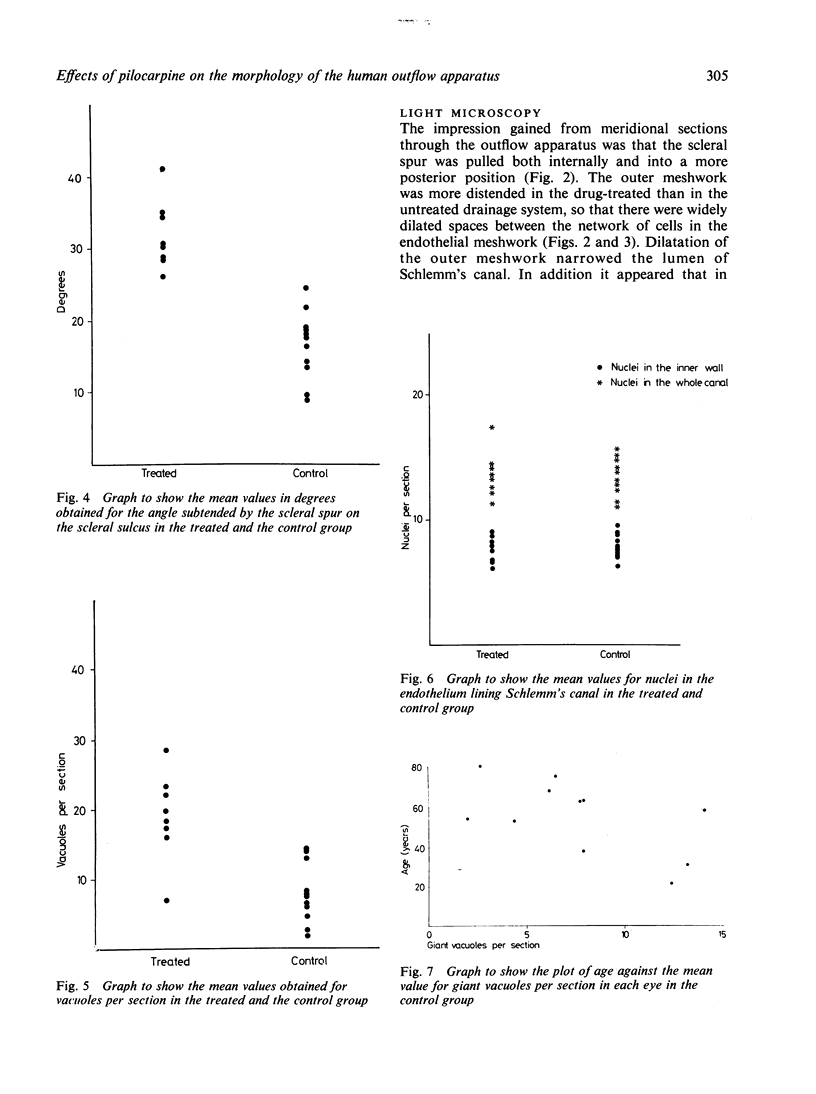
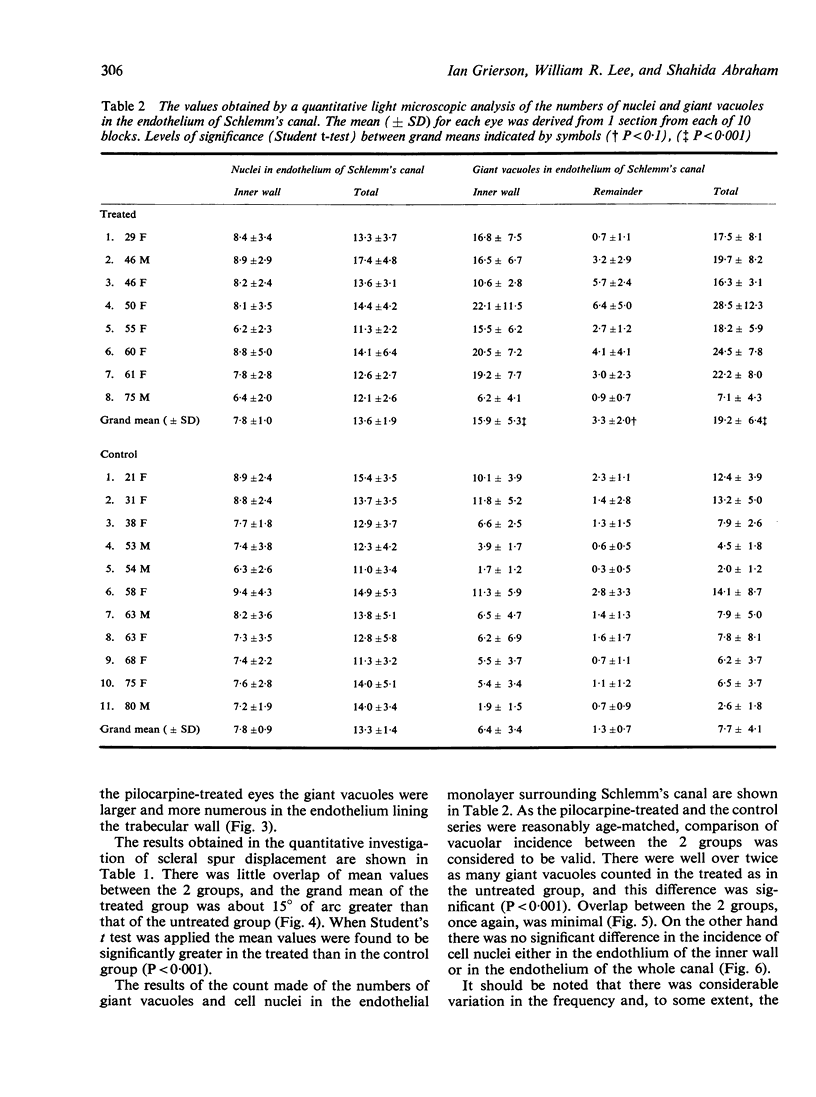
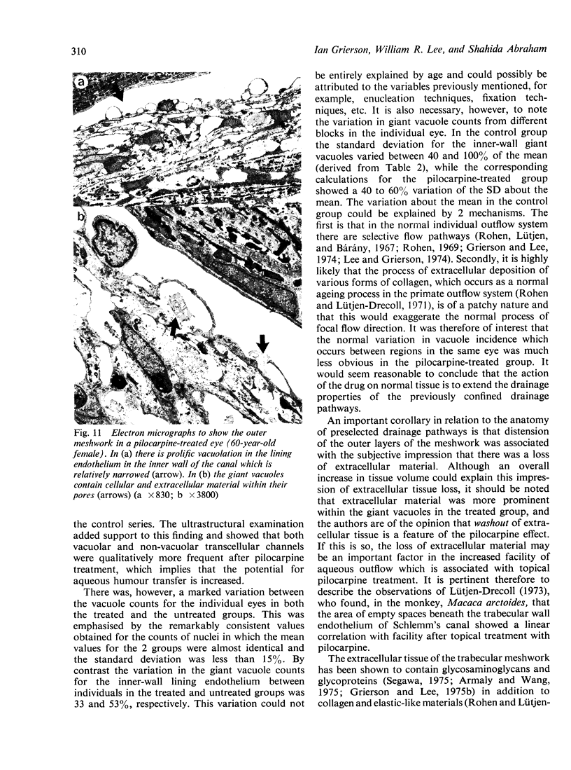
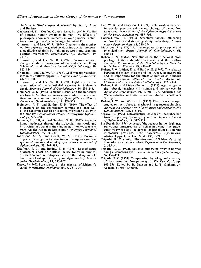
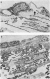
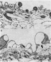
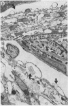
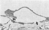
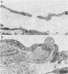
The morphology of the outflow apparatus in human eyes which had been treated topically with either 2 or 4% pilocarpine on 4 occasions at 6-hourly intervals before enucleation was compared with an untreated control series of eyes from patients in a similar age range. In the pilocarpine-treated group the scleral spur was pulled posteriorly and internally, so that the angle formed by the scleral spur and the scleral sulcus was significantly greater in the treated than the untreated series. The change in attitude of the scleral spur produced widening of the spaces between the corneoscleral trabeculae and distension of the endothelial meshwork. It was shown from counts of giant vacuoles in the endothelium of Schlemm's canal that the incidence of giant vacuoles in the pilocarpine-treated group was greater than twice that in the controls. It would appear that pilocarpine produces alterations in the configuration of the outflow apparatus which would promote the drainage of aqueous humour.
Full text
PDF
Images in this article
Selected References
These references are in PubMed. This may not be the complete list of references from this article.
- ALLEN L., BURIAN H. M. THE VALVE ACTION OF THE TRABECULAR MESHWORK. Am J Ophthalmol. 1965 Mar;59:382–389. doi: 10.1016/0002-9394(65)93733-5. [DOI] [PubMed] [Google Scholar]
- Armaly M. F., Wang Y. Demonstration of acid mucopolysaccharides in the trabecular meshwork of the Rhesus monkey. Invest Ophthalmol. 1975 Jul;14(7):507–516. [PubMed] [Google Scholar]
- Bárány E. H. The mode of action of miotics on outflow resistance. A study of pilocarpine in the vervet monkey Cercopithecus ethiops. Trans Ophthalmol Soc U K. 1966;86:539–578. [PubMed] [Google Scholar]
- FLOCKS M., ZWENG H. C. Studies on the mode of action of pilocarpine on aqueous outflow. Am J Ophthalmol. 1957 Nov;44(5 Pt 2):380–388. doi: 10.1016/0002-9394(57)93136-7. [DOI] [PubMed] [Google Scholar]
- Gaasterland D., Kupfer C., Ross K. Studies of aqueous humor dynamics in man. IV. Effects of pilocarpine upon measurements in young normal volunteers. Invest Ophthalmol. 1975 Nov;14(11):848–853. [PubMed] [Google Scholar]
- Grierson I., Lee W. R. Acid mucopolysaccharides in the outflow apparatus. Exp Eye Res. 1975 Nov;21(5):417–431. doi: 10.1016/0014-4835(75)90124-4. [DOI] [PubMed] [Google Scholar]
- Grierson I., Lee W. R. Changes in the monkey outflow apparatus at graded levels of intraocular pressure: a qualitative analysis by light microscopy and scanning electron microscopy. Exp Eye Res. 1974 Jul;19(1):21–33. doi: 10.1016/0014-4835(74)90068-2. [DOI] [PubMed] [Google Scholar]
- Grierson I., Lee W. R. Light microscopic quantitation of the endothelial vacuoles in Schlemm's canal. Am J Ophthalmol. 1977 Aug;84(2):234–246. doi: 10.1016/0002-9394(77)90857-1. [DOI] [PubMed] [Google Scholar]
- Grierson I., Lee W. R. Pressure-induced changes in the ultrastructure of the endothelium lining Schlemm's canal. Am J Ophthalmol. 1975 Nov;80(5):863–884. doi: 10.1016/0002-9394(75)90284-6. [DOI] [PubMed] [Google Scholar]
- Inomata H., Bill A., Smelser G. K. Aqueous humor pathways through the trabecular meshwork and into Schlemm's canal in the cynomolgus monkey (Macaca irus). An electron microscopic study. Am J Ophthalmol. 1972 May;73(5):760–789. doi: 10.1016/0002-9394(72)90394-7. [DOI] [PubMed] [Google Scholar]
- Johnstone M. A., Grant W. G. Pressure-dependent changes in structures of the aqueous outflow system of human and monkey eyes. Am J Ophthalmol. 1973 Mar;75(3):365–383. doi: 10.1016/0002-9394(73)91145-8. [DOI] [PubMed] [Google Scholar]
- Kaufman P. L., Bárány E. H. Loss of acute pilocarpine effect on outflow facility following surgical disinsertion and retrodisplacement of the ciliary muscle from the scleral spur in the cynomolgus monkey. Invest Ophthalmol. 1976 Oct;15(10):793–807. [PubMed] [Google Scholar]
- Lee W. R. The study of the passage of particles through the endothelium of the outflow apparatus of the monkey eye by scanning and transmission electron microscopy. Trans Ophthalmol Soc U K. 1971;91:687–705. [PubMed] [Google Scholar]
- Lütjen-Drecoll E. Structural factors influencing outflow facility and its changeability under drugs. A study in Macaca arctoides. Invest Ophthalmol. 1973 Apr;12(4):280–294. [PubMed] [Google Scholar]
- Mapstone R. Normal response to pilocarpine and phenylephrine. Br J Ophthalmol. 1977 Aug;61(8):510–511. doi: 10.1136/bjo.61.8.510. [DOI] [PMC free article] [PubMed] [Google Scholar]
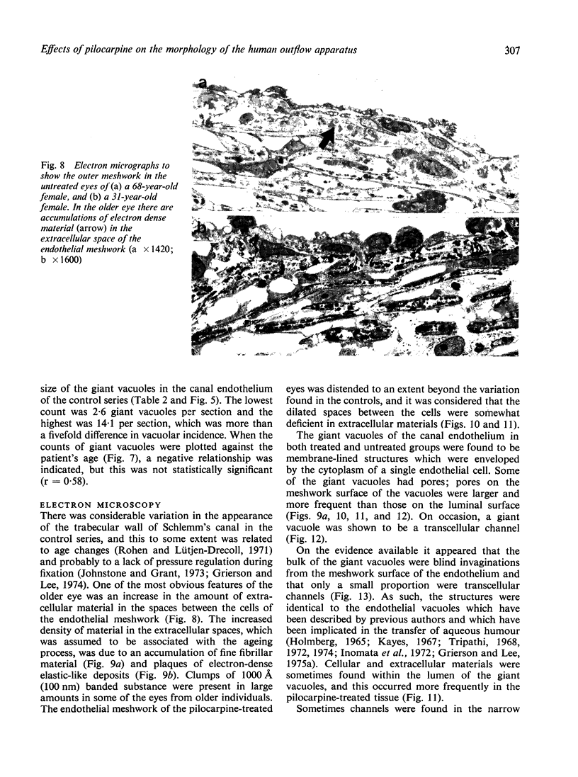
- Rohen J. W., Lütjen-Drecoll E. Age changes of the trabecular meshwork in human and monkey eyes. A light and electron microscopic study. Altern Entwickl Aging Dev. 1971;1:1–36. [PubMed] [Google Scholar]
- Rohen J. W., Lütjen E., Bárány E. The relation between the ciliary muscle and the trabecular meshwork and its importance for the effect of miotics on aqueous outflow resistance. A study in two contrasting monkey species, Macaca irus and Cercopithecus aethiops. Albrecht Von Graefes Arch Klin Exp Ophthalmol. 1967;172(1):23–47. doi: 10.1007/BF00577152. [DOI] [PubMed] [Google Scholar]
- Rohen J. W. New studies on the functional morphology of the trabecular meshwork and the outflow channels. Trans Ophthalmol Soc U K. 1970;89:431–447. [PubMed] [Google Scholar]
- Tripathi R. C. Aqueous outflow pathway in normal and glaucomatous eyes. Br J Ophthalmol. 1972 Mar;56(3):157–174. doi: 10.1136/bjo.56.3.157. [DOI] [PMC free article] [PubMed] [Google Scholar]
- Tripathi R. C. Ultrastructure of Schlemm's canal in relation to aqueous outflow. Exp Eye Res. 1968 Jul;7(3):335–341. doi: 10.1016/s0014-4835(68)80047-8. [DOI] [PubMed] [Google Scholar]