

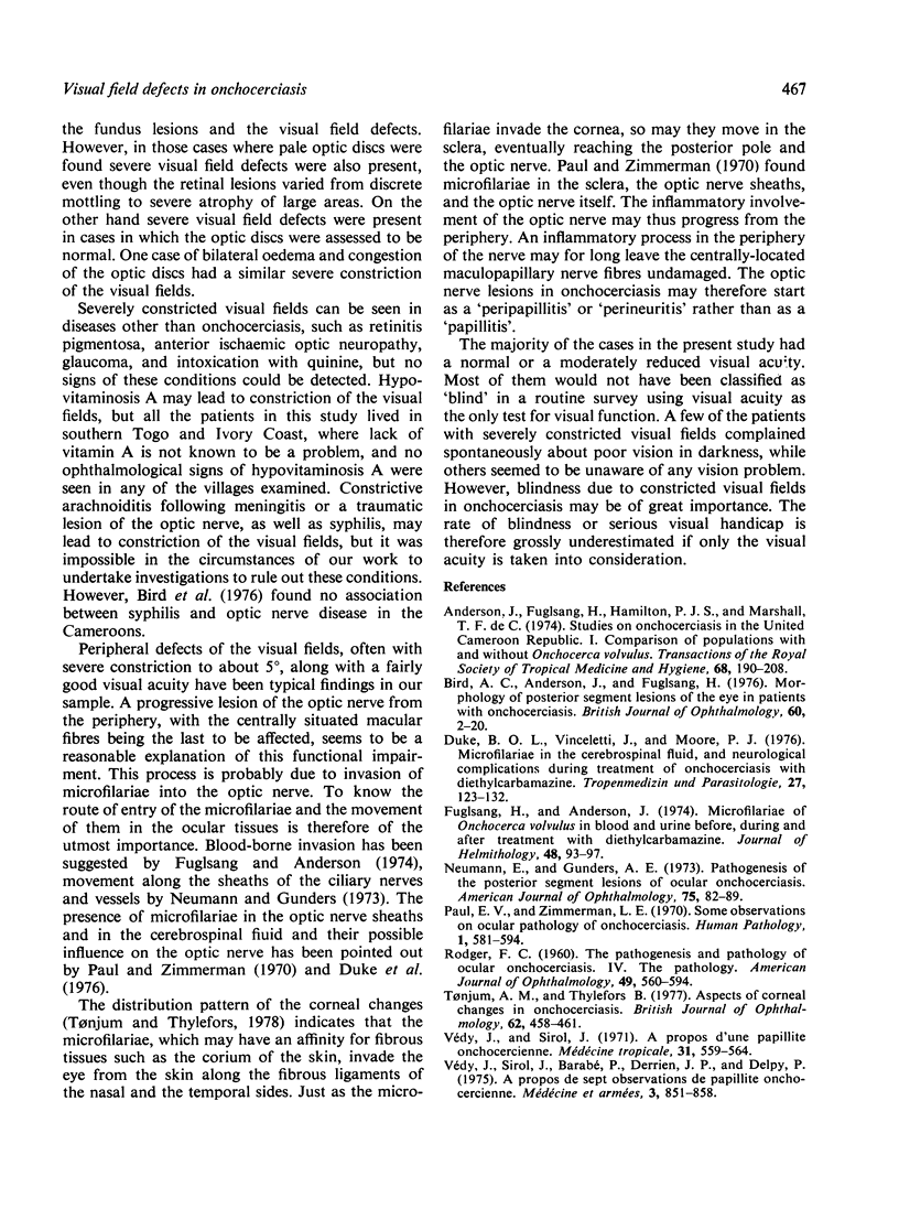
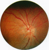
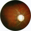
Abstract
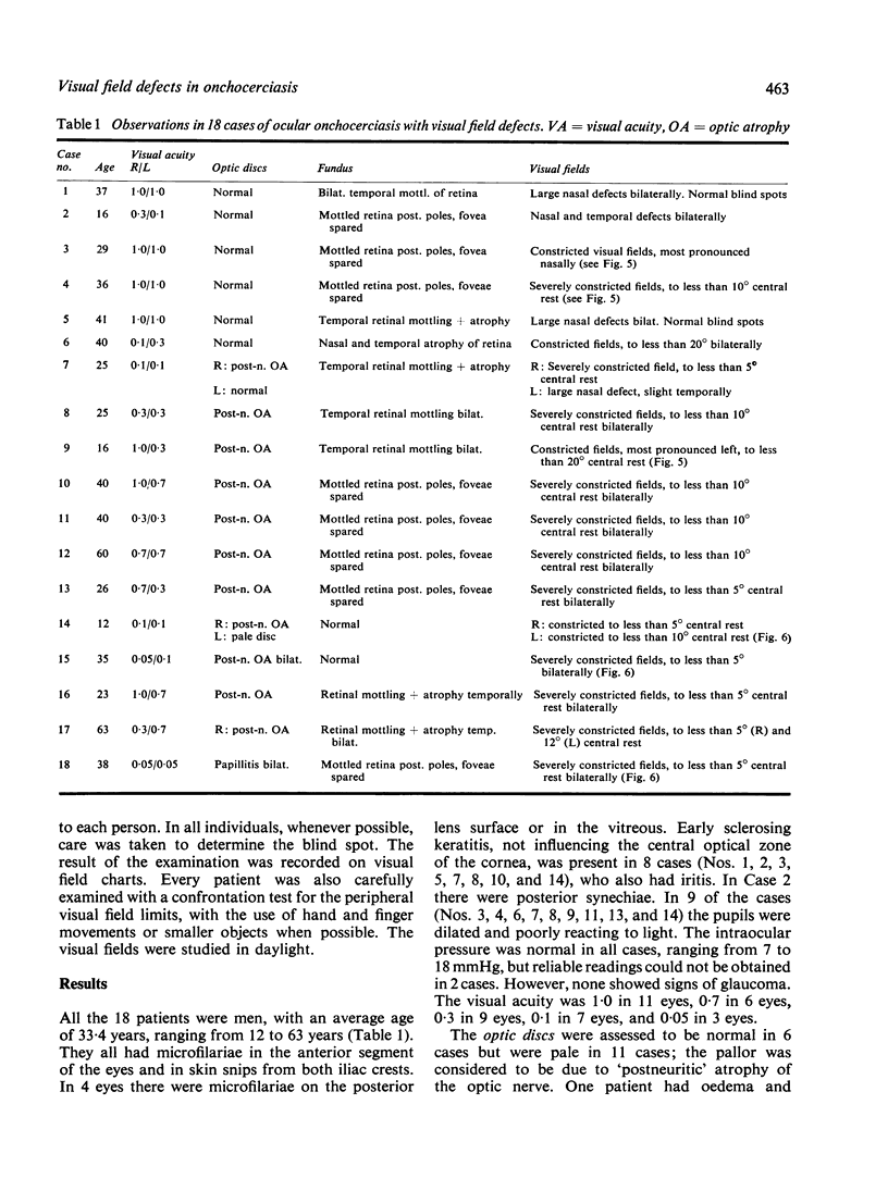
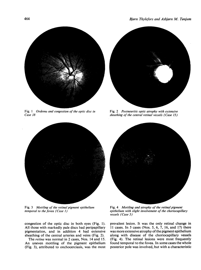
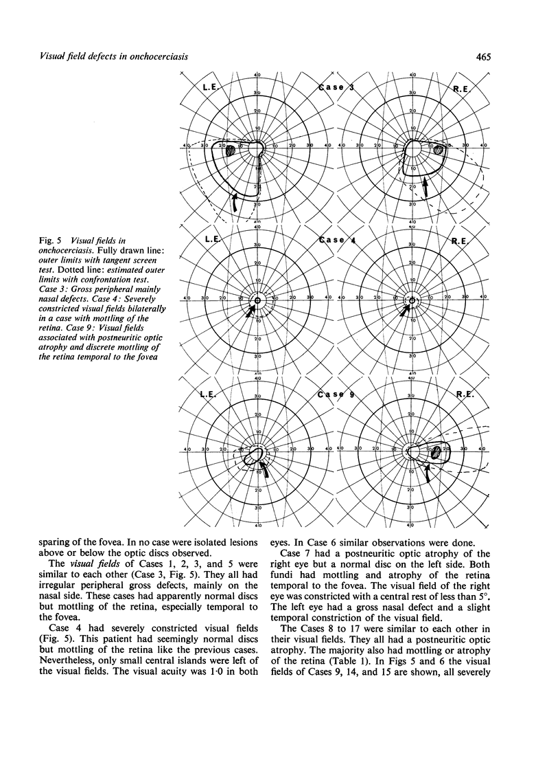
Lesions in the posterior segment of the eye in onchocerciasis may give visual field defects, but so far no detailed investigation has been done to determine the functional visual loss. Examination of the visual fields in 18 selected cases of onchocerciasis by means of a tangent screen test revealed important visual field defects associated with lesions in the posterior segment of the eye. Involvement of the optic nerve seemed to be important, giving rise to severely constricted visual fields. Cases of postneuritic optic atrophy showed a very uniform pattern of almost completely constricted visual fields, with only 5 to 10 degree central rest spared. Papillitis gave a similar severe constriction of the visual fields. The pattern of visual fields associated with optic neuropathy in onchocerciasis indicates that a progressive lesion of the optic nerve from the periphery may be responsible for the loss of vision. The visual field defects in onchocerciasis constitute a serious handicap, which must be taken into consideration when estimating the socioeconomic importance of the disease.
Full text
PDF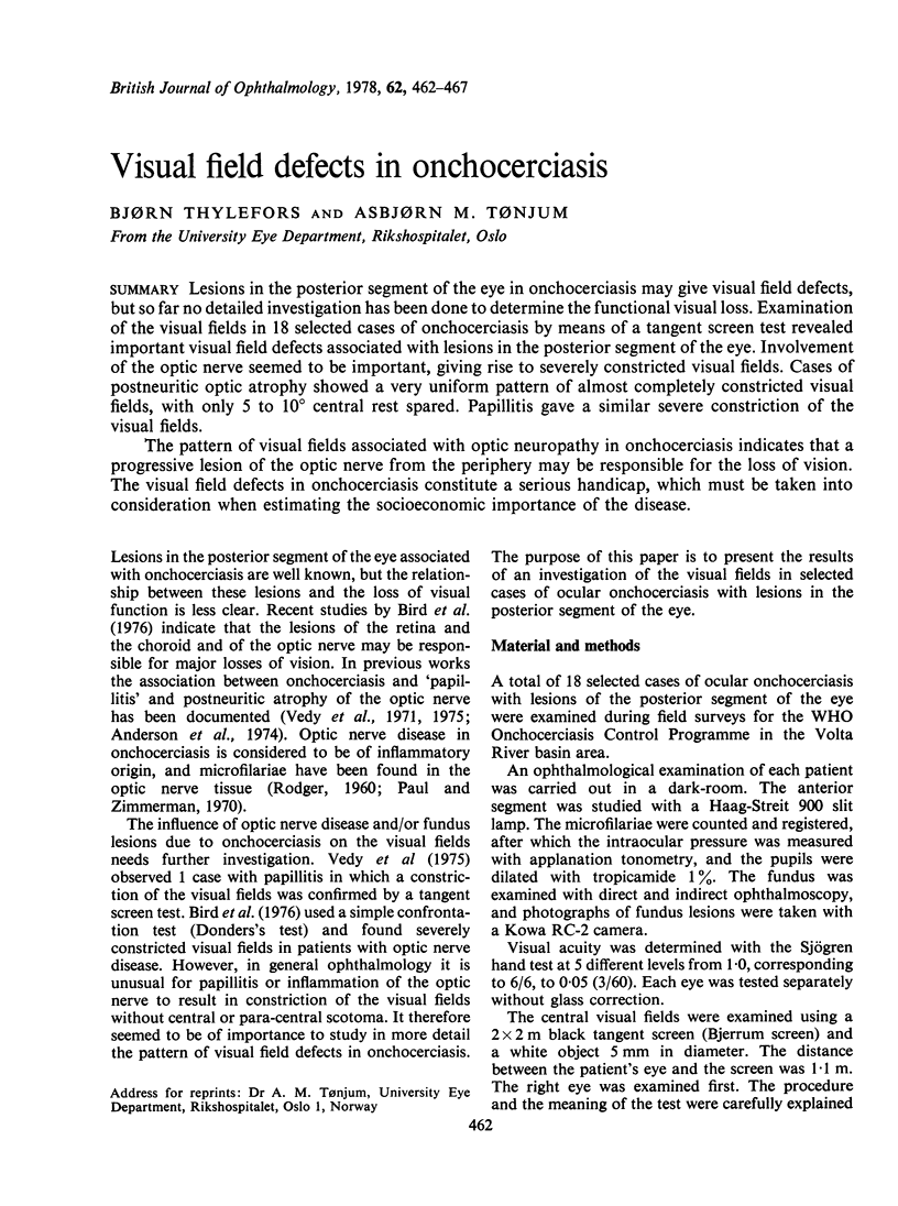

Images in this article

Selected References
These references are in PubMed. This may not be the complete list of references from this article.
- Anderson J., Fuglsang H., Hamilton P. J., de Marshall T. F. Studies on onchocerciasis in the United Cameroon Republic. I. Comparison of populations with and without Onchocerca volvulus. Trans R Soc Trop Med Hyg. 1974;68(3):190–208. doi: 10.1016/0035-9203(74)90116-3. [DOI] [PubMed] [Google Scholar]
- Bird A. C., Anderson J., Fuglsang H. Morphology of posterior segment lesions of the eye in patients with onchocerciasis. Br J Ophthalmol. 1976 Jan;60(1):2–20. doi: 10.1136/bjo.60.1.2. [DOI] [PMC free article] [PubMed] [Google Scholar]
- Duke B. O., Vincelette J., Moore P. J. Microfilariae in the cerebrospinal fluid, and neurological complications, during treatment of onchocerciasis with diethylcarbamazine. Tropenmed Parasitol. 1976 Jun;27(2):123–132. [PubMed] [Google Scholar]
- Fuglsang H., Anderson J. Microfilariae of Onchocerca volvulus in blood and urine before, during, and after treatment with diethylcarbamazine. J Helminthol. 1974 Jun;48(2):93–97. doi: 10.1017/s0022149x00022653. [DOI] [PubMed] [Google Scholar]
- Neumann E., Gunders A. E. Pathogenesis of the posterior segment lesion of ocular onchocerciasis. Am J Ophthalmol. 1973 Jan;75(1):82–89. doi: 10.1016/0002-9394(73)90656-9. [DOI] [PubMed] [Google Scholar]
- Paul E. V., Zimmerman L. E. Some observations on the ocular pathology of onchocerciasis. Hum Pathol. 1970 Dec;1(4):581–594. doi: 10.1016/s0046-8177(70)80058-2. [DOI] [PubMed] [Google Scholar]
- RODGER F. C. The pathogenesis and pathology of ocular on-chocerciasis. Part IV. The pathology. Am J Ophthalmol. 1960 Mar;49:560–594. doi: 10.1016/0002-9394(60)91658-5. [DOI] [PubMed] [Google Scholar]
- Tønjum A. M., Thylefors B. Aspects of corneal changes in onchocerciasis. Br J Ophthalmol. 1978 Jul;62(7):458–461. doi: 10.1136/bjo.62.7.458. [DOI] [PMC free article] [PubMed] [Google Scholar]
- Vedy J., Sirol J. A propos d'une papillite onchocerquienne. Med Trop (Mars) 1971 Sep-Oct;31(5):559–564. [PubMed] [Google Scholar]