Abstract
After a successful percutaneous coronary intervention to the left anterior descending artery, the guide catheter was pulled without a wire inside and so was kinked in the radial artery. It was not possible to pull or push the kinked catheter due to severe pain and fear of radial artery injury/perforation due to ‘razor effect’ of the two sharp edges of the kinked segment. Retrograde balloon-assisted tracking technique was used through femoral access, 7 F guide catheter, percutaneous transluminal coronary angioplasty wire and inflated 2.5×15 mm balloon partially outside the catheter tip to facilitate moving the kinked catheter to the aorta where unravelling was possible using a snare and a 0.035’ wire. This technique helped to keep control of both catheters, and avoid the ‘razor effect’ and radial artery injury. It could be suggested as the initial solution to sort similar problems due to its safety and efficacy.
Keywords: Interventional cardiology, Ischaemic heart disease
Background
A male patient in his 60s, an ex-smoker, had recent anterior myocardial infarction and coronary angiography was done after thrombolysis in another centre that showed tight proximal left anterior descending (LAD) artery lesion (figure 1A).
Figure 1.
Tight left anterior descending lesion (A) before stenting and (B) after stenting.
Case presentation
A patient in his 60s was referred to our centre for revascularisation and percutaneous coronary intervention was done to the culprit LAD lesion. XB 3.5 guide catheter, BMW wire and Xience alpine stent 2.75×23 mm were used, and the stent was postdilated with a non-compliant balloon 3×15 mm with a good final result (figure 1B).
Unfortunately, the guide catheter was pulled without a wire inside, so it was folded over itself and kinked in the left radial artery. A 0.35’ wire was used to cross the kinked part of the XB catheter but failed. We tried to pull it back but it was stuck in its place and any movement was causing a lot of pain to the patient. Unravelling the kinked catheter with external fixation by compression on the forearm over its distal tip was tried but this was associated with severe pain so it was decided to stop due to fear of tear of the radial artery by the sharp kinked edges.
Treatment
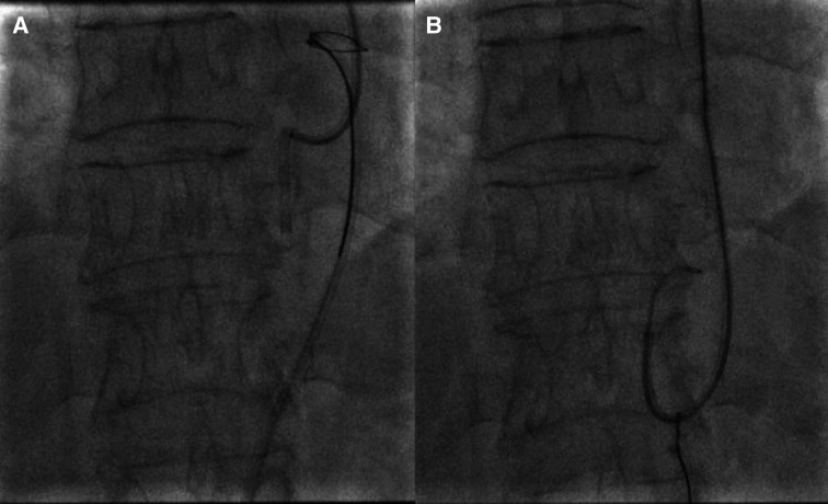
Femoral artery puncture was done and anterograde brachial angiogram with a 7 F JR guide—which was advanced to the left brachial artery—confirmed the entrapped catheter position within the left radial artery (figure 2). A Whisper 0.014’ wire through the JR4 guide was used to cross-retrograde the distal tip of the kinked guide (was not able to cross the kink itself) followed by a 2.5×15 mm balloon that was inflated at 12 atm with part of the balloon outside the XB guide tip (figure 3). The femoral JR4 guide was gently pulled with my colleague pushing the kinked XB guide and ‘razor effect’ was avoided through the tension applied with the inflated balloon. The kinked XB catheter was advanced with the help of the ‘balloon-assisted tracking technique’ without any pain (video 1).
Figure 2.

Kinked XB guide in the radial artery.
Figure 3.
Balloon-assisted tracking.
Video 1. Balloon-assisted tracking.
Once the catheter was in the descending aorta, a 6 Fr Amplatz GooseNeck Snare Kit (ev3) was used to perform catheter fixation and started the unravelling again with the help of the 0.035’ wire that was advanced through the kink in the XB guide (figure 4A). The kinked XB catheter was straightened by the 0.035’ wire and removed through the left radial sheath (figure 4B and video 2).
Figure 4.
(A) The snare was able to capture the kinked XB guide. (B) Kink unravelled using the snare and the 0.035’ wire.
Video 2. Kink unravelled with the help of the snare.
Outcome and follow-up
Final angiogram showed patent left radial artery with no injuries/perforation (figure 5) and the patient was sent home safely the following day.
Figure 5.

Final left radial angiogram.
Discussion
The transradial access is getting more popular for coronary procedures all over the world as it is associated with better safety and less bleeding than the femoral artery.1 Catheter kinking is not uncommon especially with tortuosity of the subclavian artery and in most cases could be early detected by observing the pressure tracing and unravelled by gentle rotation in the opposite direction.2
In our case, the catheter was pulled rapidly without a wire inside and so was kinked and trapped in the radial artery which prevented pulling or pushing due to severe pain associated with ‘razor effect’. It was not possible to perform external compression using external force (due to severe pain) or internal fixation with a snare (was not able to advance to the small radial artery).3 It is thought that by using the ‘balloon-assisted tracking technique’ in a retrograde way, it helped to move the trapped kinked catheter to the wider aorta and facilitated using the snare to unravel the kinked catheter and safely remove it through the radial artery to avoid unplanned vascular surgery.
Patient’s perspective.
I had the procedure done with no pain. Near the end, I felt severe pain in my left arm, and I was updated by the doctor that there is a problem in my arm and he was doing his best to sort it. After about 15 minutes without much pain, he told me that the problem was sorted, and I was discharged home safely next day. Many thanks.
Learning points.
Always remember the basics and never remove a catheter without a wire inside.
Retrograde balloon-assisted tracking is an effective and easy way to overcome trapped and kinked catheter in the radial artery safely.
This technique helped to always control both catheters.
It helped to avoid the ‘razor effect’ due to kinked catheter in a small radial artery and to avoid radial artery perforation/injury.
It could be suggested as the initial solution to sort similar problems due to its safety and efficacy.
Footnotes
Twitter: @basem_enany
Contributors: The following author was responsible for drafting of the text, sourcing and editing of clinical images, investigation results, drawing original diagrams and algorithms, and critical revision for important intellectual content—BEME. The following author gave final approval of the manuscript—BEME.
Funding: The authors have not declared a specific grant for this research from any funding agency in the public, commercial or not-for-profit sectors.
Case reports provide a valuable learning resource for the scientific community and can indicate areas of interest for future research. They should not be used in isolation to guide treatment choices or public health policy.
Competing interests: None declared.
Provenance and peer review: Not commissioned; externally peer reviewed.
Ethics statements
Patient consent for publication
Obtained.
References
- 1.Bertrand OF, Rao SV, Pancholy S, et al. Transradial approach for coronary angiography and interventions: results of the first International Transradial practice survey. JACC Cardiovasc Interv 2010;3:1022–31. 10.1016/j.jcin.2010.07.013 [DOI] [PubMed] [Google Scholar]
- 2.Moon JY, Yoo KW. Entrapment of a kinked catheter in the radial artery during Transradial coronary angiography. J Invasive Cardiol 2012;24:E3–4. [PubMed] [Google Scholar]
- 3.Ben-Dor I, Rogers T, Satler LF, et al. Reduction of catheter Kinks and knots via radial approach. Catheter Cardiovasc Interv 2018;92:1141–6. 10.1002/ccd.27623 [DOI] [PubMed] [Google Scholar]


