Abstract
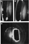
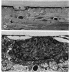
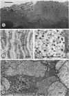
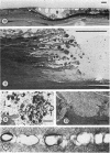
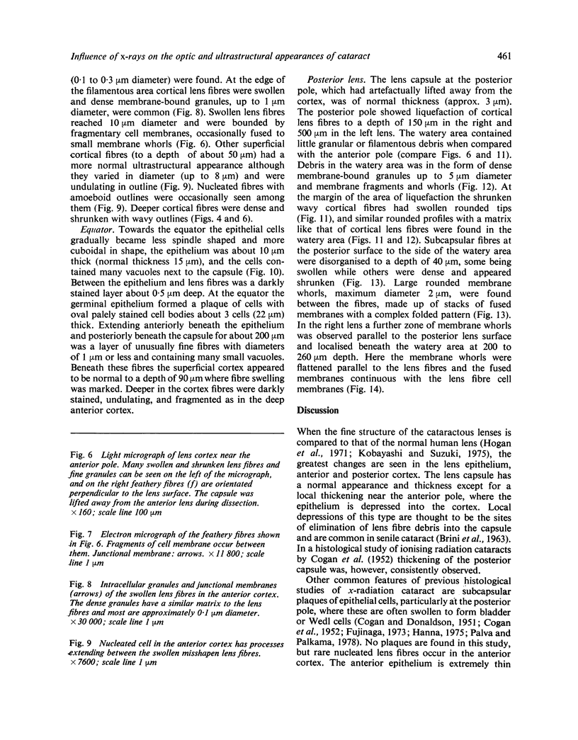
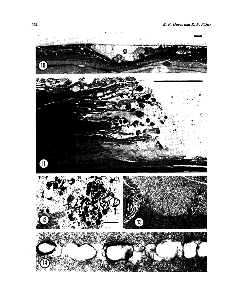
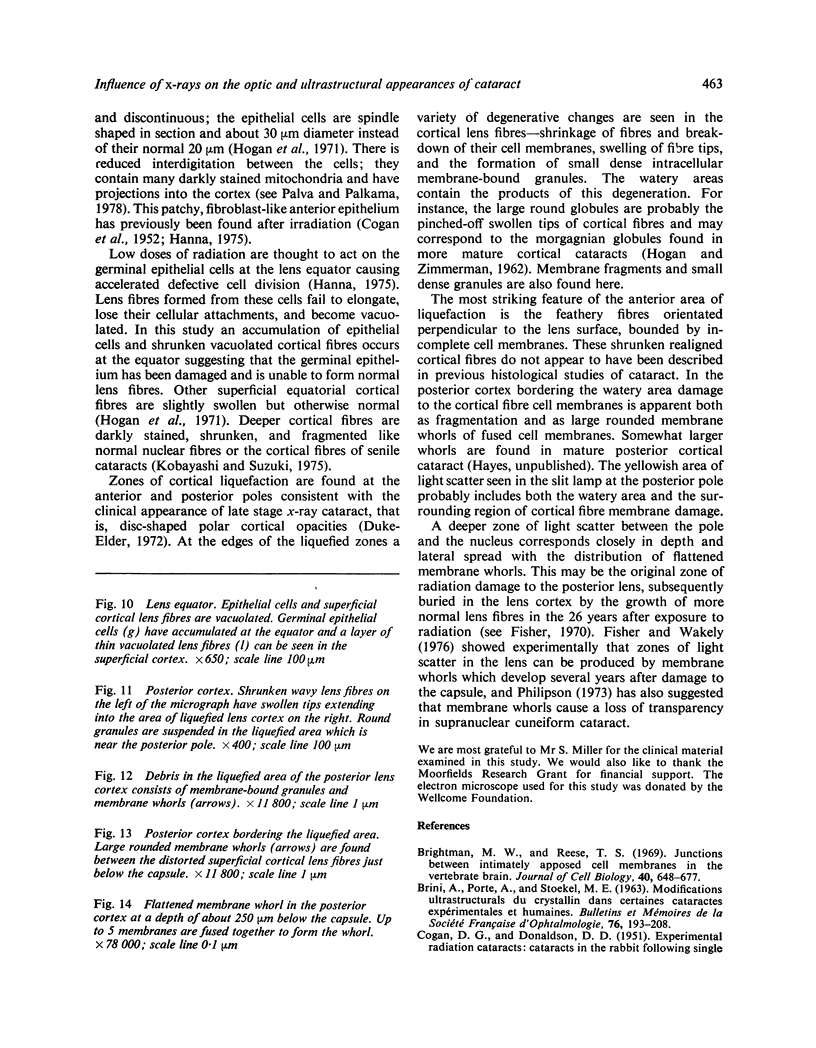
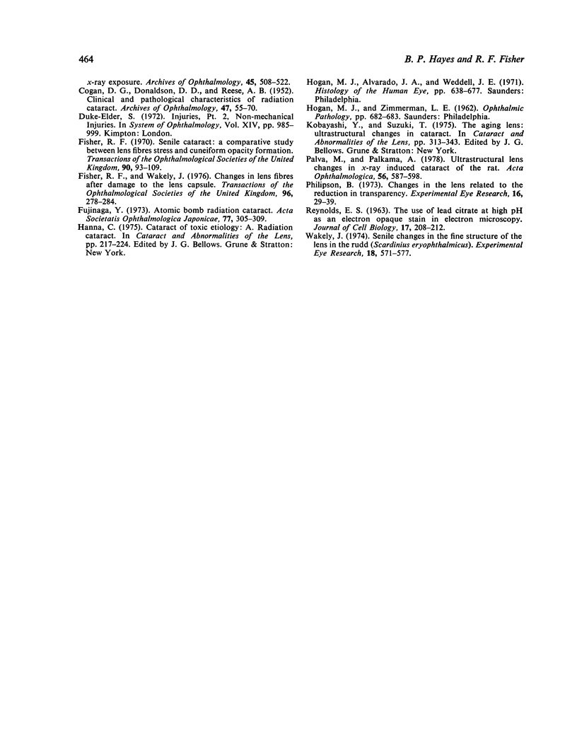
Human lenses extracted for cataract 26 years after long-term exposure to an imperfectly shielded radium source were examined by slit-lamp photography, thin-section light microscopy, and electron microscopy. Anterior epithelial cells were fibroblast-like, and germinal epithelium and vacuolated cortical fibres had accumulated at the equator. A zone of light scatter at the anterior pole corresponded to an area of breakdown of cortical lens fibres, where unusual feathery fibres were orientated perpendicular to the lens surface. Two zones of light scatter separated by a 250-microM clear interval were seen in the posterior cortex. The zone at the posterior pole corresponded to an area of fibre liquefaction and large rounded membrane whorls, while the deeper zone comprised small flattened membrane whorls. The characteristic plaques of swollen abnormal cells described in previous histological studies of x-ray cataract were not present. This and other differences probably reflect the extremely long time course and repeated subliminal doses to which the patient was exposed.
Full text
PDF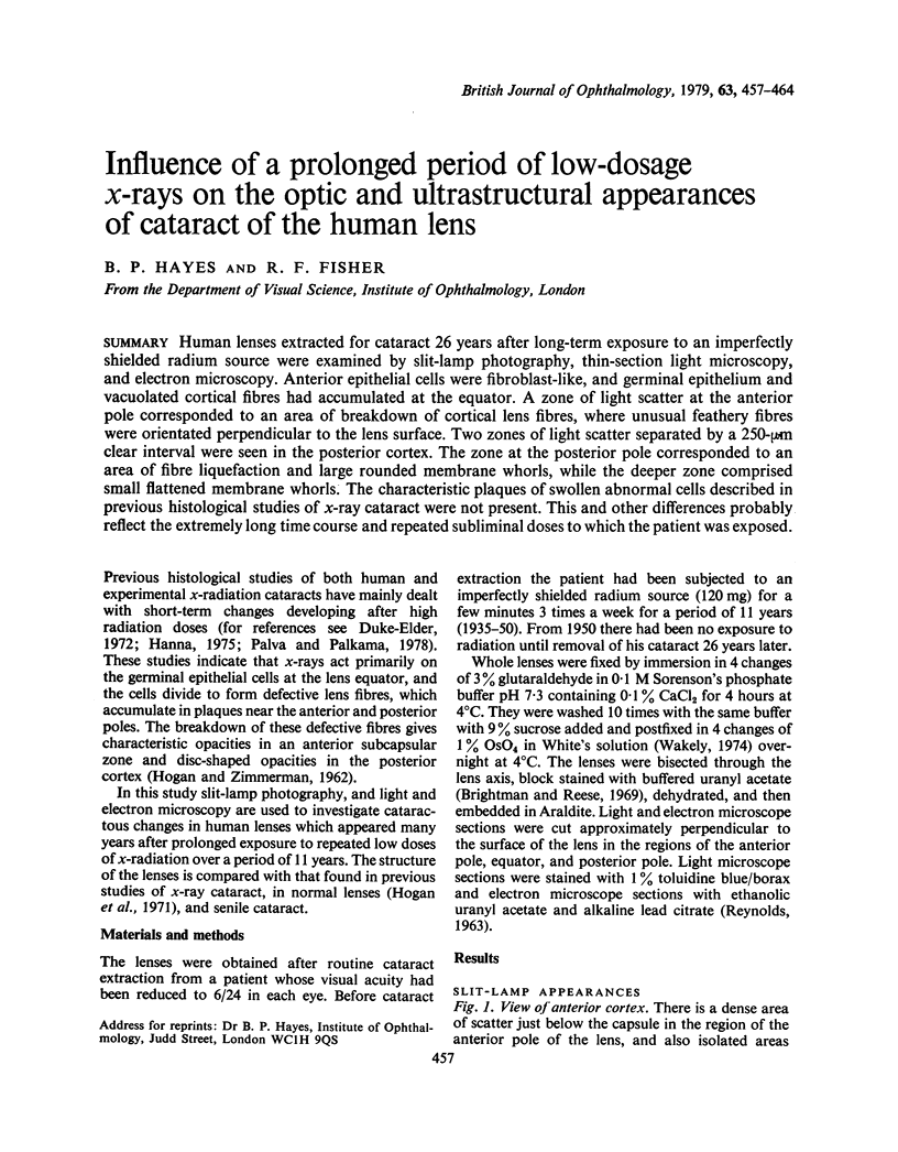
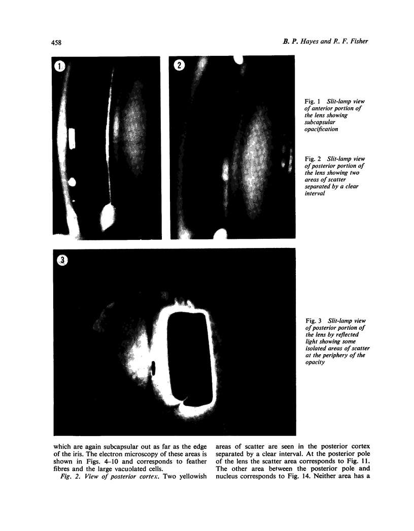
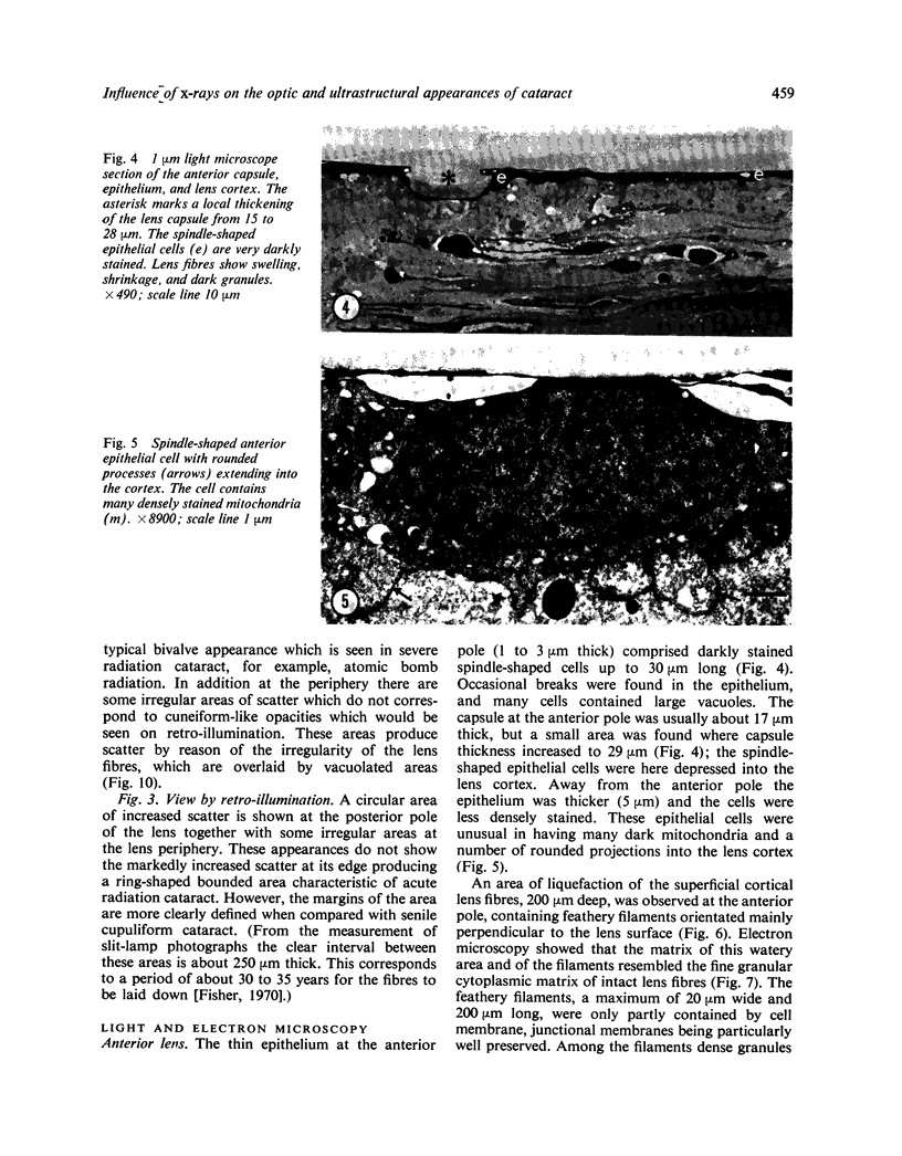
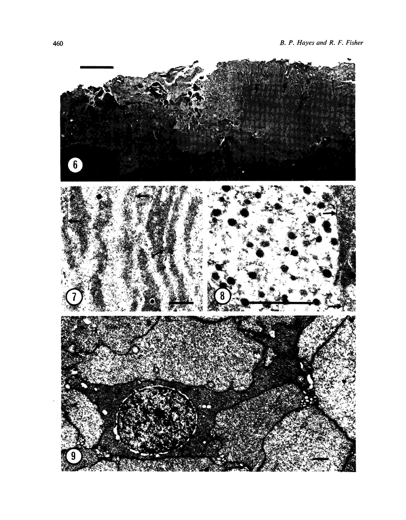
Images in this article
Selected References
These references are in PubMed. This may not be the complete list of references from this article.
- BRINI A., PORTE A., STOECKEL M. E. MODIFICATIONS ULTRASTRUCTURALES DU CRISTALLIN DANS CERTAINES CATARACTES EXP'ERIMENTALES ET HUMAINES. Bull Mem Soc Fr Ophtalmol. 1963;76:193–208. [PubMed] [Google Scholar]
- Brightman M. W., Reese T. S. Junctions between intimately apposed cell membranes in the vertebrate brain. J Cell Biol. 1969 Mar;40(3):648–677. doi: 10.1083/jcb.40.3.648. [DOI] [PMC free article] [PubMed] [Google Scholar]
- COGAN D. G., DONALDSON D. D. Experimental radiation cataracts. I. Cataracts in the rabbit following single x-ray exposure. AMA Arch Ophthalmol. 1951 May;45(5):508–522. [PubMed] [Google Scholar]
- COGAN D. G., DONALDSON D. D., REESE A. B. Clinical and pathological characteristics of radiation cataract. AMA Arch Ophthalmol. 1952 Jan;47(1):55–70. doi: 10.1001/archopht.1952.01700030058006. [DOI] [PubMed] [Google Scholar]
- Fisher R. F. Senile cataract. A comparative study between lens fibre stress and cuneiform opacity formation. Trans Ophthalmol Soc U K. 1970;90:93–109. [PubMed] [Google Scholar]
- Fisher R. F., Wakely J. Changes in lens fibres after damage to the lens capsule. Trans Ophthalmol Soc U K. 1976 Jul;96(2):278–284. [PubMed] [Google Scholar]
- Fujinaga Y. [On the atomic bomb radiation cataract]. Nippon Ganka Gakkai Zasshi. 1973;77(3):305–309. [PubMed] [Google Scholar]
- Palva M., Palkama A. Ultrastructural lens changes in X-ray induced cataract of the rat. Acta Ophthalmol (Copenh) 1978;56(4):587–598. doi: 10.1111/j.1755-3768.1978.tb01371.x. [DOI] [PubMed] [Google Scholar]
- Philipson B. Changes in the lens related to the reduction of transparency. Exp Eye Res. 1973 Jun;16(1):29–39. doi: 10.1016/0014-4835(73)90234-0. [DOI] [PubMed] [Google Scholar]
- REYNOLDS E. S. The use of lead citrate at high pH as an electron-opaque stain in electron microscopy. J Cell Biol. 1963 Apr;17:208–212. doi: 10.1083/jcb.17.1.208. [DOI] [PMC free article] [PubMed] [Google Scholar]
- Wakely J. Senile changes in the fine structure of the lens in the rudd(Scardinius eryopthalmicus). Exp Eye Res. 1974 Jun;18(6):571–577. doi: 10.1016/0014-4835(74)90063-3. [DOI] [PubMed] [Google Scholar]


