

Abstract
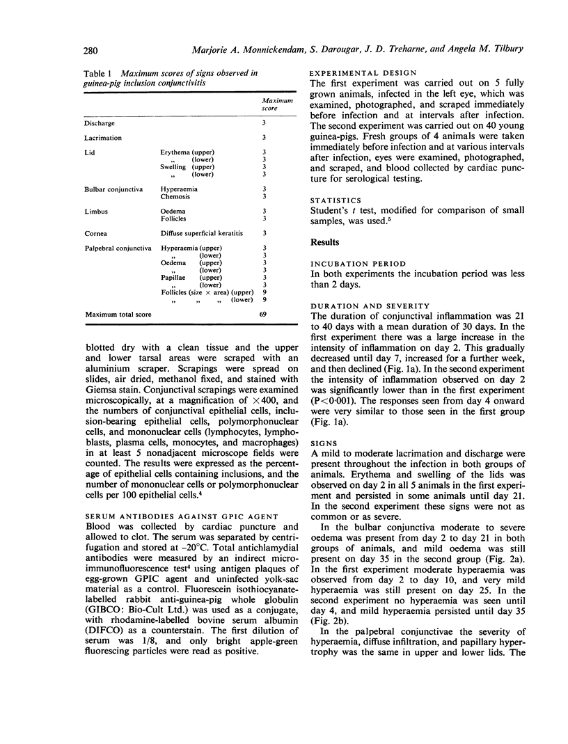
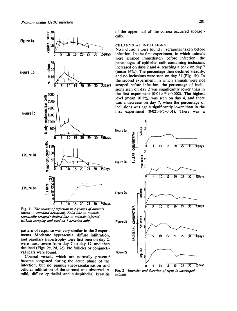
The course of primary ocular infection with guinea-pig inclusion conjunctivitis agent was followed in 2 groups of animals. One group of fully grown animals was repeatedly scraped; the other of small animals was used on 1 occasion only and scraped after clinical examination. The intensity of conjunctival inflammation was measured, conjunctival scrapings were taken, and the numbers of polymorphonuclear cells, mononuclear cells, and epithelial cells containing chlamydial inclusions were counted, and the level of antibodies in serum was measured. It was found that inflammation of the conjunctiva lasted for about 30 to 40 days, and the clinical features (oedema, hyperaemia, papillary reaction) were very similar in the 2 groups. Inclusions and polymorphonuclear cells were found for up to 21 days, and mononuclear cells were found on days 7 to 25. Serum antibodies were first detected on day 10 and reached a peak on day 21. The intensity of inflammation was significantly higher on day 2 in the animals which had been scraped. After this the severity of the inflammation and the course of disease were similar in the 2 groups.
Full text
PDF
Images in this article
Selected References
These references are in PubMed. This may not be the complete list of references from this article.
- Gordon F. B., Weiss E., Quan A. L., Dressler H. R. Observations on guinea pig inclusion conjunctivitis agent. J Infect Dis. 1966 Apr;116(2):203–207. doi: 10.1093/infdis/116.2.203. [DOI] [PubMed] [Google Scholar]
- Jawetz E., Rose L., Hanna L., Thygeson P. Experimental inclusion conjunctivitis in man: measurements of infectivity and resistance. JAMA. 1965 Nov 8;194(6):620–632. [PubMed] [Google Scholar]
- Kazdan J. J., Schachter J., Okumoto M. Inclusion conjunctivitis in the guinea pig. Am J Ophthalmol. 1967 Jul;64(1):116–124. doi: 10.1016/0002-9394(67)93351-x. [DOI] [PubMed] [Google Scholar]
- MURRAY E. S. GUINEA PIG INCLUSION CONJUNCTIVITIS VIRUS. I. ISOLATION AND IDENTIFICATION AS A MEMBER OF THE PSITTACOSIS-LYMPHOGRANULOMA-TRACHOMA GROUP. J Infect Dis. 1964 Feb;114:1–12. doi: 10.1093/infdis/114.1.1. [DOI] [PubMed] [Google Scholar]
- Monnickendam M. A., Darougar S., Treharne J. D., Tilbury A. M. Development of chronic conjunctivitis with scarring and pannus, resembling trachoma, in guinea-pigs. Br J Ophthalmol. 1980 Apr;64(4):284–290. doi: 10.1136/bjo.64.4.284. [DOI] [PMC free article] [PubMed] [Google Scholar]