Abstract
Crouch or “flexed knee” gait is a pathological gait pattern affecting many individuals with cerebral palsy. One proposed method to alleviate crouch is to provide robotic assistance to knee extension during walking. The purpose of this study was to evaluate how the magnitude of knee extensor torque affects knee kinematics, kinetics, and muscle activity. Motion capture, ground reaction force and electromyography data were collected while four participants with crouch gait from cerebral palsy walked with assistance from a novel robotic exoskeleton on an instrumented treadmill. Different magnitudes of knee extensor torque were provided during the stance (range: 0.09-0.38 Nm/kg) and swing (range: 0.09-0.29 Nm/kg) phases of the gait cycle. Using a linear regression analysis, we found that greater torque from the exoskeleton was positively associated with increased knee extension (reduction in crouch) at foot contact and mid-stance, negatively associated with the biological knee extensor moment, and positively associated with knee flexor muscle activity. Determining the relationships between exoskeleton assistance and knee kinematics and kinetics will benefit the continued investigation of robotic treatment strategies for treating crouch gait. Our findings indicate the importance of properly tuned robotic control strategies for gait rehabilitation.
I. Introduction
Crouch gait is a pathological gait pattern caused by cerebral palsy (CP), the most common group of movement disorders in children [1, 2]. Individuals with crouch gait walk with excessive lower-extremity joint flexion, which raises the metabolic cost of transport and is associated with knee pain and degeneration [3, 4]. Standard of care treatments, including orthopaedic surgery [5] and physical therapy [6], can help manage this condition, but adequate long-term correction is difficult to achieve; walking deficits worsen over time with over half of all affected individuals losing the ability to walk in adulthood [7].
Findings from a previous investigation demonstrated that a wearable robotic exoskeleton offers the potential to treat crouch gait by alleviating excessive knee joint flexion [8]. However, responses to the exoskeleton varied between individuals and optimizing how robotic assistance is provided remains a critical challenge that must be addressed prior to the implementation of exoskeletons for long-term crouch rehabilitation. In treating crouch gait, extensor assistance can be provided during the swing phase to increase knee extension prior to foot contact, and the during stance phase to assist the knee extensor muscles in supporting the body.
Optimizing robotic assistance by experimentally iterating through a large range of assistive modes and magnitudes is time consuming and challenging for individuals with movement disorders. In particular, children with CP have pathologies, such as muscle weakness, impaired selective motor control, and increased levels of spasticity, that make the effect of motorized assistance unclear. Thus, research is needed to understand the biomechanical responses to assistance and to determine how the amount of extensor toque provided during both swing and stance affect the degree of improvement in posture and rehabilitation outcomes in this population.
The goal of this study was to determine how different magnitudes of stance and swing phase knee extensor assistance provided from a robotic exoskeleton affect knee joint angles, moments, and muscle activity in individuals with crouch gait from CP. We hypothesized that increasing the level of assistive torque would lead a linear increase in knee extension and a linear decrease in the biological knee extensor moment. We also hypothesized that differences in functional outcomes across individuals would be explained by changes in muscle activity.
II. Materials and Methods
A. Study Protocol and Participant Information
This study was approved by the Institutional Review Board at the National Institutes of Health under protocol # 13-CC-0210. We recruited individuals with a diagnosis of crouch gait caused by CP between the ages of 5 and 19. Inclusion criteria included the ability to walk independently (i.e. without crutches or the aid of a therapist) for at least 30 feet and a Gross Motor Function Classification System Level of I or II. Participants were excluded if they had any medical condition other than CP that would put them at risk during this study. Written informed consent was obtained from one of the participant’s parents or from participants 18 or older, while verbal informed assent was obtained from each participant under 18. Only data from the participants who were able to walk with multiple levels of stance and swing phase extension assistance were included in the present analysis (Table I).
TABLE I.
Participant information and exoskeleton parameters.
| Subject (#) |
Age (yrs) |
Height (m) |
Body Mass (kg) |
GMFCS Levela |
MASb Score |
Baseline Condition |
Level 1 Torque Stance/Swing (Nm/kg) |
Level 2 Torque Stance/Swing (Nm/kg) |
Treadmill Speed (m/s) |
|---|---|---|---|---|---|---|---|---|---|
| P1 | 6 | 1.11 | 20.0 | II | 0 | AFOc | 0.19/0.09 | 0.28/0.14 | 0.41 |
| P2 | 11 | 1.56 | 40.8 | II | 1+ | Shod | 0.15/0.15 | 0.29/0.29 | 0.47 |
| P3 | 12 | 1.72 | 69.3 | I | 1 | Shod | 0.09/0.09 | 0.13/0.13 | 0.49 |
| P4 | 11 | 1.35 | 32.0 | II | 2 | Shod | 0.31/0.19 | 0.38/0.28 | 0.67 |
GMFCS: Gross Motor Function Classification Scale.
MAS: Modified Ashworth Scale for more affected limbs.
AFO: ankle-foot orthosis.
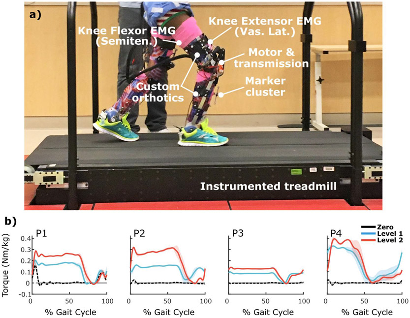
The full protocol consisted of six visits at the National Institutes of Health Clinical Center. On the first visit, we took a cast of each participant’s lower extremities for molding the custom orthotics where the exoskeleton’s electromechanical components were mounted via quick release connectors (Fig.1a). The second through fifth visits, which lasted for two-three hours each, were used to practice walking with the exoskeleton. On the sixth visit, we collected experimental gait data while the participants completed treadmill walking bouts at different levels of robotic assistance.
Figure 1.
a) Experimental setup and robotic exoskeleton. b) Knee extensor torque profiles provided by the exoskeleton during zero (black), level 1 (blue), and level 2 (red) conditions for the more affected limbs. Shading depicts ±1 standard deviation.
Kinematic data were collected using 10 infrared motion capture cameras (Vicon Motion Systems, Oxford, UK) that recorded the positions of markers placed on the torso, pelvis, and lower-extremity. We collected kinetic data from an instrumented treadmill (Bertec, Columbus, OH). Electromyography (EMG) data were also recorded from a pair of knee extensor (vastus lateralis) and knee flexor (semitendinosus) muscles (Delsys, Boston, MA).
B. Robotic Exoskeleton
We implemented a previously developed novel powered exoskeleton [9] consisting of a motor and transmission assembly that mounted laterally to custom molded thermoplastic foot, shank, and thigh orthotics (Fig. 1a). The exoskeleton had a passive, adjustable ankle joint, which, for this study, was set to free rotation. The motor and transmission assembly provided an assistive torque about the knee joint. The control strategy incorporated a finite state machine and a PID control algorithm to specify torque set-points at different intervals across the gait cycle; a reaction torque sensor was used in conjunction with a real-time control algorithm. Data from an embedded force sensor in the sole of each orthotic were used to distinguish between stance and swing. Data from an encoder were used to distinguish between early (knee flexion) and late (knee extension) swing.
C. Exoskeleton Conditions
We implemented different levels of exoskeleton assistance while the participants walked at fixed treadmill speeds (Table I, Fig. 2b). Extension assistance was provided during stance phase and the knee extension portion of swing phase. During the early swing (knee flexion) phase and the zero torque condition, the exoskeleton allowed free, frictionless rotation. For lower (level 1) and upper (level 2) exoskeleton conditions, the amounts of exoskeleton assistance were established for each participant over the course of the practice sessions; torque was gradually increased to a range that was well tolerated by each participant and that elicited improved gait mechanics as assessed visually by the research team.
Figure 2.
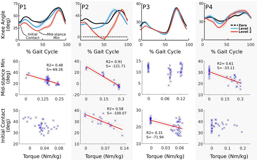
Top row) Knee angle profiles across the gait cycle during zero (black), level 1 (blue), and level 2 (red) conditions for the more affected limbs. Shading depicts ±1 standard deviation. Middle row) Exoskeleton torque plotted vs. the mean knee flexion angle during stance for individual gait cycles. Bottom row) Exoskeleton torque plotted vs. the knee angle at initial foot contact for individual gait cycles. The red lines depict the best fit line for significant relationships. R2 values are the coefficient of determination. S values are the slope of the best fit line.
D. Data and Statistical Analysis
We completed an inverse kinematics and kinetics analysis using Visual 3D to quantify joint angles and moments (C-Motion, Gaithersburg, MD, USA). Inertial properties of each custom exoskeleton were determined from CAD models (SolidWorks, Concord, MA, USA) and specified in each participant’s biomechanical model. The biological component of the knee extensor moment was computed by subtracting the measured exoskeleton torque from the total moment determined from inverse kinetics.
The EMG data were processed to create linear envelopes by band pass filtering from 15-380 Hz, rectifying, and low pass filtering at 7 Hz. The mean muscle activity during stance and swing was found by numerically integrating over the respective intervals. The time-course data were segmented from heel-strike to same side next heel-strike and normalized to percent gait cycle. Outcome measures included knee angle at initial contact, the minimum flexion angle during stance (mid-stance min) and the average knee moment and knee flexor and extensor muscle activity across the stance and swing phases. Data from each individual’s more affected (more crouched) limb are reported.
The exoskeleton was tested at two torque settings (low and high), yet variability in user interaction affected the actual amount of extension torque provided to the limbs within each phase of the gait cycle. Therefore, for each participant, linear regression analysis was used to determine the relationships between the magnitude of assistive torque and our outcome measures. For each tested relationship, we computed the coefficient of determination (R2), and, if statistically significant, the slope of the best fit line. We defined mild, moderate, and strong relationships as having R2 values between 0.25-0.49, 0.50-0.69, and 0.70-1.0, respectively.
III. Results
During data collection, all the participants completed each exoskeleton walking condition without holding onto the guardrail or requiring therapist assistance.
A. Knee Kinematics
All participants exhibited reductions in crouch either at initial contact (P3), during mid-stance (P1 and P4), or both (P2) while walking with extension assistance. In general, the relationship between exoskeleton torque and knee angle were stronger at mid-stance than at initial contact. There were mild to strong relationships between reduction in crouch at mid-stance and exoskeleton torque in 3/4 individuals (R2 = 0.48, P1; R2 = 0.61, P4; R2 = 0.91, P2) (Fig. 2 middle row). Two of four individuals exhibited mild or moderate relationships between torque and reduction in crouch at initial contact (R2 = 0.31, P3; R2 = 0.58, P2) (Fig. 2 bottom row).
B. Knee Moments
Exoskeleton assistance during walking had a significant impact on knee extensor moment in all participants. During stance, all participants exhibited moderate to strong relationships between the amount of exoskeleton torque and a reduction in the mean biological knee extensor moment (0.60<R2<0.70) (Fig. 3 middle row). During swing, all of the participants exhibited mild to moderate relationships between the extensor torque and a reduction in the mean biological extensor moment (0.31<R2<0.62) (Fig. 3 bottom row).
Figure 3.
Top row) Knee moment profiles across the gait cycle during zero (black), level 1 (blue), and level 2 (red) conditions for the more affected limbs. Shading depicts ±1 standard deviation. Middle row) Exoskeleton torque plotted vs. the mean knee moment during stance for individual gait cycles. Bottom row) Exoskeleton torque plotted vs. the mean knee moment during swing for individual gait cycles. The red lines depict the best fit line for significant relationships. R2 values are the coefficient of determination. S values are the slope of the best fit line.
C. Knee Muscle Activity
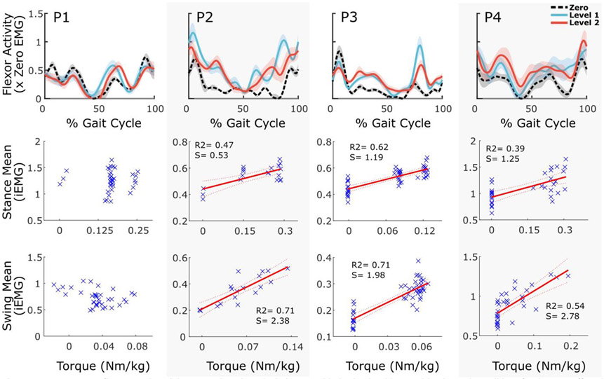
Generally, robotic extension assistance had a greater effect on antagonist (knee flexor) muscles than on agonist (knee extensor) muscles. During stance, P2, P3, and P4 exhibited moderate positive relationships between exoskeleton torque and knee flexor muscle activity (0.39<R2<0.62); during swing, the relationship was stronger for participants (0.54<R2<0.73), with higher slopes of the best fit lines (Fig. 4). P1 exhibited significant, but weak inverse relationships between exoskeleton torque and knee extensor activity during stance and swing; no other participants exhibited significant relationships during stance. However, P2 and P3 exhibited weak and moderate, respectively, positive relationships during swing (Fig 5).
Figure 4.
Top row) Knee flexor muscle activity across the gait cycle during zero (black), level 1 (blue), and level 2 (red) conditions for the more affected limbs. Shading depicts ±1 standard deviation. Middle row) Exoskeleton torque plotted vs. the mean knee flexor muscle activity during stance for individual gait cycles. Bottom row) Exoskeleton torque plotted vs. the mean knee flexor muscle activity during swing for individual gait cycles. The red lines depict the best fit line for significant relationships. R2 values are the coefficient of determination. S values are the slope of the best fit line.
Figure 5.
Top row) Knee extensor muscle activity across the gait cycle during zero (black), level 1 (blue), and level 2 (red) conditions for the more affected limbs. Shading depicts ±1 standard deviation. Middle row) Exoskeleton torque plotted vs. the mean knee extensor muscle activity during stance for individual gait cycles. Bottom row) Exoskeleton torque plotted vs. the mean knee extensor muscle activity during swing for individual gait cycles. The red lines depict the best fit line for significant relationships. R2 values are the coefficient of determination. S values are the slope of the best fit line.
IV. Discussion
In this study, we investigated the effect of assistive torque from a wearable exoskeleton on knee biomechanics in subjects with crouch gait. We partially confirm our hypothesis that increasing the magnitude of assistive torque would lead to a linear increase in knee extension angle and a linear decrease in the biological knee extensor moment. All participants exhibited positive relationships between exoskeleton extension torque and a reduction in crouch during either stance, initial contact, or both. Further, there were moderate to strong relationships between torque magnitude and reduction in the biological knee extensor moments during stance and swing phases in all participants. We also partially accept our secondary hypothesis that differences in outcomes across individuals would be explained by changes in muscle activity. The participants who exhibited greater increases in knee flexor muscle activity with increasing torque during stance exhibited weaker relationships between reduction in crouch at mid-stance and increasing torque. Our findings demonstrate that robotic extension assistance can result in improvements in posture, and that the amount of improvement is affected by the individuals’ synergistic muscle responses in addition to the level of assistance.
Knee flexor muscle activity was more closely related to the amount of exoskeleton torque compared to activity of the knee extensors. For P1, who had received a selective dorsal rhizotomy, knee flexor activity did not change with increasing exoskeleton torque. However, P1 did exhibit weak inverse relationships between exoskeleton torque and knee extensor activity during stance and swing. Interestingly, P2 and P3, the only two participants with significant relationships between increasing torque and increased knee extension at initial contact, had increased co-contraction during swing: both knee flexor and extensor activity was positively associated with increasing exoskeleton torque for these participants. This may signify the importance of harnessing volitional knee extensor activity for crouch rehabilitation.
Robotic extension assistance appears to improve posture more readily during stance than during swing. During stance, the biological knee extensor moment was negatively related to increasing torque, but remained “positive” (in the extensor direction). During swing, however, the biological knee extensor moment decreased beyond zero (increased in the flexor direction) with increasing exoskeleton torque (Fig. 3). Since a biological knee moment in the flexor direction would reduce limb extension, this may indicate that the participants responded to extensor assistance by resisting the exoskeleton’s attempt to extend the limb prior to foot contact. This is corroborated by the stronger positive relationships between knee flexor muscle activity during swing than during stance (Fig. 4). The mechanisms responsible for this behavior should be investigated further, with potential explanations including spasticity, dynamic contractures, or a resistance to open-chain perturbations to maintain step length and thereby stability.
Our results lend insights for the effective implementation of robotic assistance for crouch gait rehabilitation. For most of the participants, increasing assistive torque improved kinematic outcomes, while modestly increasing knee flexor muscle activity. However, for P3, increasing exoskeleton torque led to improvements in knee angle at initial contact, but not during mid-stance. This participant responded to greater amounts of torque during stance by increasing knee flexor activity. P3 presented with excessive knee flexion at initial contact, but the knee angle during mid-stance was closer to that in typical gait than the other three participants. Therefore, when increasing amounts of extensor torque were provided during stance, this participant may have increased their knee flexor activity to maintain baseline kinematics.
Testing a range of exoskeleton torques in order to optimize robotic assistance for patients with gait disorders is time consuming and challenging. The findings from this study can be utilized to improve the implementation of future robotic interventions by providing researchers and clinicians with insight into the range of potential relationships between the amount of assistance and biomechanical outcomes. Our findings emphasize the need for individually tuned control strategies that account for internal joint kinetics. By using the demonstrated ability to predict the baseline knee moment from kinematic parameters [10], combined with our findings from the present study of the strong and consistent relationship between the biological knee moment and assistive torque, we may be able to adjust control settings in real-time to reflect the instantaneous internal joint moment; the implementation of which will be tested in a future, larger study.
One of the primary limitations of this study was that we had a small number of participants who were only able to complete a relatively small number of exoskeleton conditions. Additionally, we restricted our investigation to linear type regression analysis for simplicity and ease of interpretation; however, some of the responses may have been better described by curvilinear relationships. The relationships between torque and our outcome measures were mostly similar between more and less (not reported) affected limbs. Still, future work should extend the biomechanical analysis to both legs, and increase the number of participants, experimental conditions (levels of assistance), and expand the regression analysis.
In conclusion, robotic extension assistance was associated with improvements in crouch at initial contract and mid-stance, reductions in the biological knee extensor moment during stance and swing, and an increase in knee flexor activity. Stronger relationships between assistive torque magnitude and knee flexor activity were correlated with diminished improvements in crouch suggesting that increasing the amount of assistance may elicit neuromuscular responses that are counter-productive for rehabilitation in some patients.
Acknowledgment
Funding for this study was provided by the Intramural Research Program of the NIH Clinical Center.
This work was supported by the Intramural Research Program at the National Institutes of Health (protocol #13-CC-0210)
Contributor Information
Zachary F. Lerner, Functional and Applied Biomechanics Section, Rehabilitation Medicine Department, Clinical Center, National Institutes of Health, Bethesda, MD, 20892, USA.; Northern Arizona University, Flagstaff, AZ 86011, USA
Diane L. Damiano, Functional and Applied Biomechanics Section, Rehabilitation Medicine Department, Clinical Center, National Institutes of Health, Bethesda, MD, 20892, USA
Thomas C. Bulea, Functional and Applied Biomechanics Section, Rehabilitation Medicine Department, Clinical Center, National Institutes of Health, Bethesda, MD, 20892, USA.
References
- [1].Odding E, Roebroeck ME, and Stam HJ, "The epidemiology of cerebral palsy: incidence, impairments and risk factors," Disabil Rehabil, vol. 28, pp. 183–91, Feb 2006. [DOI] [PubMed] [Google Scholar]
- [2].Wren TA, Rethlefsen S, and Kay RM, "Prevalence of specific gait abnormalities in children with cerebral palsy: influence of cerebral palsy subtype, age, and previous surgery," J Pediatr Orthop, vol. 25, pp. 79–83, Jan 2005. [DOI] [PubMed] [Google Scholar]
- [3].Opheim A, Jahnsen R, Olsson E, and Stanghelle J, "Walking function, pain, and fatigue in adults with cerebral palsy: a 7-year followup study," Dev Med Child Neurol, vol. 51, pp 381–8, May 2009. [DOI] [PubMed] [Google Scholar]
- [4].Rose J, Gamble JG, Burgos A, Medeiros J, and Haskell WL, "Energy expenditure index of walking for normal children and for children with cerebral palsy," Dev Med Child Neurol, vol. 32, pp. 333–40, Apr 1990. [DOI] [PubMed] [Google Scholar]
- [5].De Mattos C, Patrick Do K, Pierce R, Feng J, Aiona M, and Sussman M, "Comparison of hamstring transfer with hamstring lengthening in ambulatory children with cerebral palsy: further follow-up," J Child Orthop, vol. 8, pp. 513–20, Dec 2014. [DOI] [PMC free article] [PubMed] [Google Scholar]
- [6].Damiano DL, Vaughan CL, and Abel MF, "Muscle response to heavy resistance exercise in children with spastic cerebral palsy," Dev Med Child Neurol, vol. 37, pp. 731–9, Aug 1995. [DOI] [PubMed] [Google Scholar]
- [7].Bottos M and Gericke C, "Ambulatory capacity in cerebral palsy: prognostic criteria and consequences for intervention," Dev Med Child Neurol, vol. 45, pp. 786–90, Nov 2003. [DOI] [PubMed] [Google Scholar]
- [8].Lerner Z, Damiano D, and Bulea T, " A Robotic Exoskeleton for Treatment of Crouch Gait in Children with Cerebral Palsy: Initial Kinematic and Neuromuscular Evaluation," Proc. IEEE EMBC, 2016. [DOI] [PubMed] [Google Scholar]
- [9].Lerner Z, Damiano D, Park H, and Bulea T, "A Robotic Exoskeleton for Treatment of Crouch Gait in Children with Cerebral Palsy: Design and Initial Application," IEEE Trans Neural Syst Rehabil Eng, In Press. [DOI] [PMC free article] [PubMed] [Google Scholar]
- [10].Lerner Z, Damiano D, and Bulea T, "Estimating the Mechanical Behavior of the Knee Joint during Crouch Gait: Implications for Real-Time Motor Conrol of Robotic Knee Orthoses" IEEE Trans Neural Syst Rehabil Eng, vol. 24, pp. 621–9, Jun 2016. [DOI] [PMC free article] [PubMed] [Google Scholar]


