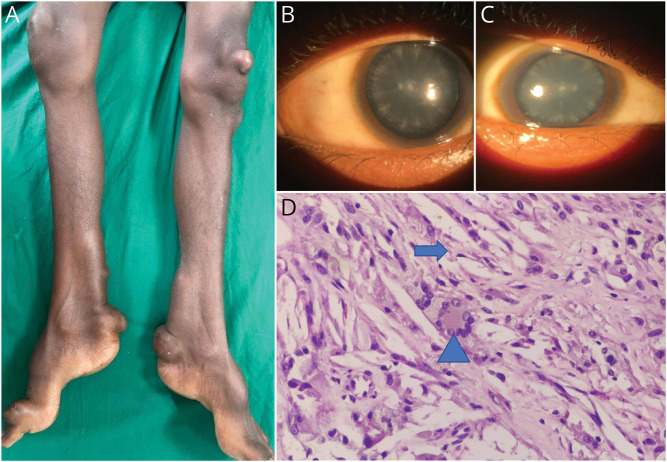
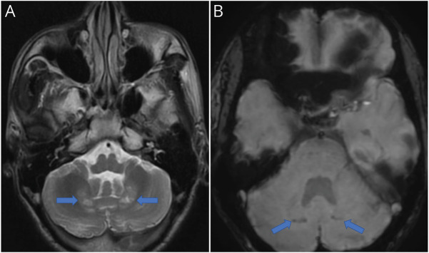
A 20-year-old male patient presented with progressive painless subcutaneous swellings over both knees, heels, and dorsum of right great toe (Figure 1A) and diminution of vision for the past 8 years. Examination revealed high-arched feet and global cognitive impairment. Slitlamp examination showed bilateral lenticular opacities (Figure 1B and C). Histopathologic examination of swelling revealed foamy histiocytes, cholesterol clefts, and touton giant cells, suggestive of xanthoma (Figure 1D). Axial T2-weighted magnetic resonance imaging of the brain showed bilateral symmetrical cerebellar dentate nuclei hyperintensities and susceptibility-weighted imaging revealed hypointensities due to mineralization, along with moderate atrophy of the cerebellar vermis (Figure 2A and B). Whole-exome sequencing revealed heterozygous pathogenic mutations on exon 3 (c.526delG) and exon 7 (c.1213C > T) in the CYP27A1 gene, suggestive of cerebrotendinous xanthomatosis (CTX). The characteristic features of CTX include progressive neurologic impairment, tendon xanthomas, cataract, and premature atherosclerosis.1 Chenodeoxycholic acid alleviates neurologic symptoms and improves prognosis.
Figure 1. Clinical Manifestations and Imaging Characteristics of Cerebrotendinous Xanthomatosis.
Tendon xanthomas (A), bilateral lenticular opacities (B and C), and histopathology of subcutaneous swelling showing cholesterol clefts (arrow) and touton giant cell (arrowhead) (D) (H&E, ×400).
Figure 2. Clinical Manifestations and Imaging Characteristics of Cerebrotendinous Xanthomatosis.
Axial T2-weighted (A) and susceptibility-weighted (B) magnetic resonance imaging of the brain revealed bilateral cerebellar dentate nuclei hyperintensities (horizontal arrows) and punctate foci of hypointensities (oblique arrows), respectively.
Footnotes
Teaching slides links.lww.com/WNL/C789
Author Contributions
R.R. Sahoo: drafting/revision of the manuscript for content, including medical writing for content; major role in the acquisition of data; analysis or interpretation of data. S. Sukriya: drafting/revision of the manuscript for content, including medical writing for content; major role in the acquisition of data; analysis or interpretation of data. G. Sudhish: drafting/revision of the manuscript for content, including medical writing for content; major role in the acquisition of data; analysis or interpretation of data. A.K. Panda: drafting/revision of the manuscript for content, including medical writing for content; major role in the acquisition of data; Analysis or interpretation of data. D. Mohapatra: drafting/revision of the manuscript for content, including medical writing for content; major role in the acquisition of data; analysis or interpretation of data. P.S. Patro: drafting/revision of the manuscript for content, including medical writing for content; major role in the acquisition of data; analysis or interpretation of data.
Study Funding
The authors report no targeted funding.
Disclosure
The authors report no relevant disclosures. Go to Neurology.org/N for full disclosures.
Reference
- 1.Nie S, Chen G, Cao X, Zhang Y. Cerebrotendinous xanthomatosis: a comprehensive review of pathogenesis, clinical manifestations, diagnosis, and management. Orphanet J Rare Dis. 2014;9(1):179. [DOI] [PMC free article] [PubMed] [Google Scholar]


