Abstract
The paradigm for activation of Ras and extracellular signal-regulated kinase (ERK)/mitogen-activated protein (MAP) kinase by extracellular stimuli via tyrosine kinases, Shc, Grb2, and Sos does not encompass an obvious role for phosphoinositide (PI) 3-kinase, and yet inhibitors of this lipid kinase family have been shown to block the ERK/MAP kinase signalling pathway under certain circumstances. Here we show that in COS cells activation of both endogenous ERK2 and Ras by low, but not high, concentrations of epidermal growth factor (EGF) is suppressed by PI 3-kinase inhibitors; since Ras activation is less susceptible than ERK2 activation, PI 3-kinase-sensitive events may occur both upstream of Ras and between Ras and ERK2. However, strong elevation of PI 3-kinase lipid product levels by expression of membrane-targeted p110α is by itself never sufficient to activate Ras or ERK2. PI 3-kinase inhibition does not affect EGF-induced receptor autophosphorylation or adapter protein phosphorylation or complex formation. The concentrations of EGF for which PI 3-kinase inhibitors block Ras activation induce formation of Shc-Grb2 complexes but not detectable EGF receptor phosphorylation and do not activate PI 3-kinase. The activation of Ras by low, but mitogenic, concentrations of EGF is therefore dependent on basal, rather than stimulated, PI 3-kinase activity; the inhibitory effects of LY294002 and wortmannin are due to their ability to reduce the activity of PI 3-kinase to below the level in a quiescent cell and reflect a permissive rather than an upstream regulatory role for PI 3-kinase in Ras activation in this system.
A wide variety of extracellular stimuli induce activation of the mitogen-activated protein (MAP) kinases extracellular signal-regulated kinase 1 (ERK1) and ERK2, which transduce proliferative or differentiation signals to the nucleus (44). The signalling pathways leading from activated growth factor receptors to ERKs have been thoroughly examined (29), and the small GTPase Ras has been shown to play a pivotal role. The mechanisms behind growth factor-induced activation of Ras are well established (32); epidermal growth factor (EGF), for example, binds to and activates its receptor tyrosine kinase, which autophosphorylates, creating binding sites for SH2-domain-containing proteins including the adapter proteins Grb2 and Shc. In addition to its SH2 domain, Grb2 also binds through its SH3 domains to the guanine nucleotide exchange factor Sos. Binding of Grb2 to phosphorylated EGF receptors results in recruitment of Sos to the plasma membrane and has been proposed as a model for activation of membrane-bound Ras (5). In addition, EGF-induced activation of Ras may be transduced via Shc, which binds to activated EGF receptors and becomes phosphorylated on tyrosine 317, creating an alternative binding site for Grb2 (34).
Once Ras has been activated by guanine nucleotide exchange factors, resulting in exchange of GTP for GDP on Ras, GTP-bound Ras interacts with and facilitates activation of the serine/threonine kinase Raf, as well as other target enzymes including phosphoinositide (PI) 3-kinase and Ral-GDP dissociation stimulator (29). Activated Raf phosphorylates and activates the downstream kinase MAP kinase/ERK kinase (MEK), which in turn phosphorylates and activates ERK (28). Ras activation has been shown to be important in activation of ERK by growth factors, but other Ras-independent pathways do exist for activating ERK, particularly protein kinase C (PKC) and calcium-mediated mechanisms (7).
While the model set out above does not display an obvious requirement for the activity of PI 3-kinase, a lipid kinase which is also activated by a wide variety of cellular stimuli (47), many reports have documented inhibition of ERK activation by pharmacological inhibitors of PI 3-kinase. These inhibitors have been reported to block ERK activation by some stimuli, such as insulin (9) and lysophosphatidic acid (LPA) and thrombin (18), but not others, such as EGF (18) or platelet-derived growth factor (PDGF) (14). The sensitivity of ERK activation to inhibition by PI 3-kinase inhibitors is in many cases dependent on cell type, and a recent report has provided convincing data that, at least in the case of PDGF, the sensitivity is a function of signal strength, with weak stimulation of ERK being dependent on PI 3-kinase but strong stimulation being independent (14).
The mechanism involved in the ability of PI 3-kinase inhibitors to block ERK activation under some circumstances remains unclear. When analyzed in detail, evidence for involvement of PI 3-kinase has been found at a number of different positions in the pathway. Perhaps the best defined is the ability of p21-activated kinase (PAK), a downstream target of PI 3-kinase via activation of Rac, to promote stimulation of the MAP kinase kinase MEK (15, 16). PAK1 phosphorylates MEK1 on serine 298, a site important for the binding of Raf-1 to MEK1. However, PI 3-kinase activity has also been reported to be required at the level of Raf-1, but not Ras, activation in the stimulation of ERK by insulin in L6 cells (9). Furthermore, PI 3-kinase inhibitors have been found to inhibit Ras activation by LPA in COS cells (18), although in this case the levels of wortmannin used (1 μM) were considerably higher than those generally thought to be specific for the PI 3-kinase family.
While most studies place the effect, if any, of PI 3-kinase inhibitors on the MAP kinase pathway downstream of Ras, there is reason to consider carefully the possibility that PI 3-kinase might also have some function upstream of Ras. It has been reported that PI 3-kinase may be able to stimulate Ras, at least in certain systems (19). This appears, at least initially, to be at odds with data from this laboratory that Ras can stimulate the activity of PI 3-kinase by direct binding to it (25, 39–41). We have therefore undertaken a detailed study of how PI 3-kinase might be involved in regulation of the Ras-MAP kinase pathway. We report here that the ability of optimal concentrations of EGF to activate Ras and ERK in the simian virus 40-transformed monkey kidney cell line COS-7 is not significantly affected by inhibition of PI 3-kinase. However, when low concentrations of EGF are used, which are probably more physiologically relevant, PI 3-kinase inhibitors do reduce the activation of both Ras and ERK. Evidence that the inhibition occurs at two points in the pathway, one upstream and one downstream of Ras, is presented. Importantly, inhibition of Ras activation by the PI 3-kinase inhibitors LY294002 and wortmannin occurs only at concentrations of EGF that are unable to activate PI 3-kinase, suggesting that it is the basal activity of PI 3-kinase that is required to support weak signal strength activation of Ras. This, plus the fact that strong activation of PI 3-kinase is not sufficient to activate Ras or ERK, demonstrates that regulation of PI 3-kinase is not involved in controlling the activity of Ras but that the basal activity of these enzymes plays a permissive role for Ras activation by weak stimuli, probably those involving Shc-Grb2-Sos, but not higher-order, complexes. PI 3-kinase lipid products may promote association of Shc-Grb2-Sos complexes with the plasma membrane; at higher concentrations of EGF, this may also be achieved by interaction with the autophosphorylated EGF receptor without requirement for PI 3-kinase.
MATERIALS AND METHODS
Expression vectors.
DNA fragments, encoding the carboxy terminus of bovine p110α with an extension corresponding to the farnesylation-palmitylation signal from H-Ras, were amplified by PCR with the sense primer 5′CACACACTCCATCAGTGGCTCAAAGACAAGAAC3′ and the antisense primer 5′CGCGGATCCTCAAGAGAGCACACACTTACAGTTCAAAGCATGCTGC3′. The PCR products were digested with ClaI and BamHI, ligated into the p110 sequence, and subcloned into the mammalian expression vector pSG5 (Stratagene) to generate p110-CAAX. The construct was confirmed by DNA sequencing. The v-Src and the Δp85 cDNA were in pSG5, and H-Ras, wild type, and N17 were in pEXV3. The Drosophila Sos (dSos) cDNA in pCMV5 was kindly provided by Michael Czech (University of Massachusetts), the Myc-tagged ERK2 in pEFHM was provided by Chris Marshall (Institute of Cancer Research), and hemagglutinin (HA)-tagged protein kinase B (PKB) in pSG5 was provided by Paul Coffer and Boudewijn Burgering (University of Utrecht).
Expression and purification of the Raf Ras-binding domain.
A bacterial culture of Escherichia coli BL21(DE3) cells harboring the plasmid pGEX KG containing the Raf Ras-binding domain (amino acids 1 to 149) fused to glutathione S-transferase (GST) was kindly provided by Barbara Marte. The culture was induced at an optical density at 600 nm of 0.4 to 0.6 for 3 h at 37°C with 1 mM isopropyl-1-thio-β-d-galactopyranoside (IPTG; Calbiochem). The cells were then lysed by sonication in phosphate-buffered saline (PBS) containing 1 mM EDTA, 1% Triton X-100, 10 μg of aprotinin per ml, and 10 μg of leupeptin per ml. The lysate was clarified by centrifugation and incubated with glutathione-agarose (Sigma) for 2 h at 4°C. The agarose beads were washed four times with buffer A (20 mM HEPES [pH 7.5], 100 mM NaCl, 10% glycerol, 0.5% Nonidet P-40, 2 mM EDTA, 10 μg of aprotinin per ml, and 10 μg of leupeptin per ml) and stored at 4°C as a 1:1 slurry in buffer A containing 0.1% NaN3.
Cell culture and transfection.
COS-7 cells were maintained in Dulbecco’s modified Eagle medium (DMEM) supplemented with 10% fetal bovine serum. For transfections, 5 × 10⁵ cells in 6-cm-diameter dishes were transfected by lipofection (Lipofectamine; Gibco BRL). Most constructs were used at 1 to 2 μg/dish, and empty pSG5 was added to make a final DNA concentration of 3 to 4 μg. Cells to be serum starved received DMEM containing 0.5% fetal bovine serum 24 h after transfection and were then incubated for another 24 h. All assays were done 48 h after transfection. To assay effects on cells attached to different substrata, 6-cm-diameter dishes were coated with two rounds of either 40 μg of collagen type IV (Sigma) per ml in PBS or 20 μg of fibronectin (Sigma) per ml in PBS and washed with PBS before 2 × 10⁶ cells/dish were plated out. To disrupt actin filaments, cells were pretreated with 5 μg of cytochalasin D (Sigma) per ml for 30 to 60 min.
Determination of GTP/GDP ratio.
Orthophosphate labelling and analysis of nucleotides bound to Ras were done essentially as described earlier (13). Briefly, 34 h after transfection cells were incubated with labelling medium (phosphate-free DMEM containing 20 mM HEPES [pH 7.5] and 0.5% dialyzed newborn calf serum) for 2 h. The cells were then incubated for 12 h with labelling medium containing 0.5 mCi of [32P]orthophosphate per dish. The cells were lysed in lysis buffer (50 mM HEPES [pH 7.5], 100 mM NaCl, 1 mM EGTA, 0.5 μg of benzamidine per ml, 5 μg of aprotinin per ml, 5 μg of leupeptin per ml, 5 μg of pepstatin A per ml, 5 μg of trypsin inhibitor per ml, and 1 mM dithiothreitol) containing 1% Triton X-114, 5 mM MgCl2, and 1 mg of bovine serum albumin per ml. Nucleus-free supernatants were mixed with 1/10 (vol/vol) 5 M NaCl, incubated at 37°C for 2 min, and then centrifuged. The lower detergent phases were redissolved in lysis buffer containing 0.5 M NaCl, 1% Triton X-100, 0.5% deoxycholate, and 0.5% sodium dodecyl sulfate (SDS) and precleared for 5 min with nonspecific rat immunoglobulin G-protein A-Sepharose (Pharmacia). Ras proteins were immunoprecipitated for 1 h with the Y13-259 antibody (Oncogene Science) coupled to protein G-Sepharose (Sigma), and the immune complexes were washed six times with 50 mM HEPES (pH 7.5)–0.5 M NaCl–0.1% Triton X-100–0.005% SDS–5 mM MgCl2. Nucleotides were eluted in 5 mM dithiothreitol–5 mM EDTA–0.2% SDS–0.5 mM GTP–0.5 mM GDP at 68°C for 20 min and separated by thin-layer chromatography on polyethyleneimine-cellulose plates developed in 0.75 M KH2PO4, pH 3.4. The percentage of GTP was analyzed with a PhosphorImager (Molecular Dynamics) and is expressed as the amount of GTP relative to GTP plus GDP.
Raf Ras-binding domain pullout.
Untransfected or transfected cells, some stimulated for 5 min with either EGF (Calbiochem) or LPA (Sigma) in the absence or presence of wortmannin (100 nM; Sigma) or LY294002 (20 μM; Affiniti Research Products), were lysed in lysis buffer containing 1% Triton X-100 and 10 mM MgCl2. Nucleus-free supernatants were incubated with GST-Raf Ras-binding domain on glutathione-agarose beads at 4°C for 30 min. The beads were then collected by centrifugation and washed three times with ice-cold PBS–0.1% Triton X-100–10 mM MgCl2. Ras proteins were separated by SDS-polyacrylamide gel electrophoresis (PAGE) and visualized by immunoblotting on nitrocellulose filters (Protran BA 83; Schleicher & Schuell) with pan-Ras antibodies (Oncogene Science) and ECL (Amersham). In some experiments, the amount of activated Ras was quantitated with enhanced chemifluorescence (Vistra Systems) and Storm (Molecular Dynamics).
Immunoprecipitation and antibodies.
Cells, some stimulated as described above, were lysed in lysis buffer containing 1% Triton X-100, and nucleus-free supernatants were incubated with the appropriate antibody at 4°C for 2 h. Protein G-Sepharose was then added, and the incubation was carried out for an additional 1 h. Immune complexes were collected by centrifugation, washed four times with PBS–0.1% Triton X-100 before being resolved by SDS-PAGE, and visualized by immunoblotting and ECL. Grb2 and EGF receptor antibodies were from Santa Cruz Biotechnology, phosphotyrosine (PY20) and Sos antibodies were from Transduction Laboratories, and Shc antibodies were from Upstate Biotechnology. The antisera PW56 and PW66 were produced by immunizing rabbits with synthetic peptides corresponding to the carboxy terminus of PKBα (RPHFPQFSYSASGTA) and to the sequence around phosphoserine 473 in activated PKB (HFPQF[phosphoserine]YSASS), respectively.
ERK2 activity assay and ERK2 phosphorylation.
Transiently transfected cells, some stimulated as described above, were lysed in lysis buffer, and Myc-tagged ERK2 was immunoprecipitated with the monoclonal antibody 9E10. Immune complexes on protein G-Sepharose beads were washed three times with PBS–0.1% Triton X-100 and once with kinase buffer A (25 mM HEPES [pH 7.5], 10 mM MgCl2, and 2 mM MnCl2). ERK2 activity was assayed in kinase buffer A containing 2 μCi of [γ-32P]ATP, 5 μM ATP, and 0.25 mg of myelin basic protein (MBP; Sigma) per ml at room temperature for 10 min. The amount of phosphorylated MBP was quantitated with a PhosphorImager. The amount of ERK2 present in the kinase assay mixtures was analyzed by immunoblotting with pan-ERK antibodies (Transduction Laboratories) and ECL. Any differences in the amounts of ERK2 present were quantitated with enhanced chemifluorescence and Storm and corrected for in the final figures. To detect ERK2 phosphorylation, 50 μg of lysates was analyzed by immunoblotting and ECL for either reduced ERK2 migration, with pan-ERK antibodies following low bis-SDS-PAGE, or direct phosphorylation, with anti-phospho-ERK antibodies (Promega).
PKB activity assay and PKB phosphorylation.
Transiently transfected cells were lysed in lysis buffer, and HA-tagged PKB was immunoprecipitated with the monoclonal antibody 12CA5. Immunoprecipitates were incubated with protein G-Sepharose, and collected immune complexes were washed three times with PBS–0.1% Triton X-100 and once with kinase buffer B (20 mM HEPES [pH 7.5], 10 mM MgCl2, and 10 mM MnCl2). PKB activity was assayed in kinase buffer B containing 10 μCi of [γ-32P]ATP, 5 μM ATP, and 0.1 mg of histone H2B (Boehringer Mannheim) per ml at room temperature for 20 min. Reaction products were resolved by SDS-PAGE, and the amount of phosphorylated H2B was quantitated with a PhosphorImager. That equal amounts of PKB were present in the kinase assays was confirmed by immunoblotting with the rabbit polyclonal antiserum PW56 and ECL. To analyze PKB phosphorylation, 50 μg of cell lysates was resolved by SDS-PAGE, and phosphorylated PKB was visualized by immunoblotting with antiserum PW66 and ECL.
RESULTS
ERK2 stimulation by low concentrations of EGF is inhibited by PI 3-kinase-inhibitory drugs.
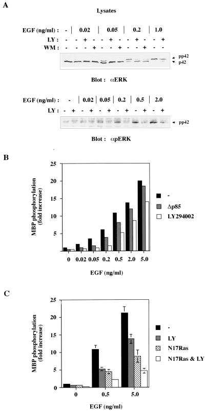
To investigate whether EGF-induced activation of ERK requires PI 3-kinase activity, untransfected COS-7 cells were serum starved for 24 h, pretreated with the PI 3-kinase inhibitor LY294002 or wortmannin, and then challenged with different concentrations of EGF. Phosphorylation of endogenous ERK2 was visualized by immunoblotting as a shift in mobility, with pan-ERK antibodies, or as direct phosphorylation with anti-phospho-ERK antibodies. Pretreatment with the PI 3-kinase inhibitors reduced EGF-induced ERK2 phosphorylation (Fig. 1A); this was most pronounced at low concentrations of EGF (0.02 and 0.05 ng/ml), where total inhibition of ERK2 phosphorylation was observed, and less significant at higher concentrations of EGF. Somewhat reduced phosphorylation of ERK2 could still be seen at 2 ng of EGF per ml.
FIG. 1.
PI 3-kinase inhibitors block ERK2 phosphorylation and activation induced by low concentrations of EGF. (A) Serum-starved COS-7 cells were either left untreated or preincubated with LY294002 (LY; 20 μM) or wortmannin (WM; 100 nM) for 5 min. The cells were then stimulated with increasing concentrations of EGF for 5 min, and 50 μg of lysates was analyzed for ERK2 phosphorylation by mobility shift assays (upper panel) or by using anti-phospho-ERK antibodies (lower panel). (B) Cells, transfected with Myc-ERK2 in the absence or presence of dominant-negative PI 3-kinase Δp85, were serum starved, and some cells were also preincubated with LY294002 (20 μM) for 5 min prior to stimulation with increasing concentrations of EGF for 5 min. The activity of immunoprecipitated ERK2 was analyzed in in vitro kinase assays with MBP as substrate. Data are expressed as fold stimulation, where basal ERK2 activity is defined as 1.0. Representative experiments are shown. (C) Cells were treated as described for panel B, except that dominant-negative N17 Ras was used instead of Δp85. Values are means ± standard errors from at least two separate experiments.
In order to examine whether inhibition of PI 3-kinase also reduced the kinase activity of ERK2, cells were transiently transfected with Myc-tagged ERK2 and stimulated with EGF in the absence or presence of LY294002. Tagged ERK2 was immunoprecipitated and subjected to an in vitro kinase reaction with MBP as substrate. Use of transfected and tagged ERK2 dramatically improved the overall sensitivity and reproducibility of these assays, compared to those of assays done on immunoprecipitated endogenous ERK. Consistent with inhibition of endogenous ERK2 phosphorylation, LY294002 completely inhibited the activation of transfected ERK2 induced by 0.02 to 0.05 ng of EGF (Fig. 1B) per ml. At higher EGF concentrations, the inhibitory effect of LY294002 was less but remained noticeable even at 5 ng of EGF per ml. To further support a role for PI 3-kinase in EGF-induced activation of ERK2, we used Δp85, a dominant-negative form of the regulatory subunit of PI 3-kinase that is unable to bind the catalytic p110 subunit (12). Δp85 is thought to inhibit endogenous PI 3-kinase activation by sequestering upstream stimulatory proteins that bind to p85; this may not inhibit stimulatory pathways feeding directly into p110, although there is evidence that Δp85 does inhibit Ras activation of PI 3-kinase (40). As shown in Fig. 1B, cotransfection of Δp85 with ERK2 also significantly reduced ERK2 activation induced by low concentrations of EGF, although less strongly than with LY294002. These data provide further evidence for a signal strength-dependent contribution of PI 3-kinase activity to growth factor-induced activation of ERK2.
Since it is known that EGF-induced activation of ERK2 can occur via Ras-independent as well as Ras-dependent pathways (7), the effect of coexpression of dominant-negative N17 Ras on the activation of epitope-tagged ERK2 was determined. At both 0.5 and 5.0 ng of EGF per ml, N17 Ras inhibited activation of ERK2 by only about 60% (Fig. 1C). Both calcium and PKC-dependent mechanisms have been suggested to provide Ras-independent pathways connecting EGF to ERK2 (7). LY294002 could further inhibit ERK2 activation above that given by N17 Ras, suggesting that at least part of the PI 3-kinase-sensitive inhibition of ERK2 activation by EGF is acting on Ras-independent pathways. When epitope-tagged wild-type Ras was cotransfected into COS-7 cells with N17 Ras, the elevation of tagged Ras-GTP levels (determined as described below) in response to 25 ng of EGF per ml for 5 min fell from 2.9- ± 0.2-fold to 1.2- ± 0.1-fold, suggesting that under these circumstances N17 Ras could fully inhibit normal Ras function.
Ras stimulation by low concentrations of EGF is inhibited by PI 3-kinase-inhibitory drugs.
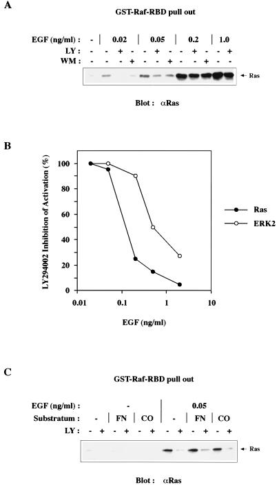
Since Ras plays a central role in the activation of ERK, the effect of inhibition of PI 3-kinase on EGF-induced Ras activation was studied. Serum-starved untransfected COS-7 cells, some pretreated with wortmannin or LY294002, were challenged with increasing concentrations of EGF. Endogenous activated GTP-bound Ras was extracted from lysates with a GST fusion protein containing the amino-terminal Ras-binding domain of Raf (49). The amount of activated Ras in the pullouts was determined by immunoblotting with pan-Ras antibodies. As shown in Fig. 2A, EGF induces a concentration-dependent activation of Ras. In agreement with an earlier report (11), we found that stimulation with about 1 to 2 ng of EGF per ml induced maximal activation of Ras. At 0.02 and 0.05 ng of EGF per ml, the percentage of activated Ras was approximately 10% and 20%, respectively, of that induced by saturating EGF concentrations. Inhibition of PI 3-kinase activity impaired Ras activation induced by low concentrations of EGF. Quantitation of Ras pullouts revealed that at 0.02 and 0.05 ng of EGF per ml, Ras activation was almost completely inhibited by 20 μM LY294002. As was the case for ERK2 phosphorylation and activation, the effect of LY294002 was much less pronounced at higher concentrations of EGF. For example, at 0.2 ng of EGF per ml LY294002 reduced Ras activation by about 25%, whereas at 1 ng of EGF per ml only about 5% inhibition was seen. Thus, at suboptimal concentrations of EGF, PI 3-kinase activity is required not only for ERK activation but also for activation of Ras. Quantitation of the ability of LY294002 to inhibit the activation of endogenous ERK2 and Ras is shown in Fig. 2B.
FIG. 2.
The PI 3-kinase inhibitor LY294002 blocks Ras activation induced by low concentrations of EGF. (A) Serum-starved COS-7 cells were either left untreated or preincubated with LY294002 (LY; 20 μM) or wortmannin (WM; 100 nM) for 5 min and then stimulated with increasing concentrations of EGF for 5 min. Active GTP-bound Ras was extracted from lysates with the GST-Raf-Ras-binding domain (RBD) coupled to glutathione agarose and analyzed by immunoblotting with pan-Ras antibodies. (B) The effect of 20 μM LY294002 on endogenous Ras GTP-loading and endogenous ERK2 activation expressed as percent inhibition of activation seen at different concentrations of EGF. (C) Ras activation is not affected by matrix composition. COS-7 cells were attached to uncoated tissue culture dishes (−) or to dishes that had been precoated with either fibronectin (FN) or collagen (CO). Serum-starved cells were then either left untreated or preincubated for 5 min with LY294002 (LY; 20 μM) before being challenged with 0.05 ng of EGF per ml for 5 min. The amount of GTP-bound Ras in lysates was determined as described for panel A. Representative experiments are shown.
A possible mechanism whereby inhibition of PI 3-kinase activity could impair Ras activation is alteration of integrin function; PI 3-kinase has indirectly been implicated in integrin activation (21, 45), and engagement of integrins has been shown to activate PI 3-kinase (20). To test whether differences in the activity states of various integrins had any effect on EGF-induced Ras activation, we analyzed cells grown on dishes coated with either collagen or fibronectin or ordinary tissue culture plastic. As shown in Fig. 2C, cells grown on each of these matrices exhibited similar levels of Ras activation at 0.05 ng of EGF per ml and the inhibitory effect of LY294002 was not affected by attachment to the different substrata. Cytochalasin D, a drug that disrupts actin filaments and causes loss of focal adhesions, also did not affect Ras activation upon EGF stimulation (data not shown). These data indicate that the effect of LY294002 on Ras activation by EGF is unlikely to be caused by changes in integrin affinity or organization of actin filaments.
Activation of PI 3-kinase is not sufficient to activate ERK2 or Ras.
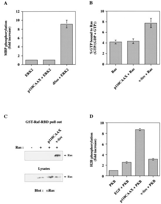
The data from Fig. 1 and 2 suggest a role for PI 3-kinase in EGF-induced activation of Ras and ERK. In order to determine whether PI 3-kinase activation is sufficient to stimulate Ras or ERK, we employed an activated form of PI 3-kinase (p110-CAAX) composed of the catalytic p110α subunit of PI 3-kinase fused to the membrane-targeting farnesylation-palmitylation signal of H-Ras (17). Analysis of PIs from COS-7 cells, transiently transfected with p110-CAAX, revealed a four- to fivefold increase in the levels of PI(3,4,5)P3 relative to control cells (data not shown). In addition, the p110-CAAX construct has recently been shown to be a potent activator of the serine/threonine kinase PKB/Akt (31) and to protect epithelial cells from apoptosis induced by cell detachment (20). We transiently expressed p110-CAAX and epitope-tagged ERK2, immunoprecipitated ERK, and measured its kinase activity. Whereas cotransfection of Drosophila Sos (dSos) increased ERK2 activity ninefold, we could not detect any activation of ERK2 by p110-CAAX (Fig. 3A). Moreover, p110-CAAX was also unable to potentiate ERK2 activation at low concentrations of EGF (data not shown). These results are consistent with recent data showing no effect on ERK activity by various activated forms of p110α (24, 31).
FIG. 3.
An activated form of PI 3-kinase, p110-CAAX, is unable to induce activation of ERK2 or Ras. (A) COS-7 cells were transfected with either Myc-ERK2 alone or in combination with p110-CAAX or Drosophila Sos (dSos). The activity of immunoprecipitated ERK2 was analyzed as described for Fig. 1B. Data are expressed as fold stimulation over basal ERK2 activity. Values are means ± standard errors from at least two separate experiments. (B) Cells, transfected with either Ras alone or in combination with p110-CAAX or v-Src, were incubated for 12 h in medium containing low serum and [32P]orthophosphate. Ras proteins were immunoprecipitated, and bound nucleotides were eluted, resolved by thin-layer chromatography, and quantitated. The amount of Ras-GTP is expressed as percentage of total guanyl nucleotides bound to Ras. Values are means ± standard errors from at least three separate experiments. (C) Cells were transfected as described for panel B and serum starved, and the amount of GTP-bound Ras in lysates was determined as described for Fig. 2A (upper panel). Fifty micrograms of lysates (1/20 of the total amount for each point) was also analyzed for expression of transfected Ras (lower panel). A representative experiment is shown. RBD, Ras-binding domain. (D) Cells were transfected with either HA-PKB alone or in combination with p110-CAAX or v-Src. Some HA-PKB-transfected cells were stimulated with 20 ng of EGF per ml for 5 min. The activity of immunoprecipitated PKB was analyzed in in vitro kinase assays with histone H2B as substrate. Data are expressed as fold stimulation over basal PKB activity. Values are means ± standard errors from at least two separate experiments.
We next examined whether p110-CAAX had any effect on the activation state of Ras by two different assays. Cells were transiently transfected with p110-CAAX and Ras followed by [32P]orthophosphate labelling. Ras proteins were immunoprecipitated, nucleotides were eluted, and the amount of GTP was determined by thin-layer chromatography and PhosphorImager analysis. Alternatively, transiently transfected cells were subjected to pullouts with GST-Raf Ras-binding domain as described above. As shown in Fig. 3B and C, p110-CAAX did not induce any detectable GTP loading of Ras in either of the two assays, whereas v-Src was strongly stimulating. That the p110-CAAX construct was active in transfections was verified in in vitro kinase assays with immunoprecipitated tagged PKB and histone H2B as substrate (Fig. 3D). Thus, although activation of Ras by low concentrations of EGF requires PI 3-kinase activity, an activated form of PI 3-kinase is not sufficient to induce activation of Ras or the downstream kinase ERK2.
Low concentrations of EGF induce association between Grb2 and Shc.
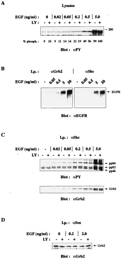
In order to examine the mechanism behind Ras activation induced by EGF, the components upstream of Ras were studied. Cells were stimulated with various concentrations of EGF, and lysates were analyzed for EGF receptor phosphorylation by immunoblotting with phosphotyrosine antibodies. As shown in Fig. 4A, no receptor autophosphorylation was detected at 0.02 ng/ml and only a weak increase in receptor phosphorylation was observed at 0.05 ng of EGF per ml. To induce significant EGF receptor phosphorylation, concentrations of 0.2 ng of EGF per ml or higher had to be used. Pretreatment of cells with LY294002 did not affect autophosphorylation of EGF receptors at any concentration of EGF. To study how stimulation with low concentrations of EGF would affect association between EGF receptors and Grb2 or Shc, cells were lysed, Grb2 or Shc was immunoprecipitated, and complexes were analyzed by immunoblotting with EGF receptor antibodies. As seen in Fig. 4B, stimulation of cells with 0.05 ng of EGF per ml was not sufficient to induce any detectable association between the EGF receptor and Grb2 or Shc. In fact, 10-fold-higher concentrations of EGF had to be used before any association between the EGF receptor and Grb2 became apparent. A longer exposure of the film also revealed a weak association between the EGF receptor and Shc at 0.5 ng/ml (data not shown). These results are consistent with data in Fig. 4A showing that 0.2 to 0.5 ng of EGF per ml is required for significant EGF receptor autophosphorylation to occur. The association between Grb2, Shc, and the EGF receptor was not affected by treatment with LY294002 (data not shown).
FIG. 4.
The PI 3-kinase inhibitor LY294002 does not block EGF-induced EGF receptor (EGFR) autophosphorylation, Shc phosphorylation, or association between Shc and Grb2. Serum-starved COS-7 cells, some preincubated for 5 min with LY294002 (LY; 20 μM), were stimulated with increasing concentrations of EGF for 5 min. (A) Fifty micrograms of lysate was analyzed for EGF receptor autophosphorylation by immunoblotting with phosphotyrosine antibodies. Numbers are expressed as percentages of receptor phosphorylation seen at 5 ng of EGF per ml. (B) EGF-induced association between EGF receptors and Grb2 or Shc was analyzed by anti-Grb2 or anti-Shc immunoprecipitation followed by immunoblotting with EGF receptor antibodies. (C) EGF-induced Shc phosphorylation and association between Shc and Grb2 were analyzed by anti-Shc immunoprecipitation followed by immunoblotting with phosphotyrosine and Grb2 antibodies, respectively. (D) Association between Grb2 and Sos was analyzed by anti-Sos immunoprecipitation followed by immunoblotting with Grb2 antibodies. Representative experiments are shown.
Phosphorylation of Shc and subsequent association with Grb2 remained an alternative possibility for activation of Ras. We therefore stimulated cells with EGF, immunoprecipitated Shc proteins, and analyzed tyrosine phosphorylation of Shc and association with Grb2. Low concentrations of EGF induced a small amount of tyrosine phosphorylation of Shc proteins (Fig. 4C, upper panel) and association between Shc and Grb2 (Fig. 4C, lower panel). Pretreatment of cells with LY294002 did not block EGF-induced phosphorylation of Shc or association between Shc and Grb2. Since a considerable amount of Grb2 is constitutively associated with the exchange factor Sos, one possibility would be that this association depended on PI-3 kinase activity. To clarify this, the interaction between Grb2 and Sos was analyzed under various conditions by immunoprecipitation and immunoblotting. As expected, and as shown in Fig. 4D, similar amounts of Grb2 were found associated with Sos in unstimulated cells, compared to EGF-stimulated cells, and LY294002 treatment did not disrupt this association. Taken together, these data provide circumstantial evidence that phosphorylation of Shc and subsequent association between Shc and Grb2, rather than binding of these adapter molecules to EGF receptors, might be the mechanism behind Ras activation induced by low concentrations of EGF. At concentrations of 0.5 ng of EGF per ml and higher, direct binding of Grb2 or Shc to activated EGF receptors is likely to contribute to maximal Ras activation.
Basal PI 3-kinase activity contributes to EGF-induced Ras activation.
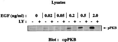
The concentrations of PI 3-kinase inhibitors used in this report together with data derived from using the dominant-negative PI 3-kinase, Δp85, indicate that the PI 3-kinase activity contributing to Ras activation might come from a class IA PI 3-kinase. In order to examine PI 3-kinase activation at different EGF concentrations, we analyzed PKB phosphorylation on serine 473 as a simple and sensitive readout for any changes in PI 3-kinase activity. As seen in Fig. 5, EGF stimulated a marked increase in PKB phosphorylation, and, consistent with earlier data (2, 6), phosphorylation was effectively blocked by LY294002 treatment. However, we were unable to detect any increase in PKB phosphorylation at concentrations below 0.2 ng of EGF per ml. A small amount of basal phosphorylation on PKB serine 473 is present even in serum-starved, unstimulated cells: this phosphorylation disappeared when cells were pretreated with LY294002 (Fig. 5). In addition, LY294002-sensitive hyperphosphorylation of the serine/threonine kinase p70S6K, another kinase downstream of PI 3-kinase, was also observed in COS-7 cells which had been serum starved for 48 h (data not shown), suggesting that there was an appreciable basal level of PI 3-kinase activity in these cells. Furthermore, direct measurement of the lipid product phosphatidylinositol(3,4,5)trisphosphate (PIP3) revealed a constitutive basal level of this lipid in serum-starved COS-7 cells [0.16% ± 0.03% of the PI(4,5)P2] that rapidly disappears (to 0.04% ± 0.02%) following treatment with 20 μM LY294002 for 5 min. Taken together, these data are compatible with the model that the PI 3-kinase activity required for Ras activation by low concentrations of EGF is not induced by EGF itself: it is likely that a serum-deprivation-resistant basal PI 3-kinase activity is required for Ras activation by low EGF concentrations.
FIG. 5.
Low concentrations of EGF do not induce phosphorylation on serine 473 in PKB. Serum-starved COS-7 cells were left untreated or preincubated for 5 min with LY294002 (LY; 20 μM) before being challenged with increasing concentrations of EGF for 5 min. Fifty micrograms of lysates was analyzed for PKB phosphorylation by immunoblotting with anti-phospho-PKB antibodies. A representative experiment is shown.
LY294002 blocks LPA-induced Ras and ERK2 activation in a signal strength-dependent manner.
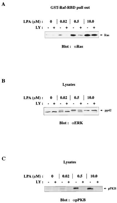
To assess the role of PI 3-kinase in G protein-coupled receptor (GPCR)-induced activation of Ras, we stimulated cells with different concentrations of LPA and analyzed the effects on the Ras-ERK pathway. Ras pullouts from cell extracts revealed that LPA stimulated Ras activation in an LY294002-sensitive manner (Fig. 6A). Interestingly, concentrations as low as 0.005 to 0.01 μM LPA induced detectable levels of Ras activation (data not shown). However, and in contrast to previous data (18), LY294002 only completely inhibited Ras activation induced by low to moderate concentrations of LPA and did not block Ras activation stimulated by 2 μM LPA or higher. The sensitivity of LPA-induced Ras activation to LY294002 appears to be dependent on the signal strength and thus resembles EGF-induced Ras activation. As shown in Fig. 6B, LPA-induced ERK2 phosphorylation was also reduced in a signal strength-dependent manner. In this case, the reduction in ERK phosphorylation correlated better with the LY294002 effect on LPA-induced Ras activation and did not show the differential effects seen with EGF. An interesting observation was that low concentrations of LPA, unlike EGF, stimulated phosphorylation of PKB (Fig. 6C). It is therefore possible that Ras activation, induced at these low concentrations of LPA, is mediated not by basal PI 3-kinase activity but rather by an LPA-activated PI 3-kinase, as has been described recently (18). However, since Ras activation by higher concentrations of LPA is unaffected by LY294002 treatment, it is also possible that related mechanisms lie behind the LY294002-sensitive activation of Ras induced by GPCRs and receptor tyrosine kinases.
FIG. 6.
Low concentrations of LPA induce LY294002-sensitive Ras activation and ERK2 phosphorylation and phosphorylation on serine 473 in PKB. Serum-starved COS-7 cells were either left untreated or preincubated with LY294002 (LY; 20 μM) for 5 min and then stimulated with increasing concentrations of LPA for 5 min. (A) The amount of active Ras in lysates was determined as described for Fig. 2A. RBD, Ras-binding domain. (B) LPA-induced ERK2 phosphorylation analyzed by mobility shift assay. (C) LPA-induced PKB phosphorylation, analyzed as described for Fig. 5. Representative experiments are shown.
DISCUSSION
Confusion has arisen recently about the role of PI 3-kinase in the regulation of the Ras-MAP kinase pathway. In part, this is undoubtedly due to the fact that many different cell types and many different stimuli have been studied, with sometimes apparently contradictory results. In this paper, we have concentrated on the use of EGF in COS cells, with the strength of stimulation being varied by using a wide range of concentrations of the ligand; recently, strength of signal has been implicated as being important in whether PDGF-induced ERK activation is sensitive to PI 3-kinase inhibitors (14). Full stimulation of Ras and ERK by EGF correlates well with optimal mitogenesis (11) but occurs at levels (about 0.5 to 2 ng/ml) very far below those commonly used to stimulate cells in culture (up to 50 ng/ml). Mitogenesis in response to EGF cannot be studied in COS cells as they are partially transformed; however, in normal cultured cells, mitogenesis, Ras activation, and ERK activation all correlate with occupation of the minor high-affinity class of EGF receptors. While high concentrations of EGF cause Ras and ERK stimulation that is not much inhibited by drugs such as LY294002 or wortmannin, or the dominant-negative PI 3-kinase mutant, Δp85, lower concentrations near the amount needed for mitogenesis cause activation of both Ras and ERK that is significantly sensitive to PI 3-kinase inhibition. The fact that the inhibition of ERK is much more marked at intermediate concentrations of EGF (0.2 to 1 ng/ml) than is the inhibition of Ras, which is most clearly seen at very low EGF concentrations (0.02 to 0.1 ng/ml [Fig. 2B]), suggests that the inhibition occurs at two levels, one upstream of Ras and one between Ras and ERK.
The involvement of PI 3-kinase in moderate EGF dose signalling downstream of Ras but upstream of ERK could occur at the level of regulation of Raf, MEK, or ERK. A reasonable possibility is that PI 3-kinase activity promotes Rac activation and thus stimulates PAK, which is able to phosphorylate MEK and may stabilize its interaction with Raf (15). Under conditions of relatively low signal input, this stabilization of the Raf-MEK complex may be essential for effective activation of MEK and hence ERK. At higher levels of EGF, sufficiently strong activation of Raf may alleviate the requirement for this cross talk between PI 3-kinase and MAP kinase activation. However, overexpression of dominant-negative N17 Rac does not appear to be able to inhibit EGF-induced ERK activation in COS-7 cells, suggesting that Rac is not a critical component here (data not shown). A dominant-negative PKB (the pleckstrin homology domain alone) does have some inhibitory effect: this construct probably acts by sequestering PIs, and so the data may not suggest a role for PKB itself in this process of ERK activation (data not shown). Alternatively, redundant signalling pathways may begin to operate at stronger signal strengths: a PKC-mediated pathway via Raf to ERK activation exists (42) and may be used at EGF concentrations sufficient to phosphorylate phospholipase Cγ, an event that requires higher levels of EGF than does Shc phosphorylation (46).
The role of PI 3-kinase activity upstream of Ras in the stimulation of Ras by low levels of EGF is less easily explained. We find that expression of an activated form of PI 3-kinase, p110α-CAAX, does not activate either Ras or ERK, and so PI 3-kinase activity is not sufficient for, but only supportive of, signalling to Ras and ERK (Fig. 3). PI 3-kinase inhibitors do not affect the phosphorylation of the EGF receptor or Shc, or formation of complexes of Shc with Grb2, EGF receptor with Shc, or EGF receptor with Grb2, and so do not cause obvious perturbation of the known signalling pathways leading to Ras activation by Sos (Fig. 4). However, during this analysis we noted that EGF concentrations that cause PI 3-kinase inhibitor-sensitive Ras activation stimulate Shc phosphorylation and Shc-Grb2 complex formation without the autophosphorylation of the EGF receptor; the ability of the EGF receptor to phosphorylate Shc without forming stable complexes with it has been reported previously (11, 46). More significantly, we found that concentrations of EGF that cause PI 3-kinase inhibitor-sensitive activation of Ras actually fail to activate PI 3-kinase itself, at least as measured by the activation state of the PI 3-kinase target PKB (30). LY294002 and wortmannin, pharmacological inhibitors of PI 3-kinase, were found to reduce the level of activity of PI 3-kinase to below the basal level found in a serum-starved cell, both as determined by measuring the activity of the downstream kinase PKB (Fig. 5) or p70S6K (data not shown), which is known to correlate well with PI 3-kinase activity, and by direct measurement of the lipid products PIP3 and PI(3,4)P2. Ras activation at low levels of EGF therefore relies on basal, but not stimulated, PI 3-kinase function: PI 3-kinase does not provide a physiologically regulated signal input to Ras but rather provides an enabling function that is continually present apart from under conditions of pharmacological inhibition. Since EGF receptor-inhibitory drugs such as AG1478 fail to reduce the basal activity of PKB, it is unlikely that the constitutive PI 3-kinase activity is due to the basal activity of the EGF receptor itself (data not shown).
The only circumstance other than inhibitory drug treatment that has been reported where basal PI 3-kinase activity levels drop markedly is that of loss of adhesion to extracellular matrix (20, 22). This may in part account for the strong reduction in growth factor signalling to ERK in detached cells, although a number of different explanations and points of impact on the pathway have been reported (43). The ability of PI 3-kinase to influence MEK activation via PAK may also be important here (38). In the case of PI 3-kinase inhibitor suppression of ERK activation by intermediate doses of EGF, these doses (e.g., 0.2 ng of EGF per ml) are inducing PKB activation and presumably PI 3-kinase activation: the effect of the inhibitors on the component of ERK activation that is operating at a level downstream of Ras may therefore represent a genuine regulatory input of growth factor-controlled PI 3-kinase signals. It should also be noted that EGF couples to ERK activation by both Ras-dependent and Ras-independent mechanisms (7). From the use of a dominant-negative Ras mutant (Fig. 1D), it would appear that at least part of the inhibitory effect of LY294002 on EGF-induced ERK activation acts on Ras-independent pathways.
The nature of the manner in which basal levels of PI 3-kinase activity can aid Ras activation by low EGF concentrations is unknown. The only detectable complex formation induced by these levels of EGF is that of Shc with Grb2-Sos; it is possible that basal PIP3 and PI(3,4)P2 promote the interaction of Shc or Sos with the plasma membrane, aiding colocalization with Ras. It is a possibility, though not directly demonstrated, that the SH2 or phosphotyrosine binding domain of Shc could interact with these PIs (36). Alternatively, the pleckstrin homology domain of Sos could interact with PIP3 and PI(3,4)P2: this has been reported in vitro, although the domain also interacts well with PI(4,5)P2, which is present at much higher concentrations in the plasma membrane (8, 35). According to the standard model for Ras activation (32), it can be assumed that at higher concentrations of EGF, which promote EGF receptor autophosphorylation and complex formation with Shc, Grb2, and Sos, any requirement for membrane localization is provided directly by receptor binding, removing any necessity for PI 3-kinase-produced lipids. Additionally, at higher concentrations of EGF quite different signalling pathways may also come into play: it has recently been shown that activation of PKC leads to stimulation of Ras by an unknown mechanism in COS cells, as had previously been found for T cells (13, 27). Higher concentrations of EGF, which stimulate phospholipase Cγ and PKC in these cells, may utilize multiple pathways to activate Ras. However, it should be noted that the PKC pathway to Ras activation does not appear to function in fibroblasts (7). Since PI 3-kinase activity has also been implicated upstream of PKC (1, 50), probably at the level of phosphorylation by phosphoinositide-dependent protein kinase 1 (3, 4), it is also possible that in cells such as COS cells PI 3-kinase function could be required to maintain PKC activity upstream of Ras stimulation. In T cells, Ras–GTPase-activating protein has been implicated in the regulation of Ras by PKC (13); however, we could find no evidence that PI 3-kinase inhibitors influenced the activity of GTPase-activating proteins in COS cells (52).
Agonists that stimulate ERK activation via heterotrimeric GPCRs, such as those for LPA, thrombin, and α2-adrenergic agonists, mediate ERK activation via release of Gβγ subunits and activation of Ras (48). Depending on cell type, several different tyrosine kinases have been proposed to serve as intermediates between Gβγ subunits and Ras activation, including the Src family kinases Pyk2 and Syk. Transactivation of the EGF receptor has also been put forward as a mechanism for GPCR-induced Ras activation (10). Several of the intermediate steps just upstream of Ras are, in fact, identical between GPCRs and receptor tyrosine kinases and include phosphorylation of Shc, subsequent association between Shc and Grb2, and Sos activation (48). Interestingly, PI 3-kinase activity has also been implicated in LPA-induced activation of Ras (18); in this case, it may be that LPA-induced Ras activation equates to activation by low doses of EGF, possibly through EGF receptor transactivation. However, in Fig. 7 it is shown that higher doses of LPA will overcome the inhibitory effect of LY294002 on Ras activation, just as was seen for EGF. Another possible link between GPCRs and ERK activation has been reported to be PI 3-kinase γ, a neutrophil-specific member of the p110 PI 3-kinase family (26). It is unclear how this enzyme induces ERK activation in COS cell cotransfections, since PI 3-kinase α fails to do so despite causing considerably stronger elevation of PIP3: it is apparent that the ability of PI 3-kinase γ to activate ERK must be quite separate from its lipid kinase activity. This has very recently been confirmed (33), with a protein kinase activity specific to p110γ being most likely to be the key regulatory activity involved. The identity of the substrates for p110γ protein kinase activity is unknown, with the only identified target being p110γ itself; one possible mechanism is for p110γ to act as some sort of scaffolding protein for the MAP kinase pathway, whose activity could be regulated by autophosphorylation. It is currently unclear how broadly relevant such a mechanism might be in the regulation of the MAP kinase pathway since the expression of p110γ appears to be very restricted; however, it is possible that other type IB PI 3-kinases with similar functions that are more widely expressed might exist. Since p110γ does not bind p85, it is unlikely to be able to account for the effects of Δp85 seen in Fig. 1C.
FIG. 7.
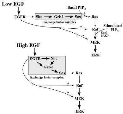
Model for the role of PI 3-kinase in the activation of Ras by EGF. At high concentrations of EGF, stable complexes of Sos form with Grb2, Shc, and the EGF receptor; their activation of Ras and subsequently ERK is not influenced by PI 3-kinase. At low concentrations of EGF, activation of Ras is by Shc-Grb2-Sos complexes which are not EGF receptor associated. Low concentrations of EGF do not stimulate PI 3-kinase activity, but their activation of Ras is dependent on basal PI 3-kinase activity. Another point at which PI 3-kinase may influence ERK activation by low or intermediate concentrations of EGF lies at the level of MEK activation. Ras-independent pathways also link EGF receptor to ERK activation: these may be sensitive to PI 3-kinase inhibition. See the text for details. Thick arrows indicate stable complex formation; thin arrows indicate transient interactions involved in signal transduction or indirect effects.
The experiments reported here indicate that basal PI 3-kinase activity can contribute to the activation of Ras by weak stimuli but that growth factor-regulated PI 3-kinase activity is not required for the activation of Ras. However, PI 3-kinase can also act as a downstream effector of Ras (25, 39–41); low-level activation of Ras is not sufficient to activate PI 3-kinase as a downstream effector (see, for example, Fig. 5), but stronger growth factor-induced stimulation of endogenous Ras (37, 51) or expression of oncogenic Ras (31, 39) can switch on PI 3-kinase. Endogenous Ras function is also required for optimal stimulation of PI 3-kinase by growth factors (23, 39). The work reported in this paper demonstrates that mitogen stimulation of PI 3-kinase does not play a role in the regulation of endogenous Ras protein in mammalian cells, although it can act at a point further down the pathway, upstream of ERK-MAP kinase. Pharmacological inhibition of PI 3-kinase reduces the level of activity of this enzyme to below levels found in quiescent cells, and under these artificial circumstances Ras activation by low doses of EGF can be inhibited. This report goes some way toward clarifying the issue of whether Ras can act upstream of PI 3-kinase or PI 3-kinase can act upstream of Ras, showing that PI 3-kinase does not play a role upstream of Ras in the cell system used here.
ACKNOWLEDGMENTS
We thank Barbara Marte, Michael Czech, Chris Marshall, Boudewijn Burgering, and Paul Coffer for kindly providing DNA constructs. Thanks also go to Patricia Warne, Pablo Rodriguez-Viciana, and Peter Parker for assistance and comments.
S.W. was supported by a European Molecular Biology Organisation Long-Term Fellowship. This work was supported by the Imperial Cancer Research Fund.
REFERENCES
- 1.Akimoto K, Takahashi R, Moriya S, Nishioka N, Takayanagi J, Kimura K, Fukui Y, Osada S, Mizuno K, Hirai S, Kazlauskas A, Ohno S. EGF or PDGF receptors activate atypical PKCλ through phosphatidylinositol 3-kinase. EMBO J. 1996;15:788–798. [PMC free article] [PubMed] [Google Scholar]
- 2.Alessi D R, Andjelkovic M, Caudwell B, Cron P, Morrice N, Cohen P, Hemmings B A. Mechanism of activation of protein kinase B by insulin and IGF-1. EMBO J. 1996;15:6541–6551. [PMC free article] [PubMed] [Google Scholar]
- 3.Bos J L. Ras-like GTPases. Biochim Biophys Acta. 1997;1333:M19–M31. doi: 10.1016/s0304-419x(97)00015-2. [DOI] [PubMed] [Google Scholar]
- 4.Bos J L, Franke B, M’Rabet L, Reedquist K, Zwartkruis F. In search of a function for the Ras-like GTPase Rap1. FEBS Lett. 1997;410:59–62. doi: 10.1016/s0014-5793(97)00324-4. [DOI] [PubMed] [Google Scholar]
- 5.Buday L, Downward J. EGF regulates p21ras through the formation of a complex of receptor, Grb2 adapter protein and Sos nucleotide exchange factor. Cell. 1993;73:611–620. doi: 10.1016/0092-8674(93)90146-h. [DOI] [PubMed] [Google Scholar]
- 6.Burgering B M T, Coffer P J. Protein kinase B (c-akt) in phosphatidylinositol 3-kinase signal transduction. Nature. 1995;376:599–602. doi: 10.1038/376599a0. [DOI] [PubMed] [Google Scholar]
- 7.Burgering B M T, de Vries-Smits A M M, Medema R H, van Weeren P C, Tertoolen L G J, Bos J L. Epidermal growth factor induces phosphorylation of extracellular signal-regulated kinase 2 via multiple pathways. Mol Cell Biol. 1993;13:7248–7256. doi: 10.1128/mcb.13.12.7248. [DOI] [PMC free article] [PubMed] [Google Scholar]
- 8.Chen R H, Corbalan-Garcia S, Bar-Sagi D. The role of the PH domain in the signal-dependent membrane targeting of Sos. EMBO J. 1997;16:1351–1359. doi: 10.1093/emboj/16.6.1351. [DOI] [PMC free article] [PubMed] [Google Scholar]
- 9.Cross D A E, Alessi D R, Vandenheede J R, McDowell H E, Hundal H S, Cohen P. The inhibition of glycogen synthase kinase 3 by insulin or insulin-like growth-factor-1 in the rat skeletal muscle cell line L6 is blocked by wortmannin, but not by rapamycin: evidence that wortmannin blocks activation of the mitogen activated protein kinase pathway in L6 cells between Ras and Raf. Biochem J. 1994;303:21–26. doi: 10.1042/bj3030021. [DOI] [PMC free article] [PubMed] [Google Scholar]
- 10.Daub H, Weiss F U, Wallasch C, Ullrich A. Role of transactivation of the EGF receptor in signalling by G-protein-coupled receptors. Nature. 1996;379:557–560. doi: 10.1038/379557a0. [DOI] [PubMed] [Google Scholar]
- 11.de Vries-Smits A M, Pronk G J, Medema J P, Burgering B M, Bos J L. Shc associates with an unphosphorylated form of the p21ras guanine nucleotide exchange factor mSOS. Oncogene. 1995;10:919–925. [PubMed] [Google Scholar]
- 12.Dhand R, Hara K, Hiles I, Bax B, Gout I, Panayotou G, Fry M J, Yonezawa K, Kasuga M, Waterfield M D. PI 3-kinase: structural and functional analysis of intersubunit interactions. EMBO J. 1994;13:511–521. doi: 10.1002/j.1460-2075.1994.tb06289.x. [DOI] [PMC free article] [PubMed] [Google Scholar]
- 13.Downward J, Graves J D, Warne P H, Rayter S, Cantrell D A. Stimulation of p21ras upon T-cell activation. Nature. 1990;346:719–723. doi: 10.1038/346719a0. [DOI] [PubMed] [Google Scholar]
- 14.Duckworth B C, Cantley L C. Conditional inhibition of the mitogen-activated protein kinase cascade by wortmannin: dependence on signal strength. J Biol Chem. 1997;272:27665–27670. doi: 10.1074/jbc.272.44.27665. [DOI] [PubMed] [Google Scholar]
- 15.Frost J A, Steen H, Shapiro P, Lewis T, Ahn N, Shaw P E, Cobb M H. Cross-cascade activation of ERKs and ternary complex factors by Rho family proteins. EMBO J. 1997;16:6426–6438. doi: 10.1093/emboj/16.21.6426. [DOI] [PMC free article] [PubMed] [Google Scholar]
- 16.Frost J A, Xu S, Hutchison M R, Marcus S, Cobb M H. Actions of Rho family small G proteins and p21-activated protein kinases on mitogen-activated protein kinase family members. Mol Cell Biol. 1996;16:3707–3713. doi: 10.1128/mcb.16.7.3707. [DOI] [PMC free article] [PubMed] [Google Scholar]
- 17.Hancock J F, Magee A I, Childs J E, Marshall C J. All ras proteins are polyisoprenylated but only some are palmitoylated. Cell. 1989;57:1167–1177. doi: 10.1016/0092-8674(89)90054-8. [DOI] [PubMed] [Google Scholar]
- 18.Hawes B E, Luttrell L M, van Biesen T, Lefkowitz R J. Phosphatidylinositol 3-kinase is an early intermediate in the Gβγ-mediated mitogen-activated protein kinase signalling pathway. J Biol Chem. 1996;271:12133–12136. doi: 10.1074/jbc.271.21.12133. [DOI] [PubMed] [Google Scholar]
- 19.Hu Q, Klippel A, Muslin A J, Fantl W J, Williams L T. Ras-dependent induction of cellular responses by constitutively active phosphatidylinositol-3 kinase. Science. 1995;268:100–102. doi: 10.1126/science.7701328. [DOI] [PubMed] [Google Scholar]
- 20.Khwaja A, Rodriguez-Viciana P, Wennstrom S, Warne P H, Downward J. Matrix adhesion and Ras transformation both activate a phosphoinositide 3-OH kinase and protein kinase B/Akt cellular survival pathway. EMBO J. 1997;16:2783–2793. doi: 10.1093/emboj/16.10.2783. [DOI] [PMC free article] [PubMed] [Google Scholar]
- 21.Kinashi T, Escobedo J A, Williams L T, Takatsu K, Springer T A. Receptor tyrosine kinase stimulates cell-matrix adhesion by phosphatidylinositol 3 kinase and phospholipase C-gamma 1 pathways. Blood. 1995;86:2086–2090. [PubMed] [Google Scholar]
- 22.King W G, Mattaliano M D, Chan T O, Tsichlis P N, Brugge J S. Phosphatidylinositol 3-kinase is required for integrin-stimulated AKT and Raf-1/mitogen-activated protein kinase pathway activation. Mol Cell Biol. 1997;17:4406–4418. doi: 10.1128/mcb.17.8.4406. [DOI] [PMC free article] [PubMed] [Google Scholar]
- 23.Klinghoffer R A, Duckworth B, Valius M, Cantley L, Kazlauskas A. Platelet-derived growth factor-dependent activation of phosphatidylinositol 3-kinase is regulated by receptor binding of SH2-domain-containing proteins which influence Ras activity. Mol Cell Biol. 1996;16:5905–5914. doi: 10.1128/mcb.16.10.5905. [DOI] [PMC free article] [PubMed] [Google Scholar]
- 24.Klippel A, Reinhard C, Kavanaugh W M, Apell G, Escobedo M-A, Williams L T. Membrane localization of phosphatidylinositol 3-kinase is sufficient to activate multiple signal-transducing kinase pathways. Mol Cell Biol. 1996;16:4117–4127. doi: 10.1128/mcb.16.8.4117. [DOI] [PMC free article] [PubMed] [Google Scholar]
- 25.Kotani K, Yonezawa K, Hara K, Ueda H, Kitamura Y, Sakaue H, Ando A, Chavaneau A, Calas B, Grigorescu F, Nishiyama M, Waterfield M D, Kasuga M. Involvement of phosphoinositide 3-kinase in insulin or IGF-1 induced membrane ruffling. EMBO J. 1994;13:2313–2321. doi: 10.1002/j.1460-2075.1994.tb06515.x. [DOI] [PMC free article] [PubMed] [Google Scholar]
- 26.Lopez-Ilasaca M, Crespo P, Pellici P G, Gutkind J S, Wetzker R. Linkage of G protein-coupled receptors to the MAPK signaling pathway through PI 3-kinase gamma. Science. 1997;275:394–397. doi: 10.1126/science.275.5298.394. [DOI] [PubMed] [Google Scholar]
- 27.Marais R, Light Y, Mason C, Paterson H, Olson M F, Marshall C J. Requirement of Ras-GTP-Raf complexes for activation of Raf-1 by protein kinase C. Science. 1998;280:109–112. doi: 10.1126/science.280.5360.109. [DOI] [PubMed] [Google Scholar]
- 28.Marshall C J. MAP kinase kinase kinase, MAP kinase kinase and MAP kinase. Curr Opin Genet Dev. 1994;4:82–89. doi: 10.1016/0959-437x(94)90095-7. [DOI] [PubMed] [Google Scholar]
- 29.Marshall C J. Ras effectors. Curr Opin Cell Biol. 1996;8:197–204. doi: 10.1016/s0955-0674(96)80066-4. [DOI] [PubMed] [Google Scholar]
- 30.Marte B M, Downward J. PKB/Akt: connecting PI 3-kinase to cell survival and beyond. Trends Biochem Sci. 1997;22:355–358. doi: 10.1016/s0968-0004(97)01097-9. [DOI] [PubMed] [Google Scholar]
- 31.Marte B M, Rodriguez-Viciana P, Wennström S, Warne P H, Downward J. PI 3-kinase and PKB/Akt act as an effector pathway for R-Ras. Curr Biol. 1997;7:63–70. doi: 10.1016/s0960-9822(06)00028-5. [DOI] [PubMed] [Google Scholar]
- 32.McCormick F. Signal transduction: how receptors turn Ras on. Nature. 1993;363:15–16. doi: 10.1038/363015a0. [DOI] [PubMed] [Google Scholar]
- 33.McLeod S J, Ingham R J, Bos J L, Kurosaki T, Gold M R. Activation of the rap1 GTPase by the B cell antigen receptor. J Biol Chem. 1998;273:29218–29223. doi: 10.1074/jbc.273.44.29218. [DOI] [PubMed] [Google Scholar]
- 34.Pelicci G, Lanfrancone L, Grignani F, McGlade J, Cavallo F, Forni G, Nicoletti I, Grignani F, Pawson T, Pelicci P G. A novel transforming protein (SHC) with an SH2 domain is implicated in mitogenic signal transduction. Cell. 1992;70:93–104. doi: 10.1016/0092-8674(92)90536-l. [DOI] [PubMed] [Google Scholar]
- 35.Rameh L E, Arvidsson A K, Carraway K L, Couvillon A D, Rathbun G, Crompton A, VanRenterghem B, Czech M P, Ravichandran K S, Burakoff S J, Wang D S, Chen C S, Cantley L C. A comparative analysis of the phosphoinositide binding specificity of pleckstrin homology domains. J Biol Chem. 1997;272:22059–22066. doi: 10.1074/jbc.272.35.22059. [DOI] [PubMed] [Google Scholar]
- 36.Rameh L E, Chen C S, Cantley L C. Phosphatidylinositol (3,4,5)P3 interacts with SH2 domains and modulates PI 3-kinase association with tyrosine-phosphorylated proteins. Cell. 1995;83:821–830. doi: 10.1016/0092-8674(95)90195-7. [DOI] [PubMed] [Google Scholar]
- 37.Rausch O, Marshall C J. Tyrosine 763 of the murine granulocyte colony-stimulating factor receptor mediates Ras-dependent activation of the JNK/SAPK mitogen-activated protein kinase pathway. Mol Cell Biol. 1997;17:1170–1179. doi: 10.1128/mcb.17.3.1170. [DOI] [PMC free article] [PubMed] [Google Scholar]
- 38.Renshaw M W, Ren X D, Schwartz M A. Growth factor activation of MAP kinase requires cell adhesion. EMBO J. 1997;16:5592–5599. doi: 10.1093/emboj/16.18.5592. [DOI] [PMC free article] [PubMed] [Google Scholar]
- 39.Rodriguez-Viciana P, Warne P H, Dhand R, Vanhaesebroeck B, Gout I, Fry M J, Waterfield M D, Downward J. Phosphatidylinositol-3-OH kinase as a direct target of Ras. Nature. 1994;370:527–532. doi: 10.1038/370527a0. [DOI] [PubMed] [Google Scholar]
- 40.Rodriguez-Viciana P, Warne P H, Khwaja A, Marte B M, Pappin D, Das P, Waterfield M D, Ridley A, Downward J. Role of phosphoinositide 3-OH kinase in cell transformation and control of the actin cytoskeleton by Ras. Cell. 1997;89:457–467. doi: 10.1016/s0092-8674(00)80226-3. [DOI] [PubMed] [Google Scholar]
- 41.Rodriguez-Viciana P, Warne P H, Vanhaesebroeck B, Waterfield M D, Downward J. Activation of phosphoinositide 3-kinase by interaction with Ras and by point mutation. EMBO J. 1996;15:2442–2451. [PMC free article] [PubMed] [Google Scholar]
- 42.Schonwasser D C, Marais R M, Marshall C J, Parker P J. Activation of the mitogen-activated protein kinase/extracellular signal-regulated kinase pathway by conventional, novel, and atypical protein kinase C isotypes. Mol Cell Biol. 1998;18:790–798. doi: 10.1128/mcb.18.2.790. [DOI] [PMC free article] [PubMed] [Google Scholar]
- 43.Schwartz M A, Schaller M D, Ginsberg M H. Integrins: emerging paradigms of signal transduction. Annu Rev Cell Dev Biol. 1995;11:549–600. doi: 10.1146/annurev.cb.11.110195.003001. [DOI] [PubMed] [Google Scholar]
- 44.Seger R, Krebs E G. The MAPK signaling cascade. FASEB J. 1995;9:726–735. [PubMed] [Google Scholar]
- 45.Shimizu Y, Mobley J L, Finkelstein L D, Chan A S. A role for phosphatidylinositol 3-kinase in the regulation of beta 1 integrin activity by the CD2 antigen. J Cell Biol. 1995;131:1867–1880. doi: 10.1083/jcb.131.6.1867. [DOI] [PMC free article] [PubMed] [Google Scholar]
- 46.Soler C, Alvarez C V, Beguinot L, Carpenter G. Potent SHC tyrosine phosphorylation by epidermal growth factor at low receptor density or in the absence of receptor autophosphorylation sites. Oncogene. 1994;9:2207–2215. [PubMed] [Google Scholar]
- 47.Stephens L R, Jackson T R, Hawkins P T. Agonist-stimulated synthesis of phosphatidylinositol(3,4,5)-trisphosphate: a new intracellular signalling system. Biochim Biophys Acta. 1993;1179:27–75. doi: 10.1016/0167-4889(93)90072-w. [DOI] [PubMed] [Google Scholar]
- 48.Sugden P H, Clerk A. Regulation of the ERK subgroup of MAP kinase cascades through G protein-coupled receptors. Cell Signal. 1997;9:337–351. doi: 10.1016/s0898-6568(96)00191-x. [DOI] [PubMed] [Google Scholar]
- 49.Taylor S J, Shalloway D. Cell cycle-dependent activation of Ras. Curr Biol. 1996;6:1621–1627. doi: 10.1016/s0960-9822(02)70785-9. [DOI] [PubMed] [Google Scholar]
- 50.Toker A, Meyer M, Reddy K K, Falck J R, Aneja R, Aneja S, Parra A, Burns D J, Ballas L M, Cantley L C. Activation of protein kinase C family members by the novel phosphoinositides PI-3,4-P2 and PI-3,4,5-P3. J Biol Chem. 1994;269:32358–32367. [PubMed] [Google Scholar]
- 51.van Weering D H, de Rooij J, Marte B, Downward J, Bos J L, Burgering B M. Protein kinase B activation and lamellipodium formation are independent phosphoinositide 3-kinase-mediated events differentially regulated by endogenous Ras. Mol Cell Biol. 1998;18:1802–1811. doi: 10.1128/mcb.18.4.1802. [DOI] [PMC free article] [PubMed] [Google Scholar]
- 52.Wennström, S., and J. Downward. Unpublished observations.


