



Abstract
Background
Molecular defects in the SHOX gene including deletions, duplications or pathogenic point mutations are responsible for well-known pathologies involving short stature as a clinical manifestation: Léri–Weill dyschondrosteosis, Langer mesomelic dysplasia, Turner syndrome or idiopathic short stature. Duplications flanking the SHOX gene (upstream or downstream of the intact SHOX gene involving conserved non-coding cis-regulatory DNA elements - CNEs) have been described but their clinical involvement is still difficult to understand.
Results
We describe two cases with short stature and normal GH-IGF1 status. Multiplex ligation-dependent probe amplification (MLPA) and array comparative genomic hybridization (arrayCGH) identified in both cases heterozygous duplications involving downstream regions of SHOX gene, within CNEs (CNE8, CNE9 and CNE4, CNE5, CNE6, ECR1, CNE8, CNE9 and surrounding areas, respectively). One of the cases showed a maternally inherited duplication. Although every case has several particularities, we consider that duplications in these non-coding regions of SHOX gene may explain the short stature phenotype.
Conclusion
To our knowledge, these are the first Romanian-reported cases of ISS with a large duplication of downstream SHOX enhancers CNEs region. The spectrum of phenotypic consequences and the exact mechanism of the presumed clinical expression of these genetic alterations still needs to be evaluated and described.
Keywords: idiopathic short stature, SHOX, regulatory element, deletion, duplication, CNE
INTRODUCTION
The short-stature homeobox-containing gene (SHOX) is located on both sexual chromosomes (Xp22.33 and Yp11.2) within the pseudoautosomal region 1 (PAR1), where each chromosome contains the same DNA region.
SHOX is expressed in osteoblasts, bone marrow fibroblasts and hypertrophic chondrocytes in the human embryo since the second month of gestation and in the growth plate in the postnatal period; its protein controls indirectly the chondrocyte proliferation, differentiation and apoptosis by regulating other genes. SHOX interacts with the SOX trio (SOX9, SOX5 and SOX6 genes) and thus regulates aggregan, with an important role in chondrogenesis and cartilage matrix synthesis (1,2). It also blocks FGFR3, a negative regulator of chondrogenesis and stimulates NPR2/NPPC activation, thus controlling chondrocyte proliferation and differentiation and synthesis of cartilage matrix (3-5).
SHOX haploinsufficiency is responsible for Léri–Weill dyschondrosteosis (LWD; OMIM #127300) and for the short stature and skeletal anomalies in Turner syndrome (6-8). Genetic alterations of both alleles of the SHOX gene cause Langer mesomelic dysplasia (LMD; OMIM #249700), characterized by disproportionate severe short stature with mesomelic and rhizomelic shortening of the upper and lower limbs. (8,9). Both heterozygous deletion or point mutations (homozygous or compound heterozygous status) of SHOX are described in the literature in 1.5 - 15% of idiopathic short stature cases (ISS, OMIM 300582)(10,11).
SHOX gene has 7 exons (1-6 a,b) and its transcripts are represented by SHOXa encoded by exons 1-5 and 6a, and SHOXb, encoded by exons 1-5 and 6b (12). As for most developmental genes, the minimal promoter region of SHOX requires the assistance of cis-regulatory elements in PAR1 (13-15)
Thus, SHOX is surrounded by some conserved non-coding DNA elements (CNEs) [alias, evolutionarily conserved sequences (ECS), evolutionarily conserved regions (ECR)] with either enhancer or repressor functions, called regulatory elements of genes, as they are essential for controlling gene expression at different levels in different tissues (15,16).
Four of these regulatory CNEs are described downstream and have been demonstrated to have transcriptional activity: CNE4, CNE5, CNE7/ECR1 and CNE9/ECS4(14,17,18). Another 2 regions with regulatory activity but not always highly conserved across species have been recently described: ZED (Zeugopodal Enhancer Downstream of SHOX), located at X:827,128–827,691 (19) and a potential additional regulatory element at approximately X:970,000(20,21).
Three CNEs with regulatory function are situated upstream: CNE-5, CNE-3 and CNE-2 (22) another three CNEs, situated also downstream, are not shown to have any regulatory function upon SHOX transcription (CNE2, CNE3, and CNE6).
CNVs (copy number variations) that disrupt CNEs could potentially have phenotypic effects similar to those of changes in the gene itself. Moreover, changes in non-coding regions may reduce gene expression and cause incorrect splicing of messenger RNA (mRNA) or misfolding of polypeptide strands (23, 24). SHOX downstream deletions can lead to the haploinsufficiency of the gene. Other dosage anomalies such as duplication affecting SHOX downstream and upstream areas are more difficult to interpret and are rarely reported in the literature.
MATERILAS AND METHODS
Cases description
Case 1. A 7 years and 1-month-old Caucasian girl was admitted to the Pediatric Endocrinology Department for short stature. The child has been adopted since her first month of life, so few familial auxologic or health status data were available. She was prematurely born at 28 weeks of gestation with a birth weight of 1400 g (+1.67 SDS), 41 cm length (+1.59 SDS), head circumference and Apgar scores unavailable. As the pregnancy was not monitored and the mother had a precarious socio-economic situation, the infant was tested for congenital syphilis and she had a positive serum Venereal Diseases Research Laboratory (VDRL); she was treated with intravenous aqueous penicillin G, as protocols; no other medical data from newborn period or small infancy were recorded. At age 3 years malabsorption and digestive pathology were excluded.
Early motor development in infancy was slightly delayed: she held her head up at 7 months of age, sat unassisted at 10 months, and walked independently at 2 years. Speech development was also delayed, with first words at 3 years. She received physiotherapy from the age of 6 months, and she participated in special education considering her speaking disability.
The patient has been repeatedly evaluated in our department at age 4, age 4 years 11 months and 5 years and 11 months respectively, for short stature – her height was at -2.2 to -2.4 SDS and the BMI was < 1% on all occasions. She had normal biological parameters, except for mild normocytic normochromic anaemia with normal iron load, normal thyroid function and normal IGF1.
Clinical examination revealed: height (H) 112.6 cm (-2.02 SD, Romanian References), weight 15 kg, BMI 11.8 Kg/sqm (-3.89 SD, Romanian References)(25) average growth velocity 6.1 cm/year (0.16 SD) (Prader A); sitting height (SH) 61 cm, SH/H ratio 54.2%, arm span 107.8 cm, arm span /total height 95.7%, no skeletal abnormalities (Rappold score 2)(26); mild craniofacial dysmorphism with moderate microcephaly, high forehead, upslanting palpebral fissures, retrognathism, high-arched palate, no pigmented skin spots; pubertal stage B1/P1 (Tanner-Marshall method).
The biochemical tests revealed: mild normocytic anaemia (Hb=11.1 g/dL) with normal iron load, normal hepatic and renal function and normal phospho-calcic profile. Thyroid and adrenal function were normal. The somatotropic axis assay showed an IGF1 value at the lower limit (88.7 ng/mL, SDS -0.46) and a normal response to the clonidine test for GH secretion (21.7 ng/mL). Pituitary MRI showed normal hypothalamus and a slightly decreased pituitary gland. The bone age was 5 years, without Madelung deformity or other carpal radiological abnormalities.
Both cardiologic and ophthalmologic examinations were normal. Psychologic examination at age 7.1 years revealed that the proband had a development delay regarding socialization (4.4 years), language (4.2 years), auto-service (4.2 years), cognition (4.4 years), motor skills (5.3 years) and IQ of 75.
Case 2. The second patient was a Caucasian boy full-term born after an uncomplicated pregnancy, with an Apgar score of 9, a birth weight of 2600 g (−1.6 SDS) and a length of 48 cm (−0.7 SDS). He was the only child of non-consanguineous parents. The mother’s height was 149.5 cm (-2.5 SDS) and the father’s height was 172 cm (-0.7 SDS), with a parental target height of 169 cm (-1.18 SDS). Maternal ancestors (maternal grandmother and maternal aunt) had also short stature, with an adult height of 150 cm (- 2,4 SDS).
The boy had normal psychomotor development in infancy and small childhood, without any learning difficulties.
After 3 years of age, his average growth velocity was 5 cm/year, with growth deceleration between age 3 (H = 92 cm, SDS -1.77) and age 6 years and 10 months (H=110 cm, SDS -2.39).
The abdominal ultrasound has been repeatedly normal, with normal echocardiography. Malabsorption and digestive pathology were excluded.
At his referral to the Pediatric Endocrinology Department (age 7 years 2 months) he had short stature (H=112.3 cm, SDS - 2.3 SDS, Romanian References (25)), with normal BMI (16.4 Kg/m2, SDS +0.4); sitting height (SH) 60.9 cm, SH/H ratio 54.2%, arm span 114.5 cm and arm span /total height 101 %. The physical examination revealed prepubertal status, broad shoulders, muscular hypertrophy and mild craniofacial dysmorphism (mild microcephaly, micrognathia, thin lips, down slanting palpebral fissures), dentin dysplasia (Rappold Score = 3) (26).
Biochemical tests were normal. His endocrine evaluation revealed normal thyroid and adrenal function, normal IGF1 (124.4 ng/mL, + 0.18 SDS) and a peak normal growth hormone value (20.5 ng/mL) at the clonidine test.
His bone age at a chronological age of 7 years and 2 months was 6.5 years (Greulich–Pyle method), and the X-ray examination ruled out any radiological deformities.
Genetic analysis
Whole blood EDTA and heparin samples were collected with approval from the Institutional Ethics Committee and family consent. Cytogenetic analysis was carried out using G-banding and the result showed a normal female karyotype 46,XX in case 1 and a normal male karyotype (46,XY) in case 2. Case´s 2 Mother refused karyotyping, as she was a fertile woman with a female phenotype.
The genomic DNA from the whole blood EDTA sample was isolated using Wizard® SV Genomic Purification System (Promega Corporation, Madison, WI, USA).
SHOX genetic analysis
Copy number variations (CNVs) analysis involving SHOX gene and SHOX-area (PAR1 enhancer regions) was performed using SALSA Multiplex ligation based amplification (MLPA) Probemix P018-G2 SHOX kit (MRC-Holland, Amsterdam, The Netherlands) following manufacturer’s protocols with a starting template DNA of 200 ng measured by DeNovix DS-11 spectrophotometer (DeNovix Inc., Wilmington, DE, USA).
SALSA MLPA Probemix P018-G2 SHOX kit includes 32 probes for the PAR1 region on chromosome Xp22.33/Yp11.32, including at least one probe for each exon of SHOX transcript variant 1, and one probe for intron 6 only present in the SHOXb splice variant. Several probes are present for SHOX regulatory regions, located upstream and downstream of SHOX. Moreover, this probemix contains multiple flanking probes targeting the X chromosome outside the SHOX area: one probe detecting the area just before the SHOX upstream regulatory regions, five probes inside the PAR1 region but downstream of the SHOX area, and seven probes outside of PAR1. Flanking probes can be used to characterize larger deletions/duplications and to distinguish SHOX deletions from a Turner syndrome karyotype. In addition, nine reference probes are included that detect autosomal chromosomal locations.
The experiment included three reference DNA samples, as recommended. Briefly, the sample DNA with sufficient buffering capacity by dilution in TE0.1(10 mM Tris-HCl pH 8.0 + 0.1 mM EDTA) is denatured (5 min, 98°C), hybridized overnight at 60°C with the SALSA probemix, then treated with Ligase-65 enzyme for 15 min at 54°C followed by incubation at 98°C for 5 min to stop the reaction. PCR amplification is then carried out with specific SALSA PCR primers. Fragment analysis of PCR products was run on a CEQ8000 Genetic Analyzer (Beckman Coulter, Fullerton, CA). Coffalyser.Net™ version v.220513.1739 was used to analyze raw data sequencing files. After intra- and intersample normalization using reference probes and reference samples, results are expressed as a final ratio (FR), with a normal copy number between 0.80-1.20, heterozygous deletion with a FR between 0.40-0.65, homozygous deletion with FR=0, heterozygous duplication for FR between 1.30-1.65 and homozygous duplication FR, 1.75-2.15. In our cases, the copy number changes were confirmed by repeating the MLPA analysis in a separate run.
Array comparative genomic hybridization (Array-CGH) analysis
For a more specific characterization of SHOX duplication lengths and positioning we performed array-CGH using SurePrint G3 Human CGH + SNP Microarray 4x180K (Agilent Technologies, Santa Clara, CA, USA) according to the manufacturers’ recommendations. Agilent SureTag DNA Labeling Kit was used to label patients’ DNA and sex-matched control DNA (Agilent's Human DNA Male and Female) with either cyanine 3 or cyanine 5. 500 ng of genomic DNA was used for the labeling reaction. DNA fragmentation was performed by double enzymatic digestion (Alu I+ Rsa I). Patients DNA and control DNA were labelled by random priming with Cy5-dUTP and Cy3-dUTP, respectively. Cyanine 3 and cyanine 5 labeled samples were purified, mixed and hybridized to Agilent microarrays from the kit at 67°C for 24h at 20 rpm.
Microarrays were scanned using an Agilent DNA Microarray Scanner. Intensities were extracted from the raw image files using Agilent's Feature Extraction software and the data analysis was performed through the Agilent CytoGenomics v5.3 software. For each spot of the array, the value of log2 ratios between the Cy5-labeled test DNA and the Cy3-labeled reference DNA were calculated. CNVs were detected using the ADM-2 algorithm with filters of minimal size of 100 kb in region and > 5 Mb of copy-number neutral loss of heterozygosity regions (All genomics coordinates were established using the human genome as defined by the UCSC build hg19 (GRCh37).
The obtained variants were evaluated for possible pathogenicity based on their presence in various databases: UCSC Genome Browser (https://genome.ucsc.edu/cgi-bin/hgGateway), Database of Genomic Variants (DGV (http://dgv.tcag.ca/dgv/app/home), and Database of Genomic Variation and Phenotype in Humans using Ensembl Resources (DECIPHER).
RESULTS
MLPA using the P018-G2 probemix revealed a large heterozygous duplication downstream of the SHOX gene in both children (table 1), of at least 150 kb in patient 1 and at least 212 kb in patient 2. The same duplication was identified in the mother of the 2nd case (the genetic analysis was performed according to the phenotype: the mother was short stature, the father had a normal height; the boy had a toddler brother with height -1.5 SDS, he was not yet evaluated, Figure 1).
Table 1.
Length and position of duplications within SHOX-area in the two cases according to genetic evaluation
| Method | Referrence | Position | Length |
|---|---|---|---|
| Case 1 | |||
| arrayCGH | hg19 (GRCh37) | arr[GRCh37]Xp22.33 (795717-961601)x3 | 165.85 kb |
| MLPA | hg19 (GRCh37) | dup het Xp22.33 (809260-963670) | 154.4 kb |
| Case 2 | |||
| arrayCGH | hg19 (GRCh37) | arr[GRCh37]Xp22.33 (689188-970558)x3 | 281.37 kb |
| MLPA | hg19 (GRCh37) | dup het Xp22.33 (751590-963680) | 212.09 kb |
Figure 1.
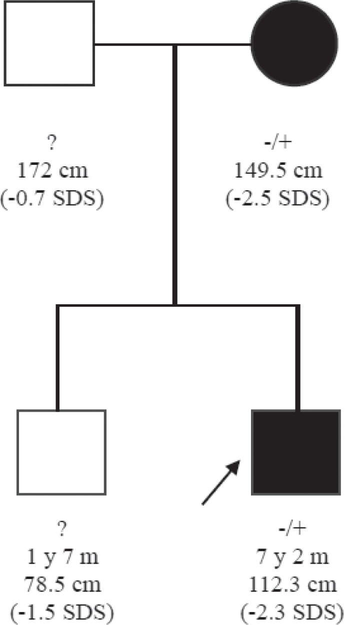
Pedigree of the second patient. The proband is indicated by arrow. -/+ indicate the wild type and the variant alleles.
The duplications were confirmed using arrayCGH (Fig. 2), which also excluded other genomic alterations as causes of short stature, tagged by the aCGH.
Figure 2.

Genomic context of highly conserved non-coding DNA elements (CNEs) in SHOX upstream and downstream regions and indication of lengths and positioning of duplications in our patients. The upper scale illustrates the distance from the Xp/Yp telomere (hg19/GRCh37) (adapted from https://www.ncbi.nlm.nih.gov/genome/gdv/browser/gene/?id=108410392).
The duplication in case 1 was located on the Xp22-PAR1 chromosome region from X:795,717 to X:961,601 and encompassed highly conserved non-coding DNA elements (CNEs) with enhancer activity: CNE8 (chrX:811,550-812,300, 751 bp), CNE9 (chrX:816,903-835,698, 18,796 bp), ZED and downstream areas (Fig. 2). In case 2, there was a larger heterozygous duplication downstream of the SHOX gene: Xp22.33 (689,188-970,558), including several regulatory elements: CNE4 (chrX:714,084-714,753, 670 bp), CNE5 (chrX:750,824-751,850, 1,027 bp), ECR1/CNE7 (chrX:780,581-781,235, 655 bp), CNE8 (chrX:811,550-812,300, 751 bp), ZED (X:827,128–827,691), CNE9 (chrX:816,903-835,698, 18,796 bp) and downstream areas (Fig. 2).
Notably, duplications in both patients encompassed an area of at least 100kb distal last known SHOX enhancer (CNE9).
DISCUSSION
Molecular defects in SHOX gene including deletions, duplications or pathogenic point mutations affecting SHOX production are responsible for well-known pathologies involving short stature as a clinical symptom: Léri–Weill dyschondrosteosis (LWD; OMIM ID#127300), Langer mesomelic dysplasia (LMD; OMIM ID#249700), Turner syndrome or idiopathic short stature (ISS; OMIM ID#300582). In these cases, SHOX defects are associated with affected bone development and impaired longitudinal body growth.
Duplications of the SHOX gene are variably associated with short, normal, or even tall stature. Normal or tall stature has been described in sex chromosome trisomy, in large structural anomalies involving duplications of PAR1 and SHOX (usually associated with gonadal dysgenesis and therefore prolonged skeletal growth), but also in isolated SHOX duplications with normal gonadal function (27,28). Duplications of the SHOX gene are also described in ISS and LWD (Table 2), as they appear to reduce gene expression by changing the distance between the promoter and the enhancer regions – this might disrupt their interaction, resulting in a transcription block, lack of gene activation, incorrect splicing of messenger RNA (mRNA) or misfolding of polypeptide strands and thus functional SHOX haploinsufficiency and short stature (29).
Table 2.
Duplications of upstream and downstream SHOX CNEs and their phenotypes - literature data (IUGR-intrauterine growth retardation, NA – not available)
| Author, year | SHOX and SHOX-area duplication size and localization | Methodology | Clinical phenotype | X Ray | ||
|---|---|---|---|---|---|---|
| Type | Localization | Size | ||||
| Iughetti, 2010 (39) | dup | exon 1 SHOX gene to enhancer | 500 kb | MLPA | ISS | Normal |
| Benito-Sanz, 2011 (20) | dup | SHOX gene 5’ region | MLPA | ISS | Normal | |
| Caliebe, 2012 (32) | dup | 647,566–915,079 bp | 267.5 kb | MLPA | IUGR; microcephaly, SS | NA |
| dup | Xp22.33 (779,805–915,079 bp); 239.4 kb proximal of SHOX | 135.3 kb | MLPA | ISS | NA | |
| Hirschfeldova, 2012 (40) | dup | CNE9 | MLPA, SHOX sequencing | LWD – proportional short stature, (-2.5 SD), short forearm, cubitus valgus, and muscular hypertrophy | NA | |
| Sandoval, 2014 (34) | dup | CNE9 | MLPA, aCGH | ISS, disproportionate | NA | |
| Fukami, 2015 (16) | dup | CNE-2 (upstream). Exons 1-6b, CNE4, CNE5 (downstream) | MLPA | ISS | Normal | |
| dup | Exons 1-6b, CNE4, CNE5, ECR1 and CNE9/ECS4 (downstream) | MLPA | ISS | Normal | ||
| dup | CNE-2, CNE-3, CNE-5 (upstream), Exons 1-3 | MLPA | ISS | Normal | ||
| dup | CNE9 | MLPA | ISS | Normal | ||
| Bunyan, 2016 (36) | dup | CNE2, CNE3, CNE4, CNE5, CNE6, ECR1,CNE8, ECS4(CNE9) and possibly X:970,000 | MLPA, aCGH | Short stature, LWD, Disproportionate limbs | bilateral Madelung deformity | |
| dup | CNE8, ECS4 (CNE9) and X:970,000 | Short stature, LWD – Disproportionate limbs | bilateral Madelung deformity | |||
| dup | CNE8, ECS4 (CNE9) and X:970,000 | Short stature, LWD Disproportionate limbs, bowing of forearms | bilateral Madelung deformity | |||
| dup | ECS4 (CNE9) X:821,993-961,522 (hg19) | 113-140 kb | ISS; short stature | Normal | ||
| dup | X:970,000 (and possibly affects ECS4) | Short stature, LWD Mesomelia | bilateral Madelung deformity | |||
| dup | CNE-3, CNE-2 | Short stature, LWD | Bilateral Madelung deformity | |||
| Shima, 2016 (37) | dup | SHOX Exons 1–3 and upstream CNE-5, CNE-3, and CNE-2 | ISS | Normal | ||
| dup | Entire SHOX gene and upstream CNE-2, downstream CNE4 and CNE5 | ISS | Normal | |||
| dup | Entire SHOX gene and downstream CNE4, CNE5, ECR1 and CNE9/ECS4 | ISS | Normal | |||
| dup | CNE9 | ISS | Normal | |||
| dup | Downstream region | ISS | Normal | |||
| Hirschfeldova, 2017 (35) | dup | Large dup SHOX gene and downstream regulatory elements | MLPA | ISS | Normal | |
| dup | CNE9 (different sizes) | MLPA | ISS | Normal | ||
| dup | Large dup Intron 6 SHOX gene, CNE3, 4, 5, 7, 8, 9 | MLPA | ISS | Normal | ||
| dup | Large dup CNE4, 5, 7, 8, 9 | MLPA | ISS | Normal | ||
| dup | CNE-2, CNE-5 (upstream) | MLPA | ISS | Normal | ||
| Capkova, 2020 (41) | dup | Downstream regulatory elements, not detailed | MLPA, FISH, Sanger Sequencing | ISS | Normal | |
| Fanelli A, 2022 (38) | dup | chrX:917,600 - 1,030,000 | aCGH and breakpoint PCR | LWD | Normal | |
Duplications in regulatory enhancer regions upstream and downstream SHOX genes are rarely found, and they have variable lengths, localizations, and phenotypic consequences, not always explained.
Duplications of upstream SHOX CNEs have been reported in the literature in patients with ISS and LWD (Table 2) (14,30-32).
Duplications of downstream SHOX CNEs - CNE9 - not including SHOX itself have first been reported in a patient with LWD in 2012 (33). After that, isolated CNE9 duplications were identified in ISS patients and patients with LWD (14, 32, 34, 35), underlying CNE9 implication in SHOX expressivity. The duplicated PAR1 area associated with short stature expanded in other studies upstream CNE9 (CNE2, CNE3, CNE4, CNE5, CNE7/ECR1, CNE8) or downstream CNE 9, to presumably highly-conserved across species regions: chrX:970,000(20) or chrX:917,600 - 1,030,000 (36) (Table 2).
A recent study evaluating the downstream SHOX-area found next to CNE9 a zeugopodal enhancer (ZED) - an 11.5 kb genomic fragment that does not contain any of the highly conserved sequences previously suspected to function as SHOX limb enhancers (CNE7, CNE8 or CNE9) (19). The authors showed that a fragment of only 563 bp is responsible for ZED enhancer activity. Although ZED’s location and specific activity qualify him as a discrete SHOX limb enhancer, several other reports of CNVs in ISS patients implicate other genomic regions in the cis-regulation of SHOX (36).
Besides isolated case reports describing the implication of downstream SHOX enhancers in ISS and LWD (Table 2), microduplications in upstream and downstream SHOX have also been described in the control normal stature population, except for: duplications encompassing the CNE7-CNE9 region (including zeugopodal enhancer situated next upstream to CNE9) and another region situated distal to CNE9, identified as chrX:970,000 (20) or as a region with high evolutionary conservation of approximately 112 kb (chrX:917,600–1,030,000) (36) – duplications of these regions are shown to be more frequent in patients compared to population samples (20, 33, 36).
Taken together (Table 2), studies have described a higher frequency of CNVs in the downstream elements of SHOX than in its upstream elements and also a higher frequency of duplications compared to deletions, especially in patients with LWD, thus sustaining the impact of downstream regulatory elements of SHOX on the growing skeleton (13, 15, 32, 35, 37, 38).
In order to evaluate the prevalence and associated phenotypic effects of the identified downstream CNVs, we also searched for duplications of these regions in the Database of Genomic Variants (DGV) and Database of Chromosomal Imbalance and Phenotype in Humans Using Ensembl Resources (Decipher). In Decipher there are 151 patients reported overlapping our downstream duplicated region for patient 2. We selected a short list of approximately 500 kb duplication regions overlapping our duplications (Table 3). The majority of these CNVs are reported as likely pathogenic and are observed in patients with short stature, intellectual disability or autism.
Table 3.
List of ≈ 500 kb duplication region overlapping our duplications (Database of Genomic Variants (DGV) and Database of Chromosomal Imbalance and Phenotype in Humans Using Ensembl Resources (Decipher), www.deciphergenomics.org) – cases associated with short stature are highlighted
| No | Decipher patient number | Duplication length | Location GRCh37/hg19 | Overlap with patient 1 | Overlap with patient 2 | Patogenicity | Inheritance | Reported phenotype |
|---|---|---|---|---|---|---|---|---|
| 1 | 501685 | 578 kb | chrX:430431-1008430 | 165.8 kb | 281.3 kb | likely pathogenic | unknown | tall stature |
| 2 | 321306 | 575.6 kb | chrX:444444-1020083 | 165.8 kb | 281.3 kb | unspecified | unknown | proportionate short stature |
| 3 | 327886 | 575.6 kb | chrX:444444-1020083 | 165.8 kb | 281.3 kb | unspecified | maternally | neurodevelopmental delay |
| 4 | 472564 | 417.9 kb | chrX:458034-875989 | 78.2 kb | 186.8 kb | pathogenic | unknown | |
| 5 | 278988 | 537.1 kb | chrX:481940-1019080 | 165.8 kb | 281.3 kb | unspecified | unknown | intellectual disability, moderate. macrocephaly |
| 6 | 378974 | 537.1 kb | chrX:481940-1019080 | 165.8 kb | 281.3 kb | uncertain | unknown | cognitive impairment |
| 7 | 279033 | 327.9 kb | chrX:481940-809860 | 12.1 kb | 120.6 kb | unspecified | unknown | intellectual disability, moderate |
| 8 | 367150 | 327.9 kb | chrX:481940-809860 | 12.1 kb | 120.6 kb | uncertain | unknown | cognitive impairment, generalized hypotonia. poor coordination. tall stature |
| 9 | 307967 | 488.8 kb | chrX:481940-970747 | 165.8 kb | 281.3 kb | pathogenic | unknown | short stature, obesity |
| 10 | 292734 | 516.2 kb | chrX:481940-998152 | 165.8 kb | 281.3 kb | uncertain | maternally | microtia |
| 11 | 501321 | 587.7 kb | chrX:505654-1232802 | 165.8 kb | 281.3 kb | pathogenic | unknown | short stature |
| 12 | 253249 | 407.3 kb | chrX:510558-917875 | 120.1 kb | 228.6 kb | unspecified | inherited from normal parent | intellectual disability |
| 13 | 472225 | 314.1 kb | chrX:515011-829150 | 31.4 kb | 139.9 kb | pathogenic | unknown | |
| 14 | 398712 | 582.72 kb | chrX:535224-1257358 | 165.8 kb | 281.3 kb | uncertain | unknown | brachydactyly, short stature |
| 15 | 262464 | 290.5 kb | chrX:545753-836276 | 38.5 kb | 147 kb | unspecified | inherited from normal parent | intellectual disability |
| 16 | 488457 | 357.6 kb | chrX:592753-950433 | 152.7 kb | 261.2 kb | pathogenic | unknown | short stature |
| 17 | 327474 | 213.4 kb | chrX:605776-819243 | 21.5 kb | 130 kb | likely pathogenic | unknown | Madelung deformity |
| 18 | 504301 | 507.6 kb | chrX:607140-1254176 | 165.8 kb | 281.3 kb | uncertain | unknown | hypotonia, persistent head lag |
| 19 | 421367 | 436.9 kb | chrX:610580-1186938 | 165.8 kb | 281.3 kb | uncertain | unknown | intellectual disability |
| 20 | 284012 | 271.3 kb | chrX:679520-950907 | 153.1 kb | 261.7 kb | unspecified | unknown | short stature |
| 21 | 476460 | 136.7 kb | chrX:714838-851541 | 53.8 kb | 162.3 kb | uncertain | unknown | |
| 22 | 501096 | 106.8 kb | chrX:715197-822052 | 26.3 kb | 106.8 kb | likely pathogenic | unknown | short stature |
| 23 | 363319 | 40 kb | chrX:782028-822052 | 24.3 kb | 142.8 kb | pathogenic | unknown | mild short stature |
| 24 | 501125 | 36.7 kb | chrX:782334-819108 | 21.3 kb | 36.7 kb | likely pathogenic | unknown | short stature |
| 25 | 501180 | 36.7 kb | chrX:782334-819108 | 21.3 kb | 36.7 kb | likely pathogenic | maternally | short stature |
| 26 | 501325 | 36.7 kb | chrX:782334-819108 | 21.3 kb | 36.7 kb | likely pathogenic | unknown | short stature |
| 27 | 501827 | 36.7 kb | chrX:782334-819108 | 21.3 kb | 36.7 kb | likely pathogenic | unknown | |
| 28 | 500817 | 39.7 kb | chrX:782334-822052 | 24.3 kb | 36.7 kb | likely pathogenic | paternally | short stature |
| 29 | 294915 | 391 kb | chrX:428.231-819.243 | 23.5 kb | 130 kb | likely pathogenic | paternally | ventriculomegaly |
| 30 | 501831 | 516.3 kb | chrX:492.059-1.008.430 | 165.8 kb | 281.3 kb | likely pathogenic | unknown | craniosynostosis |
| 31 | 292606 | 302.1 kb | chrX:517.114-819.243 | 23.5 kb | 130 kb | likely pathogenic | maternally | intrauterine growth retardation, macrocephaly |
| 32 | 433135 | 346.6 kb | chrX:561527-908212 | 112.4 kb | 219 kb | uncertain | maternally | intellectual disability, mild |
| 33 | 385376 | 365.6 kb | chrX:579005-944608 | 148.8 kb | 255.4 kb | uncertain | unknown | |
| 34 | 288720 | 225.5 kb | chrX:652203-877796 | 82 kb | 188.6 kb | likely bening | unknown | intellectual disability |
| 35 | 420946 | 364.2 kb | chrX:660.336-1.024.551 | 165.8 kb | 281.3 kb | likely pathogenic | unknown | microcephaly, proportionate short stature |
| 36 | 255494 | 177.4 kb | chrX:767.642-945.079 | 149.3 kb | 177.4 kb | unspecified | de novo | proportionate short stature |
The extensive search of databases for similar CNVs revealed that there are no other reported patients with the same length and position of duplications, as in our patients.
Both our patients had duplications involving CNE9 and ZED and partially the PAR1 region downstream CNE9 (X: 835,700 to 961,601 in patient 1, X:835,700 to 970,558 in patient 2), described to be associated with ISS and LWD in the literature (20, 36).
However, the clinical features of patient 1 cannot be solely due to this duplication and are likely to be a combination of genetic and environmental factors (pregnancy-related problems). The patient had microcephaly and mild intellectual disability, which could suggest neurobehavioral disorders associated with prenatal alcohol exposure (fetal alcohol spectrum disorders) – still the girl’s dysmorphic face had no elements to suggest fetal alcohol syndrome: her palpebral fissures were of normal length, hypertelorism was not described, the filter has preserved relief, the upper lip was not thin and alcohol consumption history was not declared. Nevertheless, developmental delay can be another manifestation of the genetic disorder – duplication of the SHOX area was found to be associated with intellectual disability, cognitive and language impairment and autism, but the association is still debatable (33, 39, 40). Regarding the diagnosis presumption of congenital syphilis, only the serological test was positive and the multidisciplinary evaluations carried out later did not reveal any other anomalies: ophthalmologic exam (no evidence of chorioretinitis, uveitis, cataract, glaucoma), abdominal ultrasound (no hepatosplenomegaly), brain IRM, long bone radiography were all normal.
The putative effect of SHOX-area duplication on the short stature of the second patient is more likely, especially since the same SHOX downstream area duplication has been described in his mother.
The cause of the SHOX-area duplications and the exact mechanism of influence upon gene expression are still unclear. Several published hypotheses have tried to explain this observation:
- PAR1 is enriched in Alu repeats (known to be associated with genetic instability) and thus it is prone to nonallelic homologous recombination (NAHR) and non-homologous end-joining (NHEJ)(14,41); these events would therefore favor micro-rearrangements of the downstream of SHOX.
- It has been shown that in PAR1 the recombination rate is nearly 17 times higher than the rest of the genome during male meiosis (42), thus enhancing the probability of unbalanced rearrangements.
- Chromosome conformation capture (3C) in chicken embryo limbs demonstrated that the downstream ECR1 and CNE9 interact with the SHOX promoter(17); extra CNE copies would therefore disrupt the transcription of the gene (31);
- SHOX duplications and their regulatory elements can change the normal chromatin structure and affect gene expression (12); the increased distance between SHOX and its regulatory elements might therefore alter its expression (14); duplications may interfere with the regulatory interactions necessary for SHOX expression, perhaps including perturbation of ZED function (19).
- Fukami et al. (14) showed that CNE9 (ECS4) is a potential binding site for different proteins (HOXA9, HOXB9, PBX1, MEIS1) and complexes (PBX1-HOXA9 and MEIS1-HOXA9) involved in skeletal development; its duplication would therefore alter the expression of these transcription factors (20, 43);
- Most duplications in the genome are reported to be in tandem. If we discuss inverted tandem duplication, this could favour the formation of loops in the single-stranded DNA blocking the activity of this region. Topologically associating domain (TAD) boundaries are DNA regions that preferentially associate with each other; duplications can disrupt these associations or can restrict the interactions of regulatory elements (44).
For our patients, we can hypothesize that the first three mechanisms could explain ISS (we cannot demonstrate non-inverted duplication or TAD implication).
As GH therapy is recommended for patients with LWD/Turner Syndrome, the correct diagnosis of the genetic alteration and its effects on stature will offer the opportunity for a trial of GH treatment in these children. Pathogenic duplications of the enhancer region regulating SHOX expression may be assimilated with SHOX deficiency; no specific cases with duplicated downstream SHOX CNEs and rhGH treatment have been so far described in the literature.
In conclusion, these are the first Romanian-reported cases of ISS with a large duplication of downstream SHOX enhancers CNE region and two of the few cases described in the literature. The spectrum of phenotypic consequences of the genetic alterations still needs to be evaluated and described, as well as the exact mechanism of the presumed clinical expression.
Acknowledgements
The authors thank the patients and their mothers for their kind cooperation.
Conflict of interest
The authors declare that they have no conflict of interest.
References
- 1.Aza-Carmona M, Shears DJ, Yuste-Checa P, Barca-Tierno V, Hisado-Oliva A, Belinchón A, Benito-Sanz S, Rodríguez JI, Argente J, Campos-Barros A, Scambler PJ, Heath KE. SHOX interacts with the chondrogenic transcription factors SOX5 and SOX6 to activate the aggrecan enhancer. Hum Mol Genet. 2011;20(8):1547–1559. doi: 10.1093/hmg/ddr032. [DOI] [PubMed] [Google Scholar]
- 2.Jee YH, Baron J, Nilsson O. New developments in the genetic diagnosis of short stature. Current Opinion in Pediatrics. 2018;30(4):541–547. doi: 10.1097/MOP.0000000000000653. [DOI] [PMC free article] [PubMed] [Google Scholar]
- 3.Yokokura T, Kamei H, Shibano T, Yamanaka D, Sawada-Yamaguchi R, Hakuno F, Takahashi SI, Shimizu T. The short-stature homeobox-containing gene (shox/SHOX) is required for the regulation of cell proliferation and bone differentiation in zebrafish embryo and human mesenchymal stem cells. Front Endocrinol (Lausanne) 2017;88:125. doi: 10.3389/fendo.2017.00125. [DOI] [PMC free article] [PubMed] [Google Scholar]
- 4.Rappold GA, Durand C, Decker E, Marchini A, Schneider KU. New roles of SHOX as regulator of target genes. Pediatr Endocrinol Rev. 2012;9(Suppl 2):733–738. [PubMed] [Google Scholar]
- 5.Vasques GA, Andrade NLM, Jorge AAL. Genetic causes of isolated short stature. Arch Endocrinol Metab. 2019;63(1):70–78. doi: 10.20945/2359-3997000000105. [DOI] [PMC free article] [PubMed] [Google Scholar]
- 6.Goldman B, Polani PE, Daker MG, Angell RR. Clinical and cytogenetic aspects of X-chromosome deletions. Clin Genet. 1982;21(1):36–52. doi: 10.1111/j.1399-0004.1982.tb02077.x. [DOI] [PubMed] [Google Scholar]
- 7.Ogata T, Goodfellow P, Petit C, Aya M, Matsuo N. Short stature in a girl with a terminal Xp deletion distal to DXYS15: Localisation of a growth gene(s) in the pseudoautosomal region. J Med Genet. 1992;29(7):455–459. [PMC free article] [PubMed] [Google Scholar]
- 8.Cormier-Daire V, Belin V, Cusin V, Viot G, Girlich D, Toutain A, Moncla A, Vekemans M, Le Merrer M, Munnich A. SHOX mutations in dyschondrosteosis (Leri-Weill syndrome) Nat Genet. 1998;19(1):67–69. doi: 10.1038/ng0198-67. [DOI] [PubMed] [Google Scholar]
- 9.Zinn AR, Wei F, Zhang L, Elder FF, Scott CI, Jr, Marttila P, Ross JL. Complete SHOX deficiency causes Langer mesomelic dysplasia. Am J Med Genet. 2002;110(2):158–163. doi: 10.1002/ajmg.10422. [DOI] [PubMed] [Google Scholar]
- 10.Binder G, Schwarze CP, Ranke MB. Identification of short stature caused by SHOX defects and therapeutic effect of recombinant human growth hormone. J Clin Endocrinol Metab. 2000;85(1):245–249. doi: 10.1210/jcem.85.1.6375. [DOI] [PubMed] [Google Scholar]
- 11.Huber C, Rosilio M, Munnich A, Cormier-Daire V. High incidence of SHOX anomalies in individuals with short stature. J Med Genet. 2006;43(9):735–739. doi: 10.1136/jmg.2006.040998. [DOI] [PMC free article] [PubMed] [Google Scholar]
- 12.Rao SS, Huntley MH, Durand NC, Stamenova EK, Bochkov ID, Robinson JT, Sanborn AL, Machol I, Omer AD, Lander ES, Aiden EL. A 3D map of the human genome at kilobase resolution reveals principles of chromatin looping. Cell. 2014;159(7):1665–1680. doi: 10.1016/j.cell.2014.11.021. [DOI] [PMC free article] [PubMed] [Google Scholar]
- 13.Benito-Sanz S, Thomas NS, Huber C, Gorbenko del Blanco D, Aza-Carmona M, Crolla JA, Maloney V, Rappold G, Argente J, Campos-Barros A, Cormier-Daire V, Heath KE. A novel class of pseudoautosomal region 1 deletions downstream of SHOX is associated with Léri-Weill dyschondrosteosis. Am J Hum Genet. 2005;77(4):533–544. doi: 10.1086/449313. [DOI] [PMC free article] [PubMed] [Google Scholar]
- 14.Fukami M, Naiki Y, Muroya K, Hamajima T, Soneda S, Horikawa R, Jinno T, Katsumi M, Nakamura A, Asakura Y, Adachi M, Ogata T, Kanzaki S, Japanese SHOX study group Rare pseudoautosomal copy-number variations involving SHOX and/or its flanking regions in individuals with and without short stature. J Hum Genet. 2015;60(9):553–556. doi: 10.1038/jhg.2015.53. [DOI] [PubMed] [Google Scholar]
- 15.Chen J, Wildhardt G, Zhong Z, Röth R, Weiss B, Steinberger D, Decker J, Blum WF, Rappold G. Enhancer deletions of the SHOX gene as a frequent cause of short stature: The essential role of a 250 kb downstream regulatory domain. 2009;46(12):834–839. doi: 10.1136/jmg.2009.067785. [DOI] [PMC free article] [PubMed] [Google Scholar]
- 16.Chatterjee S, Ahituv N. Gene Regulatory Elements, Major Drivers of Human Disease. 2017;18:45–63. doi: 10.1146/annurev-genom-091416-035537. [DOI] [PubMed] [Google Scholar]
- 17.Benito-Sanz S, Aza-Carmona M, Rodríguez-Estevez A, Rica-Etxebarria I, Gracia R, Campos-Barros A, Heath KE. Identification of the first PAR1 deletion encompassing upstream SHOX enhancers in a family with idiopathic short stature. Eur J Hum Genet. 2012;20(1):125–127. doi: 10.1038/ejhg.2011.210. [DOI] [PMC free article] [PubMed] [Google Scholar]
- 18.Sabherwal N, Bangs F, Röth R, Weiss B, Jantz K, Tiecke E, Hinkel GK, Spaich C, Hauffa BP, van der Kamp H, Kapeller J, Tickle C, Rappold G. Long-range conserved non-coding SHOX sequences regulate expression in developing chicken limb and are associated with short stature phenotypes in human patients. Hum Mol Genet. 2007;16(2):210–222. doi: 10.1093/hmg/ddl470. [DOI] [PubMed] [Google Scholar]
- 19.Skuplik I, Benito-Sanz S, Rosin JM, Bobick BE, Heath KE, Cobb J. Identification of a limb enhancer that is removed by pathogenic deletions downstream of the SHOX gene. Sci Rep. 2018;8(1):14292. doi: 10.1038/s41598-018-32565-1. [DOI] [PMC free article] [PubMed] [Google Scholar]
- 20.Bunyan DJ, Gevers E, Hobbs JI, Duncan-Flavell PJ, Howarth RJ, Holder-Espinasse M. Rare dosage abnormalities flanking the SHOX gene. Egyptian Journal of Medical Human Genetics. 2021;22(1):89. [Google Scholar]
- 21.Tsuchiya T, Shibata M, Numabe H, Jinno T, Nakabayashi K, Nishimura G, Nagai T, Ogata T, Fukami M. Compound heterozygous deletions in pseudoautosomal region 1 in an infant with mild manifestations of langer mesomelic dysplasia. Am J Med Genet A. 2014;164A(2):505–510. doi: 10.1002/ajmg.a.36284. [DOI] [PubMed] [Google Scholar]
- 22.Durand C, Bangs F, Signolet J, Decker E, Tickle C, Rappold G. Enhancer elements upstream of the SHOX gene are active in the developing limb. Eur J Hum Genet. 2010;18(5):527–532. doi: 10.1038/ejhg.2009.216. [DOI] [PMC free article] [PubMed] [Google Scholar]
- 23.Durand C, Roeth R, Dweep H, Vlatkovic I, Decker E, Schneider KU, Rappold G. Alternative splicing and nonsense-mediated RNA decay contribute to the regulation of SHOX expression. PLoS One. 2011;6(3):e18115. doi: 10.1371/journal.pone.0018115. [DOI] [PMC free article] [PubMed] [Google Scholar]
- 24.Kumar A, Jain V, Chowdhury MR, Kumar M, Kaur P, Kabra M. Pathogenic/likely pathogenic variants in the SHOX GHR and IGFALS genes among Indian children with idiopathic short stature. J Pediatr Endocrinol Metab. 2020;33(1):79–88. doi: 10.1515/jpem-2019-0234. [DOI] [PubMed] [Google Scholar]
- 25.Pascanu I, Pop R, Barbu CG, Dumitrescu CP, Gherlan I, Marginean O, Preda C, Procopiuc C, Vulpoi C, Hermanussen Development of synthetic growth charts for Romanian population. Acta Endocrinol (Buchar) 2016;12(3):309–318. doi: 10.4183/aeb.2016.309. [DOI] [PMC free article] [PubMed] [Google Scholar]
- 26.Rappold G, Blum WF, Shavrikova EP, Crowe BJ, Roeth R, Quigley CA, Ross JL, Niesler B. Genotypes and phenotypes in children with short stature: Clinical indicators of SHOX haploinsufficiency. J Med Genet. 2007;44(5):306–313. doi: 10.1136/jmg.2006.046581. [DOI] [PMC free article] [PubMed] [Google Scholar]
- 27.Ottesen AM, Aksglaede L, Garn I, Tartaglia N, Tassone F, Gravholt CH, Bojesen A, Sørensen K, Jørgensen N, Rajpert-De Meyts E, Gerdes T, Lind AM, Kjaergaard S, Juul A. Increased number of sex chromosomes affects height in a nonlinear fashion: A study of 305 patients with sex chromosome aneuploidy. Am J Med Genet A. 2010;152A(5):1206–1212. doi: 10.1002/ajmg.a.33334. [DOI] [PMC free article] [PubMed] [Google Scholar]
- 28.Das D. Growth Hormone Dynamics among Children with Mixed Gonadal Dysgenesis (45,X/46,XY) Acta Endocrinol (Buchar) 2021;17(1):117–123. doi: 10.4183/aeb.2021.117. [DOI] [PMC free article] [PubMed] [Google Scholar]
- 29.Marchini A, Ogata T, Rappold GA. A Track record on SHOX From basic research to complex models and therapy. Endocr Rev. 2016;37(4):417–448. doi: 10.1210/er.2016-1036. [DOI] [PMC free article] [PubMed] [Google Scholar]
- 30.Caliebe J, Broekman S, Boogaard M, Bosch CA, Ruivenkamp CA, Oostdijk W, Kant SG, Binder G, Ranke MB, Wit JM, Losekoot M. IGF1, IGF1R and SHOX mutation analysis in short children born small for gestational age and short children with normal birth size (idiopathic short stature) Horm Res Paediatr. 2012;77(4):250–260. doi: 10.1159/000338341. [DOI] [PubMed] [Google Scholar]
- 31.Bunyan DJ, Taylor EJ, Maloney VK, Blyth M. Homozygosity for a novel deletion downstream of the SHOX gene provides evidence for an additional long range regulatory region with a mild phenotypic effect. Am J Med Genet A. 2014;164A(11):2764–2768. doi: 10.1002/ajmg.a.36724. [DOI] [PubMed] [Google Scholar]
- 32.Sandoval GTV, Jaimes GC, Barrios MC, Cespedes C, Velasco HM. Shox gene and conserved noncoding element deletions/ duplications in colombian patients with idiopathic short stature. Mol Genet Genomic Med. 2014;2(2):95–102. doi: 10.1002/mgg3.39. [DOI] [PMC free article] [PubMed] [Google Scholar]
- 33.Hirschfeldova K, Solc R. Comparison of SHOX and associated elements duplications distribution between patients (Lėri-Weill dyschondrosteosis/idiopathic short stature) and population sample. Gene. 2017;627:164–168. doi: 10.1016/j.gene.2017.06.034. [DOI] [PubMed] [Google Scholar]
- 34.Bunyan DJ, Baffico M, Capone L, Vannelli S, Iughetti L, Schmitt S, Taylor EJ, Herridge AA, Shears D, Forabosco A, Coviello DA. Duplications upstream and downstream of SHOX identified as novel causes of Leri-Weill dyschondrosteosis or idiopathic short stature. Gene. 2017;627:164–168. doi: 10.1002/ajmg.a.37524. [DOI] [PubMed] [Google Scholar]
- 35.Shima H, Tanaka T, Kamimaki T, Dateki S, Muroya K, Horikawa R, Kanno J, Adachi M, Naiki Y, Tanaka H, Mabe H, Yagasaki H, Kure S, Matsubara Y, Tajima T, Kashimada K, Ishii T, Asakura Y, Fujiwara I, Soneda S, Nagasaki K, Hamajima T, Kanzaki S, Jinno T, Ogata T, Fukami M, Japanese SHOX study group Systematic molecular analyses of SHOX in Japanese patients with idiopathic short stature and Leri-Weill dyschondrosteosis. J Hum Genet. 2016;61(7):585–591. doi: 10.1038/jhg.2016.18. [DOI] [PubMed] [Google Scholar]
- 36.Fanelli A, Vannelli S, Babu D, Mellone S, Cucci A, Monzani A, Al Essa W, Secco A, Follenzi A, Bellone S, Prodam F, Giordano M. Copy number variations residing outside the SHOX enhancer region are involved in Short Stature and Léri-Weill dyschondrosteosis. Mol Genet Genomic Med. 2022;10(1):e1793. doi: 10.1002/mgg3.1793. [DOI] [PMC free article] [PubMed] [Google Scholar]
- 37.Fukami M, Dateki S, Kato F, Hasegawa Y, Mochizuki H, Horikawa R, Ogata T. Identification and characterization of cryptic SHOX intragenic deletions in three Japanese patients with Léri-Weill dyschondrosteosis. Hum Genet. 2008;53(5):454–459. doi: 10.1007/s10038-008-0269-z. [DOI] [PubMed] [Google Scholar]
- 38.Capkova P, Capkova Z, Rohon P, Adamová K, Zapletalova J. Short stature and SHOX (Short stature homeobox) variants-efficacy of screening using various strategies. PeerJ. 2020;8:e10236. doi: 10.7717/peerj.10236. [DOI] [PMC free article] [PubMed] [Google Scholar]
- 39.Hirschfeldova K, Solc R, Baxova A, Zapletalova J, Kebrdlova V, Gaillyova R, Prasilova S, Soukalova J, Mihalova R, Lnenicka P, Florianova M, Stekrova J. SHOX gene defects and selected dysmorphic signs in patients of idiopathic short stature and Léri-Weill dyschondrosteosis. Gene. 2012;491(2):123–127. doi: 10.1016/j.gene.2011.10.011. [DOI] [PubMed] [Google Scholar]
- 40.Solc R, Hirschfeldova K, Kebrdlova V, Baxova A. Analysis of common SHOX gene sequence variants and ~4.9-kb PAR1 deletion in ISS patients. J Genet. 2014;93(2):505–508. doi: 10.1007/s12041-014-0375-y. [DOI] [PubMed] [Google Scholar]
- 41.Benito-Sanz S, Royo JL, Barroso E, Paumard-Hernández B, Barreda-Bonis AC, Liu P, Gracía R, Lupski JR, Campos-Barros Á, Gómez-Skarmeta JL, Heath KE. Identification of the first recurrent PAR1 deletion in Léri-Weill dyschondrosteosis and idiopathic short stature reveals the presence of a novel SHOX enhancer. J Med Genet. 2012;49(7):442–450. doi: 10.1136/jmedgenet-2011-100678. [DOI] [PubMed] [Google Scholar]
- 42.Hinch AG, Altemose N, Noor N, Donnelly P, Myers SR. Recombination in the Human Pseudoautosomal Region PAR1. PLoS Genet. 2014;10(7):e1004503. doi: 10.1371/journal.pgen.1004503. [DOI] [PMC free article] [PubMed] [Google Scholar]
- 43.Richardson ME, Chong H, Mu W, Conner BR, Hsuan V, Willett S, Lam S, Tsai P, Pesaran T, Chamberlin AC, Park MS, Gray P, Karam R, Elliott A. DNA breakpoint assay reveals a majority of gross duplications occur in tandem reducing VUS classifications in breast cancer predisposition genes. Genet Med. 2019;21(3):683–693. doi: 10.1038/s41436-018-0092-7. [DOI] [PMC free article] [PubMed] [Google Scholar]
- 44.Lupiáñez DG, Kraft K, Heinrich V, Krawitz P, Brancati F, Klopocki E, Horn D, Kayserili H, Opitz JM, Laxova R, Santos-Simarro F, Gilbert-Dussardier B, Wittler L, Borschiwer M, Haas SA, Osterwalder M, Franke M, Timmermann B, Hecht J, Spielmann M, Visel A, Mundlos S. Disruptions of topological chromatin domains cause pathogenic rewiring of gene-enhancer interactions. Cell. 2015;161(5):1012–1025. doi: 10.1016/j.cell.2015.04.004. [DOI] [PMC free article] [PubMed] [Google Scholar]