In the crystal, pairs of molecules are linked by C—H⋯O hydrogen bonds, forming dimers with
 (14) ring motifs. Molecules are connected via C—H⋯π interactions forming a three-dimensional network.
Keywords: crystal structure; 2,4-dimethylfuran; chalcones; hydrogen bond; C—H⋯π interactions; Hirshfeld surface analysis
Abstract
The title compound, C15H14O2, adopts an E configuration about the C=C double bond. The furan ring is inclined to the phenyl ring by 12.03 (9)°. In the crystal, pairs of molecules are linked by C—H⋯O hydrogen bonds, forming dimers with R 2 2(14) ring motifs. The molecules are connected via C—H⋯π interactions, forming a three dimensional network. No π–π interactions are observed.
1. Chemical context
Various C—C, C—N, C—S and C—O bond-formation reactions are keystones in organic synthesis. The application of such reactions has been expanded considerably, extending these approaches in different branches of chemistry, including green, medicinal, pharmaceutical and natural products chemistry, material science, supramolecular chemistry (Asadov et al., 2003 ▸; Çelik et al., 2023 ▸; Chalkha et al., 2023 ▸; Gurbanov et al., 2020 ▸; Zubkov et al., 2018 ▸). α,β-Unsaturated ketones containing aryl–aryl or aryl–alkyl groups at both ends are known as chalcones or enones. There have been several important examples of enone derivatives used as target products and also as synthetic intermediates. Many natural compounds containing enone moieties, such as cyanthiwigin U, (+)-cepharamine, phorbol and grandisine G, have been the object of a total synthesis (Cuthbertson & Taylor, 2013 ▸; Kawamura et al., 2016 ▸). These compounds have been obtained by many solvent-assisted or solvent-free methods. The enone moiety is a widespread structural motif of various synthetic biologically active compounds, possessing enzyme inhibitory, anticancer and antimicrobial activity (Poustforoosh et al., 2022 ▸; Tapera et al., 2022 ▸; Sarkı et al., 2023 ▸).
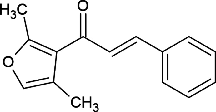
In a continuation of our investigations in heterocyclic systems exhibiting biological activity and in the framework of ongoing structural studies (Maharramov et al., 2021 ▸, 2022 ▸), we report herein the crystal structure and Hirshfeld surface analysis of the title compound, (E)-1-(2,4-dimethylfuran-3-yl)-3-phenylprop-2-en-1-one.
2. Structural commentary
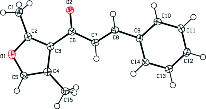
As seen as Fig. 1 ▸, the title compound adopts an E configuration about the C=C double bond. The whole molecule is nearly planar. The furan ring (O1/C2–C5) is inclined to the phenyl ring (C9–C14) by 12.03 (9)°. The torsion angles are C2—C3—C6—O2 = 14.5 (2), C2—C3—C6—C7 = −164.79 (15), C3—C6—C7—C8 = −173.80 (15), C6—C7—C8—C9 = 179.30 (15) and C7—C8—C9—C10 = 172.52 (16)°. The geometrical parameter values of the the title compound are in agreement with those reported for similar compounds in the Database survey section.
Figure 1.
The molecular structure of the title compound, showing the atom labelling and displacement ellipsoids drawn at the 50% probability level.
3. Supramolecular features and Hirshfeld surface analysis
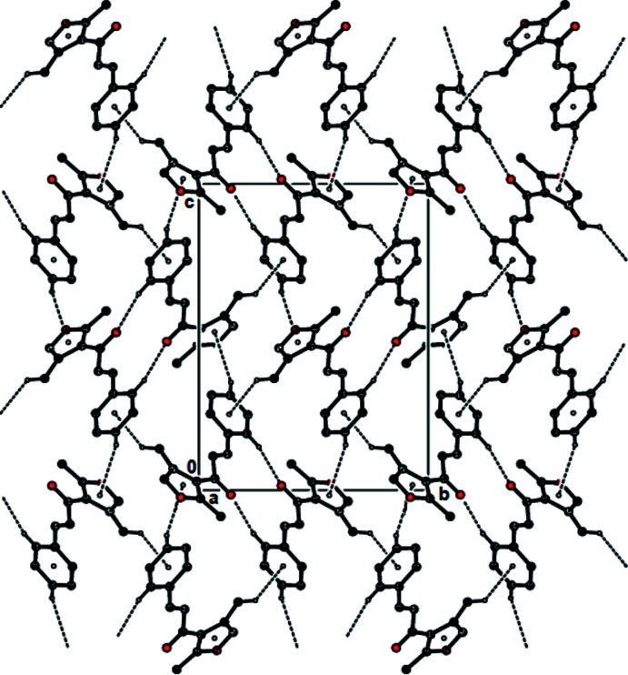
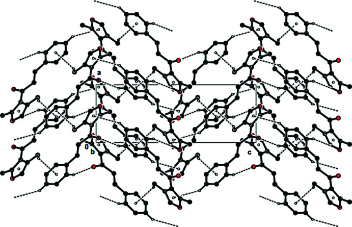
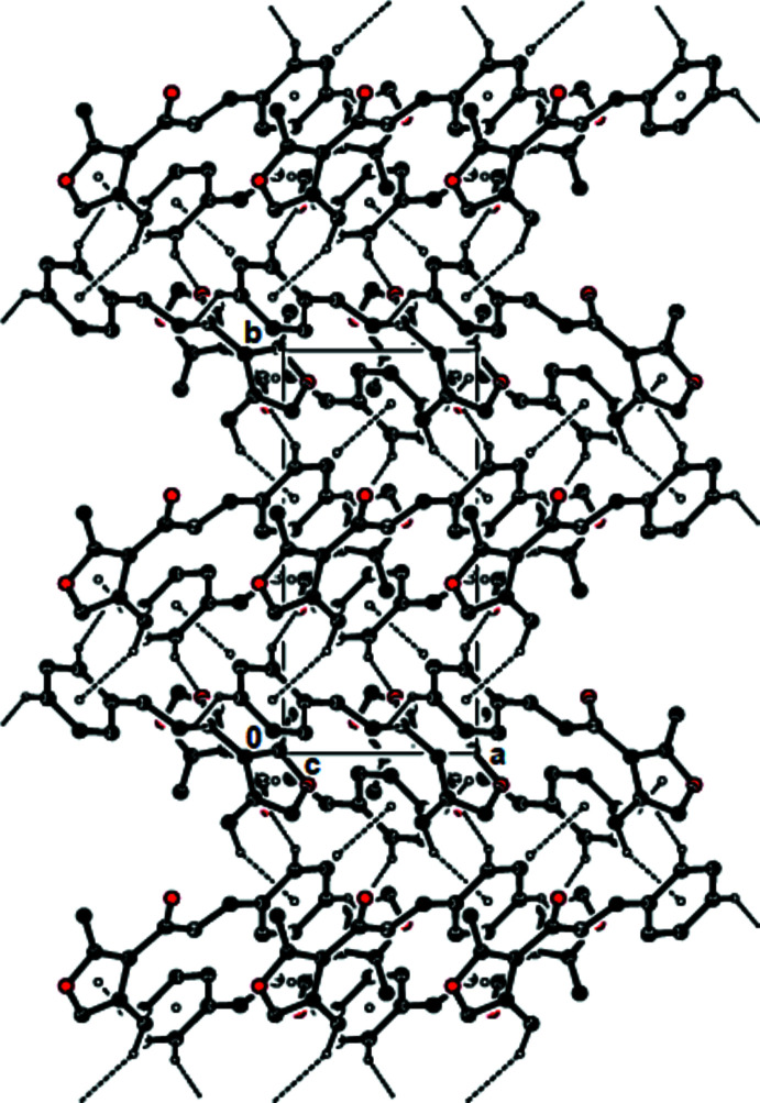
In the crystal, pairs of molecules are linked by C—H⋯O hydrogen bonds, forming dimers with
 (14) ring motifs (Bernstein et al., 1995 ▸; Table 1 ▸; Figs. 2 ▸ and 3 ▸). The molecules are connected via C—H⋯π interactions, forming a three-dimensional network (Table 1 ▸; Fig. 4 ▸). No π–π interactions are observed.
Table 1. Hydrogen-bond geometry (Å, °).
Cg1 and Cg2 are the centroids of the furan (O1/C2–C5) and phenyl (C9–C14) rings, respectively.
| D—H⋯A | D—H | H⋯A | D⋯A | D—H⋯A |
|---|---|---|---|---|
| C10—H10⋯O2i | 0.95 | 2.52 | 3.413 (2) | 157 |
| C12—H12⋯Cg1ii | 0.95 | 2.91 | 3.7339 (19) | 146 |
| C15—H15B⋯Cg2iii | 0.98 | 2.85 | 3.6366 (19) | 137 |
Symmetry codes: (i)
 ; (ii)
 ; (iii)
 .
.
Figure 2.
View of the C—H⋯O hydrogen bonds and C—H⋯π interactions of the title compound down the a axis. Only the H atoms involved in these interactions have been included.
Figure 3.
View of the C—H⋯O hydrogen bonds and C—H⋯π interactions of the title compound down the b axis. Only the H atoms involved in these interactions have been included.
Figure 4.
View of the C—H⋯O hydrogen bonds and C—H⋯π interactions of the title compound down the c axis. Only the H atoms involved in these interactions have been included.
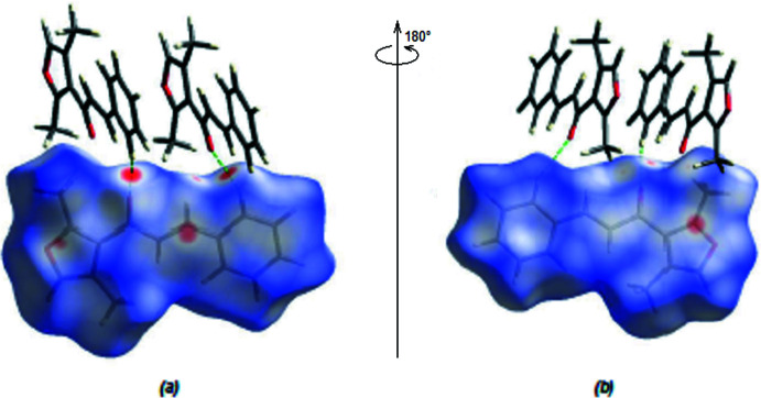
CrystalExplorer17.5 (Spackman et al., 2021 ▸) was used to compute Hirshfeld surfaces of the title molecule and two-dimensional fingerprints. The d norm mappings for the title compound were performed in the range −0.1518 (red) to +1.1567 (blue) a.u. On the d norm surfaces, bright-red spots indicate the locations of the C—H⋯O interactions and O⋯C/C⋯O contacts (Tables 1 ▸ and 2 ▸; Fig. 5 ▸ a,b).
Table 2. Summary of short interatomic contacts (Å) in the title compound.
| H1B⋯H8 | 2.57 | −1 + x, y, z |
| H1C⋯H13 | 2.52 |
 − x, 1 − y,
 + z
+ z
|
| H1B⋯H8 | 2.47 | −
 + x,
+ x,
 − y, 1 − z
|
| H1A⋯H10 | 2.39 | −
 + x,
+ x,
 − y, 1 − z
|
| C12⋯H5 | 2.94 | 1 − x,
 + y,
+ y,
 − z
− z
|
| H13⋯H1A | 2.49 |
 − x, 1 − y, −
 + z
+ z
|
| C15⋯H11 | 3.05 | 2 − x, −
 + y,
+ y,
 − z
− z
|
Figure 5.
(a) Front and (b) back sides of the three-dimensional Hirshfeld surface of the title compound mapped over d norm, with a fixed colour scale of −0.1518 to +1.1567 a.u.
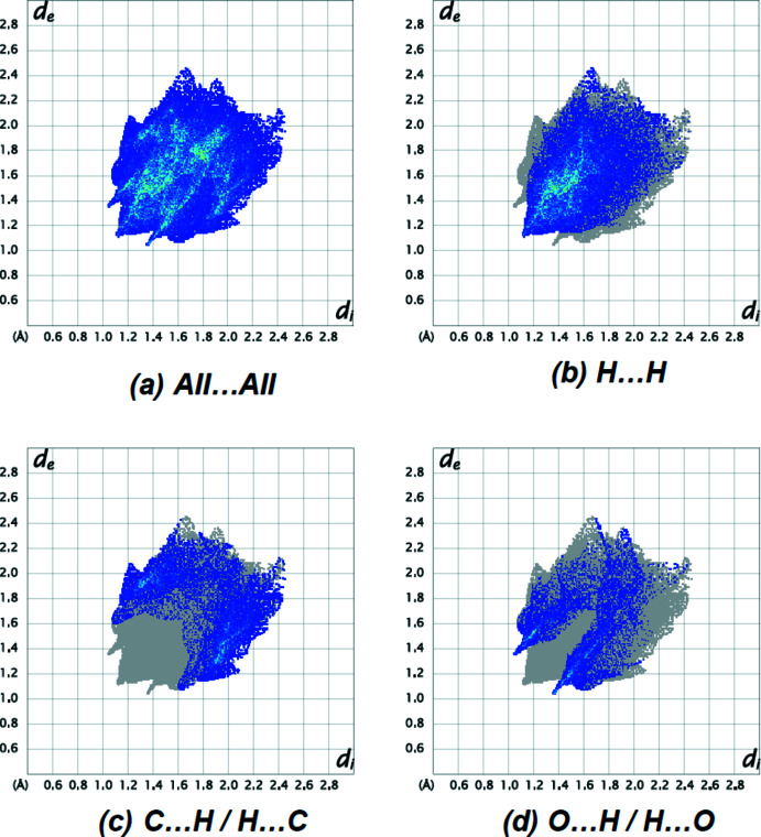
The most important interatomic contact is H⋯H (51.1%; Fig. 6 ▸ b) as it makes the highest contribution to the crystal packing. The C⋯H/H⋯C (Fig. 6 ▸ c; 25.3%), O⋯H/H⋯O (Fig. 6 ▸ d; 15.9%), C⋯C (5.1%) and O⋯C/C⋯O (2.5%) contacts have little directional influence on the molecular packing.
Figure 6.
The two-dimensional fingerprint plots of the title compound, showing (a) all interactions, and delineated into (b) H⋯H, (c) C⋯H/H⋯C and (d) O⋯H/H⋯O interactions. [d e and d i represent the distances from a point on the Hirshfeld surface to the nearest atoms outside (external) and inside (internal) the surface, respectively].
4. Database survey
A search of the Cambridge Structural Database (CSD, Version 5.43, last update November 2022; Groom et al., 2016 ▸) for the ‘1-(furan-3-yl)-3-phenylprop-2-en-1-one’ skeleton of the title compound yielded one hit, 1-(3-furyl)-3-[3-(trifluoromethyl)phenyl]prop-2-en-1-one (CSD refcode KUDNAA; Bąkowicz et al., 2015 ▸). When the positions of the furan and phenyl rings are switched, 1-(3-chlorophenyl)-3-(3-furyl)prop-2-en-1-one (NUQFOW; Zingales et al. 2015 ▸), (E)-3-(2-furyl)-1-phenylprop-2-en-1-one (NOTCUW01; Vázquez-Vuelvas et al. 2015 ▸) are the most similar structures.
In KUDNAA, molecules are linked by intermolecular C—H⋯O interactions, forming zigzag chains with C(5) motifs along the b-axis direction. In addition, molecules are connected by face-to-face π–π stacking interactions [centroid–centroid distances = 3.926 (3) and 3.925 (2) Å] between the opposing benzene and furan rings of the molecules along the c-axis direction. In NUQFOW, the molecule exhibits a non-planar geometry, the furan ring being inclined to the benzene ring by 50.52 (16)°. In the crystal of NUQFOW, molecules stack along the a-axis; however, there are no significant intermolecular interactions present. In NOTCUW01, the molecule also adopts an E configuration about the C=C double bond and the furan and phenyl rings are inclined to one another by 24.07 (7)°. In the crystal of NOTCUW01, molecules are connected by weak C—H⋯O hydrogen bonds and C—H⋯π interactions, forming ribbons extending along the c-axis direction.
5. Synthesis and crystallization
To a solution of 1-(2,4-dimethylfuran-3-yl)ethan-1-one (2 g, 14.5 mmol) in ethanol (10 mL), were added 10 mL of aqueous solution of sodium hydroxide (0.65 g, 16.3 mmol) and the mixture was stirred at room temperature for 2 h. Then benzaldehyde (1.73 g, 16.3 mmol) was added to the vigorously stirred reaction mixture and it was left overnight. The precipitated crystals were separated by filtration and recrystallized from an ethanol/water (1:1) solution (yield 90%; m.p. 349–350 K).
1H NMR (300 MHz, DMSO-d 6, ppm): 2.1 (s, 3H, CH3); 2.5 (s, 3H, CH3); 7.2 (d, 1H, =CH, 3 J H–H = 15.8 Hz); 7.3 (s, 1H, fur.), 7.4 (m, 3H, arom.), 7.5 (d, 1H, =CH, 3 J H–H = 15.8 Hz); 7.8 (m, 2H, arom.). 13C NMR (75 MHz, DMSO-d 6, ppm): 10.3 (CH3), 15.0 (CH3), 120.4 (Cquat.), 122.9 (Cquat.), 126.3 (=CH), 128.9 (CH, arom.), 129.4 (CH, arom.), 130.9 (CH, arom.), 134.8 (Cquat.), 139.0 (CH, furan), 142.8 (=CH), 158.2 (Cquat.), 187.7 (CO).
6. Refinement
Crystal data, data collection and structure refinement details are summarized in Table 3 ▸. All C-bound H atoms were placed at calculated positions and refined using a riding model, with C—H = 0.95 and 0.98 Å, and with U iso(H) = 1.2 or 1.5U eq(C).
Table 3. Experimental details.
| Crystal data | |
| Chemical formula | C15H14O2 |
| M r | 226.26 |
| Crystal system, space group | Orthorhombic, P212121 |
| Temperature (K) | 100 |
| a, b, c (Å) | 5.84787 (5), 12.18109 (9), 16.24568 (15) |
| V (Å3) | 1157.24 (2) |
| Z | 4 |
| Radiation type | Cu Kα |
| μ (mm−1) | 0.68 |
| Crystal size (mm) | 0.24 × 0.20 × 0.18 |
| Data collection | |
| Diffractometer | XtaLAB Synergy, Dualflex, HyPix |
| Absorption correction | Multi-scan (CrysAlis PRO; Rigaku OD, 2021 ▸) |
| T min, T max | 0.579, 1.000 |
| No. of measured, independent and observed [I > 2σ(I)] reflections | 12000, 2410, 2382 |
| R int | 0.031 |
| (sin θ/λ)max (Å−1) | 0.634 |
| Refinement | |
| R[F 2 > 2σ(F 2)], wR(F 2), S | 0.030, 0.081, 1.06 |
| No. of reflections | 2410 |
| No. of parameters | 157 |
| H-atom treatment | H-atom parameters constrained |
| Δρmax, Δρmin (e Å−3) | 0.18, −0.24 |
| Absolute structure | Flack x determined using 940 quotients [(I +)−(I −)]/[(I +)+(I −)] (Parsons et al., 2013 ▸) |
| Absolute structure parameter | 0.16 (7) |
Supplementary Material
Crystal structure: contains datablock(s) I. DOI: 10.1107/S2056989023006084/vm2287sup1.cif
Structure factors: contains datablock(s) I. DOI: 10.1107/S2056989023006084/vm2287Isup2.hkl
Supporting information file. DOI: 10.1107/S2056989023006084/vm2287Isup3.cml
CCDC reference: 2280559
Additional supporting information: crystallographic information; 3D view; checkCIF report
Acknowledgments
Authors’ contributions are as follows. Conceptualization, ANK and IGM; methodology, ANK, FNN and IGM; investigation, ANK, MA and AİS; writing (original draft), MA and ANK; writing (review and editing of the manuscript), MA and ANK; visualization, MA, ANK and IGM; funding acquisition, VNK, AB and ANK; resources, AB, VNK and RMR; supervision, ANK and MA.
supplementary crystallographic information
Crystal data
| C15H14O2 | Dx = 1.299 Mg m−3 |
| Mr = 226.26 | Cu Kα radiation, λ = 1.54184 Å |
| Orthorhombic, P212121 | Cell parameters from 10780 reflections |
| a = 5.84787 (5) Å | θ = 4.5–77.7° |
| b = 12.18109 (9) Å | µ = 0.68 mm−1 |
| c = 16.24568 (15) Å | T = 100 K |
| V = 1157.24 (2) Å3 | Prism, colourless |
| Z = 4 | 0.24 × 0.20 × 0.18 mm |
| F(000) = 480 |
Data collection
| XtaLAB Synergy, Dualflex, HyPix diffractometer | 2382 reflections with I > 2σ(I) |
| Radiation source: micro-focus sealed X-ray tube | Rint = 0.031 |
| φ and ω scans | θmax = 77.8°, θmin = 4.5° |
| Absorption correction: multi-scan (CrysAlisPro; Rigaku OD, 2021) | h = −7→6 |
| Tmin = 0.579, Tmax = 1.000 | k = −15→15 |
| 12000 measured reflections | l = −20→20 |
| 2410 independent reflections |
Refinement
| Refinement on F2 | Hydrogen site location: inferred from neighbouring sites |
| Least-squares matrix: full | H-atom parameters constrained |
| R[F2 > 2σ(F2)] = 0.030 | w = 1/[σ2(Fo2) + (0.0426P)2 + 0.3199P] where P = (Fo2 + 2Fc2)/3 |
| wR(F2) = 0.081 | (Δ/σ)max < 0.001 |
| S = 1.06 | Δρmax = 0.18 e Å−3 |
| 2410 reflections | Δρmin = −0.24 e Å−3 |
| 157 parameters | Extinction correction: SHELXL, Fc*=kFc[1+0.001xFc2λ3/sin(2θ)]-1/4 |
| 0 restraints | Extinction coefficient: 0.0046 (4) |
| Primary atom site location: difference Fourier map | Absolute structure: Flack x determined using 940 quotients [(I+)-(I-)]/[(I+)+(I-)] (Parsons et al., 2013) |
| Secondary atom site location: difference Fourier map | Absolute structure parameter: 0.16 (7) |
Special details
| Experimental. CrysAlisPro 1.171.41.117a (Rigaku OD, 2021) Empirical absorption correction using spherical harmonics, implemented in SCALE3 ABSPACK scaling algorithm. |
| Geometry. All esds (except the esd in the dihedral angle between two l.s. planes) are estimated using the full covariance matrix. The cell esds are taken into account individually in the estimation of esds in distances, angles and torsion angles; correlations between esds in cell parameters are only used when they are defined by crystal symmetry. An approximate (isotropic) treatment of cell esds is used for estimating esds involving l.s. planes. |
Fractional atomic coordinates and isotropic or equivalent isotropic displacement parameters (Å2)
| x | y | z | Uiso*/Ueq | ||
| O1 | −0.1250 (2) | 0.42159 (10) | 0.52334 (8) | 0.0225 (3) | |
| O2 | 0.4218 (2) | 0.63977 (10) | 0.51486 (7) | 0.0208 (3) | |
| C1 | −0.0327 (3) | 0.59606 (14) | 0.58548 (11) | 0.0211 (4) | |
| H1A | −0.1936 | 0.5903 | 0.6018 | 0.032* | |
| H1B | −0.0064 | 0.6672 | 0.5590 | 0.032* | |
| H1C | 0.0647 | 0.5898 | 0.6343 | 0.032* | |
| C2 | 0.0234 (3) | 0.50662 (13) | 0.52698 (10) | 0.0174 (3) | |
| C3 | 0.1991 (3) | 0.49066 (13) | 0.47138 (10) | 0.0162 (3) | |
| C4 | 0.1518 (3) | 0.38801 (13) | 0.43028 (10) | 0.0183 (4) | |
| C5 | −0.0431 (3) | 0.34981 (13) | 0.46379 (11) | 0.0197 (3) | |
| H5 | −0.1150 | 0.2828 | 0.4488 | 0.024* | |
| C6 | 0.3897 (3) | 0.56901 (13) | 0.46209 (10) | 0.0165 (3) | |
| C7 | 0.5382 (3) | 0.56193 (14) | 0.38864 (10) | 0.0184 (3) | |
| H7 | 0.5012 | 0.5122 | 0.3456 | 0.022* | |
| C8 | 0.7247 (3) | 0.62533 (13) | 0.38247 (10) | 0.0169 (3) | |
| H8 | 0.7528 | 0.6742 | 0.4269 | 0.020* | |
| C9 | 0.8896 (3) | 0.62758 (13) | 0.31489 (10) | 0.0162 (3) | |
| C10 | 1.0585 (3) | 0.70888 (13) | 0.31532 (11) | 0.0193 (3) | |
| H10 | 1.0611 | 0.7615 | 0.3584 | 0.023* | |
| C11 | 1.2225 (3) | 0.71374 (14) | 0.25365 (11) | 0.0217 (4) | |
| H11 | 1.3360 | 0.7695 | 0.2547 | 0.026* | |
| C12 | 1.2207 (3) | 0.63690 (14) | 0.19019 (11) | 0.0206 (4) | |
| H12 | 1.3331 | 0.6400 | 0.1480 | 0.025* | |
| C13 | 1.0538 (3) | 0.55571 (14) | 0.18888 (11) | 0.0214 (4) | |
| H13 | 1.0518 | 0.5033 | 0.1456 | 0.026* | |
| C14 | 0.8901 (3) | 0.55079 (14) | 0.25044 (11) | 0.0207 (3) | |
| H14 | 0.7771 | 0.4948 | 0.2490 | 0.025* | |
| C15 | 0.2820 (3) | 0.32838 (14) | 0.36480 (11) | 0.0222 (4) | |
| H15A | 0.2637 | 0.3667 | 0.3122 | 0.033* | |
| H15B | 0.2231 | 0.2534 | 0.3598 | 0.033* | |
| H15C | 0.4444 | 0.3260 | 0.3796 | 0.033* |
Atomic displacement parameters (Å2)
| U11 | U22 | U33 | U12 | U13 | U23 | |
| O1 | 0.0189 (6) | 0.0244 (6) | 0.0242 (6) | −0.0017 (5) | 0.0021 (5) | 0.0025 (5) |
| O2 | 0.0195 (6) | 0.0223 (6) | 0.0207 (6) | −0.0032 (5) | 0.0029 (5) | −0.0048 (5) |
| C1 | 0.0186 (7) | 0.0235 (8) | 0.0211 (8) | 0.0027 (7) | 0.0039 (7) | 0.0001 (6) |
| C2 | 0.0161 (7) | 0.0175 (7) | 0.0187 (8) | 0.0009 (6) | −0.0019 (6) | 0.0034 (6) |
| C3 | 0.0155 (7) | 0.0171 (7) | 0.0161 (7) | 0.0005 (6) | −0.0013 (6) | 0.0017 (6) |
| C4 | 0.0190 (8) | 0.0175 (8) | 0.0184 (7) | 0.0015 (6) | −0.0016 (6) | 0.0009 (6) |
| C5 | 0.0190 (7) | 0.0160 (7) | 0.0241 (8) | −0.0030 (6) | −0.0008 (7) | −0.0005 (6) |
| C6 | 0.0155 (7) | 0.0167 (7) | 0.0172 (7) | 0.0020 (6) | −0.0014 (6) | 0.0011 (6) |
| C7 | 0.0189 (7) | 0.0185 (7) | 0.0177 (7) | −0.0006 (6) | 0.0009 (6) | −0.0025 (6) |
| C8 | 0.0181 (7) | 0.0154 (7) | 0.0172 (7) | 0.0021 (6) | −0.0007 (6) | −0.0011 (6) |
| C9 | 0.0155 (7) | 0.0170 (7) | 0.0160 (7) | 0.0009 (6) | 0.0002 (6) | 0.0010 (6) |
| C10 | 0.0192 (8) | 0.0182 (7) | 0.0204 (8) | −0.0017 (7) | 0.0003 (7) | −0.0035 (6) |
| C11 | 0.0192 (8) | 0.0208 (8) | 0.0250 (8) | −0.0056 (7) | 0.0037 (7) | −0.0011 (7) |
| C12 | 0.0193 (8) | 0.0224 (8) | 0.0200 (8) | 0.0008 (7) | 0.0044 (7) | 0.0006 (7) |
| C13 | 0.0225 (8) | 0.0218 (8) | 0.0199 (8) | −0.0018 (7) | 0.0024 (7) | −0.0047 (7) |
| C14 | 0.0193 (7) | 0.0201 (8) | 0.0226 (8) | −0.0055 (7) | 0.0023 (7) | −0.0042 (6) |
| C15 | 0.0222 (8) | 0.0187 (8) | 0.0258 (9) | −0.0009 (7) | 0.0014 (7) | −0.0060 (7) |
Geometric parameters (Å, º)
| O1—C2 | 1.352 (2) | C8—C9 | 1.461 (2) |
| O1—C5 | 1.389 (2) | C8—H8 | 0.9500 |
| O2—C6 | 1.230 (2) | C9—C10 | 1.399 (2) |
| C1—C2 | 1.482 (2) | C9—C14 | 1.404 (2) |
| C1—H1A | 0.9800 | C10—C11 | 1.388 (2) |
| C1—H1B | 0.9800 | C10—H10 | 0.9500 |
| C1—H1C | 0.9800 | C11—C12 | 1.392 (2) |
| C2—C3 | 1.382 (2) | C11—H11 | 0.9500 |
| C3—C4 | 1.444 (2) | C12—C13 | 1.390 (2) |
| C3—C6 | 1.475 (2) | C12—H12 | 0.9500 |
| C4—C5 | 1.346 (2) | C13—C14 | 1.385 (2) |
| C4—C15 | 1.496 (2) | C13—H13 | 0.9500 |
| C5—H5 | 0.9500 | C14—H14 | 0.9500 |
| C6—C7 | 1.478 (2) | C15—H15A | 0.9800 |
| C7—C8 | 1.340 (2) | C15—H15B | 0.9800 |
| C7—H7 | 0.9500 | C15—H15C | 0.9800 |
| C2—O1—C5 | 106.96 (13) | C7—C8—H8 | 116.4 |
| C2—C1—H1A | 109.5 | C9—C8—H8 | 116.4 |
| C2—C1—H1B | 109.5 | C10—C9—C14 | 118.27 (15) |
| H1A—C1—H1B | 109.5 | C10—C9—C8 | 118.39 (14) |
| C2—C1—H1C | 109.5 | C14—C9—C8 | 123.32 (15) |
| H1A—C1—H1C | 109.5 | C11—C10—C9 | 120.97 (15) |
| H1B—C1—H1C | 109.5 | C11—C10—H10 | 119.5 |
| O1—C2—C3 | 109.92 (14) | C9—C10—H10 | 119.5 |
| O1—C2—C1 | 116.68 (14) | C10—C11—C12 | 120.04 (16) |
| C3—C2—C1 | 133.39 (16) | C10—C11—H11 | 120.0 |
| C2—C3—C4 | 106.33 (14) | C12—C11—H11 | 120.0 |
| C2—C3—C6 | 122.53 (15) | C13—C12—C11 | 119.66 (16) |
| C4—C3—C6 | 131.14 (15) | C13—C12—H12 | 120.2 |
| C5—C4—C3 | 105.95 (15) | C11—C12—H12 | 120.2 |
| C5—C4—C15 | 123.41 (16) | C14—C13—C12 | 120.33 (15) |
| C3—C4—C15 | 130.63 (15) | C14—C13—H13 | 119.8 |
| C4—C5—O1 | 110.84 (14) | C12—C13—H13 | 119.8 |
| C4—C5—H5 | 124.6 | C13—C14—C9 | 120.75 (16) |
| O1—C5—H5 | 124.6 | C13—C14—H14 | 119.6 |
| O2—C6—C3 | 119.83 (15) | C9—C14—H14 | 119.6 |
| O2—C6—C7 | 120.92 (15) | C4—C15—H15A | 109.5 |
| C3—C6—C7 | 119.25 (14) | C4—C15—H15B | 109.5 |
| C8—C7—C6 | 120.31 (15) | H15A—C15—H15B | 109.5 |
| C8—C7—H7 | 119.8 | C4—C15—H15C | 109.5 |
| C6—C7—H7 | 119.8 | H15A—C15—H15C | 109.5 |
| C7—C8—C9 | 127.15 (15) | H15B—C15—H15C | 109.5 |
| C5—O1—C2—C3 | −0.31 (17) | C2—C3—C6—C7 | −164.79 (15) |
| C5—O1—C2—C1 | 178.43 (14) | C4—C3—C6—C7 | 15.8 (3) |
| O1—C2—C3—C4 | 0.56 (18) | O2—C6—C7—C8 | 6.9 (2) |
| C1—C2—C3—C4 | −177.90 (17) | C3—C6—C7—C8 | −173.80 (15) |
| O1—C2—C3—C6 | −179.00 (14) | C6—C7—C8—C9 | 179.30 (15) |
| C1—C2—C3—C6 | 2.5 (3) | C7—C8—C9—C10 | 172.52 (16) |
| C2—C3—C4—C5 | −0.59 (18) | C7—C8—C9—C14 | −8.9 (3) |
| C6—C3—C4—C5 | 178.92 (17) | C14—C9—C10—C11 | 0.1 (3) |
| C2—C3—C4—C15 | −179.55 (17) | C8—C9—C10—C11 | 178.70 (16) |
| C6—C3—C4—C15 | 0.0 (3) | C9—C10—C11—C12 | −0.1 (3) |
| C3—C4—C5—O1 | 0.42 (18) | C10—C11—C12—C13 | 0.2 (3) |
| C15—C4—C5—O1 | 179.47 (15) | C11—C12—C13—C14 | −0.2 (3) |
| C2—O1—C5—C4 | −0.08 (18) | C12—C13—C14—C9 | 0.2 (3) |
| C2—C3—C6—O2 | 14.5 (2) | C10—C9—C14—C13 | −0.1 (3) |
| C4—C3—C6—O2 | −164.92 (16) | C8—C9—C14—C13 | −178.66 (16) |
Hydrogen-bond geometry (Å, º)
Cg1 and Cg2 are the centroids of the furan (O1/C2–C5) and phenyl (C9–C14) rings, respectively.
| D—H···A | D—H | H···A | D···A | D—H···A |
| C8—H8···O2 | 0.95 | 2.44 | 2.792 (2) | 102 |
| C10—H10···O2i | 0.95 | 2.52 | 3.413 (2) | 157 |
| C12—H12···Cg1ii | 0.95 | 2.91 | 3.7339 (19) | 146 |
| C15—H15B···Cg2iii | 0.98 | 2.85 | 3.6366 (19) | 137 |
Symmetry codes: (i) x+1/2, −y+3/2, −z+1; (ii) −x+3/2, −y+1, z−1/2; (iii) −x+1, y−1/2, −z+1/2.
Funding Statement
This paper was supported by Baku State University and the RUDN University Strategic Academic Leadership Program.
References
- Asadov, K. A., Gurevich, P. A., Egorova, E. A., Burangulova, R. N. & Guseinov, F. N. (2003). Chem. Heterocycl. Compd. 39, 1521–1522.
- Bąkowicz, J., Galica, T. & Turowska-Tyrk, I. (2015). Z. Kristallogr. 230, 131–137.
- Bernstein, J., Davis, R. E., Shimoni, L. & Chang, N.-L. (1995). Angew. Chem. Int. Ed. Engl. 34, 1555–1573.
- Çelik, M. S., Çetinus, A., Yenidünya, A. F., Çetinkaya, S. & Tüzün, B. (2023). J. Mol. Struct. 1272, 134158.
- Chalkha, M., Ameziane el Hassani, A., Nakkabi, A., Tüzün, B., Bakhouch, M., Benjelloun, A. T., Sfaira, M., Saadi, M., Ammari, L. E. & Yazidi, M. E. (2023). J. Mol. Struct. 1273, 134255.
- Cuthbertson, J. D. & Taylor, R. J. K. (2013). Angew. Chem. Int. Ed. 52, 1490–1493. [DOI] [PubMed]
- Farrugia, L. J. (2012). J. Appl. Cryst. 45, 849–854.
- Groom, C. R., Bruno, I. J., Lightfoot, M. P. & Ward, S. C. (2016). Acta Cryst. B72, 171–179. [DOI] [PMC free article] [PubMed]
- Gurbanov, A. V., Kuznetsov, M. L., Mahmudov, K. T., Pombeiro, A. J. L. & Resnati, G. (2020). Chem. Eur. J. 26, 14833–14837. [DOI] [PubMed]
- Kawamura, S., Chu, H., Felding, J. & Baran, P. S. (2016). Nature, 532, 90–93. [DOI] [PMC free article] [PubMed]
- Maharramov, A. M., Shikhaliyev, N. G., Zeynalli, N. R., Niyazova, A. A., Garazade, Kh. A. & Shikhaliyeva, I. M. (2021). UNEC J. Eng. Appl. Sci. 1, 5–11.
- Maharramov, A. M., Suleymanova, G. T., Qajar, A. M., Niyazova, A. A., Ahmadova, N. E., Shikhaliyeva, I. M., Garazade, Kh. A., Nenajdenko, V. G. & Shikaliyev, N. G. (2022). UNEC J. Eng. Appl. Sci. 2, 64–73.
- Parsons, S., Flack, H. D. & Wagner, T. (2013). Acta Cryst. B69, 249–259. [DOI] [PMC free article] [PubMed]
- Poustforoosh, A., Hashemipour, H., Tüzün, B., Azadpour, M., Faramarz, S., Pardakhty, A., Mehrabani, M. & Nematollahi, M. H. (2022). Curr. Microbiol. 79, 241. [DOI] [PMC free article] [PubMed]
- Rigaku OD (2021). CrysAlis PRO. Rigaku Oxford Diffraction, Yarnton, England.
- Sarkı, G., Tüzün, B., Ünlüer, D. & Kantekin, H. (2023). Inorg. Chim. Acta, 545, 121113.
- Sheldrick, G. M. (2015a). Acta Cryst. A71, 3–8.
- Sheldrick, G. M. (2015b). Acta Cryst. C71, 3–8.
- Spackman, P. R., Turner, M. J., McKinnon, J. J., Wolff, S. K., Grimwood, D. J., Jayatilaka, D. & Spackman, M. A. (2021). J. Appl. Cryst. 54, 1006–1011. [DOI] [PMC free article] [PubMed]
- Spek, A. L. (2020). Acta Cryst. E76, 1–11. [DOI] [PMC free article] [PubMed]
- Tapera, M., Kekeçmuhammed, H., Tüzün, B., Sarıpınar, E., Koçyiğit, M., Yıldırım, E., Doğan, M. & Zorlu, Y. (2022). J. Mol. Struct. 1269, 133816.
- Vázquez-Vuelvas, O. F., Enríquez-Figueroa, R. A., García-Ortega, H., Flores-Alamo, M. & Pineda-Contreras, A. (2015). Acta Cryst. E71, 161–164. [DOI] [PMC free article] [PubMed]
- Zingales, S. K., Wallace, M. Z. & Padgett, C. W. (2015). Acta Cryst. E71, o707. [DOI] [PMC free article] [PubMed]
- Zubkov, F. I., Mertsalov, D. F., Zaytsev, V. P., Varlamov, A. V., Gurbanov, A. V., Dorovatovskii, P. V., Timofeeva, T. V., Khrustalev, V. N. & Mahmudov, K. T. (2018). J. Mol. Liq. 249, 949–952.
Associated Data
This section collects any data citations, data availability statements, or supplementary materials included in this article.
Supplementary Materials
Crystal structure: contains datablock(s) I. DOI: 10.1107/S2056989023006084/vm2287sup1.cif
Structure factors: contains datablock(s) I. DOI: 10.1107/S2056989023006084/vm2287Isup2.hkl
Supporting information file. DOI: 10.1107/S2056989023006084/vm2287Isup3.cml
CCDC reference: 2280559
Additional supporting information: crystallographic information; 3D view; checkCIF report


