Abstract
Background and Objectives
Patients with multiple sclerosis (PwMS) receiving extended dosing of rituximab (RTX) have exhibited no return of disease activity, which suggests that maintenance of deep depletion of circulating B cells is not necessary to maintain the efficacy of RTX in MS.
Methods
This was a prospective monocentric observational study including all consecutive PwMS who started or continued RTX after 2019, when the medical staff decided to extend the dosing interval up to 24 months for all patients. Circulating B-cell subsets were monitored regularly and systematically in case of relapse. The first extended interval was analyzed.
Results
We included 236 PwMS (81% with relapsing-remitting MS; mean [SD] age 43 [12] years; median [range] EDSS score 4 [0–8]; mean relapse rate during the year before RTX start 1.09 [0.99]; 41.5% with MRI activity). The median number of RTX infusions before extension was 4 (1–13). At the time of the analysis, the median delay in dosing was 17 months (8–39); the median proportion of circulating CD19+ B cells was 7% (0–25) of total lymphocytes and that of CD27+ memory B cells was 4% (0–16) of total B cells. The mean annual relapse rate did not differ before and after the extension: 0.03 (0.5) and 0.04 (0.15) (p = 0.51). Similarly, annual relapse rates did not differ before and after extension in patients with EDSS score ≤3 (n = 79) or disease duration ≤5 years (n = 71) at RTX onset. During the “extended dosing” period, MRI demonstrated no lesion accrual in 228 of the 236 patients (97%). Five patients experienced clinical relapse, which was confirmed by MRI. In these patients, the level of B-cell subset reconstitution at the time of the relapse did not differ from that for patients with the same extension window.
Discussion
The efficacy of RTX outlasted substantial reconstitution of circulating B cells in PwMS, which suggests that renewal of the immune system underlies the prolonged effect of RTX in MS. These findings suggest that extended interval dosing of RTX that leads to a significant reconstitution of circulating B cells is safe in PwMS, could reduce the risk of infection, and could improve vaccine efficacy.
Introduction
B-cell–depleting therapies have been recently and widely used in patients with multiple sclerosis (PwMS), with outstanding control of inflammation. Nonetheless, recent studies have shown that prolonged maintenance therapy with rituximab (RTX), the first B-cell–depleting therapy used in MS, consisting of 6-month dosing intervals, is associated with the highest risk of infection among all therapies for MS.1 In addition, B-cell–depleting therapies reduce the efficacy of most vaccines, which could limit the mitigation of the risk of infection.2,3 In this regard, the COVID-19 pandemic highlighted these safety concerns because B-cell–depleting therapies are associated with both increased risk of severe COVID-194 and reduced COVID-19 vaccine efficacy.5-8
Hypogammaglobulinemia is frequent after several years of maintenance therapy with B-cell–depleting therapies and could be the main factor contributing to the risk of infection.9-12 Mechanisms leading to hypogammaglobulinemia are not well understood. Nonetheless, the risk of hypogammaglobulinemia increases with time on B-cell–depleting maintenance therapy.12 Importantly, the reversibility of hypogammaglobulinemia after B-cell–depleting therapy withdrawal could be related to the kinetics of CD27+ memory B-cell repopulation,13,14 which highly depends on the time of maintenance therapy. Thus, one way to reduce the risk of hypogammaglobulinemia and subsequent infections could be to increase the dosing interval to lead to substantial CD27+ memory B-cell repopulation. In addition, increasing the dosing interval could improve the vaccine efficacy, as recently evidenced with the COVID-19 vaccine.15 Importantly, reducing the overall dose of B-cell–depleting therapies to improve safety would be relevant if a potential return of disease activity after extension is rare. This situation would be true if deep depletion of CD27+ memory B cells is not required to obtain full control of MS activity. This seems to be the case, as suggested by different studies including the phase II RTX trial16 and the ocrelizumab (OCR) phase II extension trial17 in relapse-remitting MS (RRMS), which found no return of disease activity within the 12 and 18 months after the last infusion. More recently, studies involving the RTX/OCR dosing interval longer than 6 months have confirmed these findings, which suggest that B-cell repopulation, known to generally occur after 8 months, is not immediately associated with disease reactivation.18-21 However, because most studies did not perform B-cell monitoring, we do not know whether the potential maintenance of efficacy of RTX/OCR after 6 months is due to slow B-cell repopulation in most patients or persists after significant B-cell repopulation, thus suggesting a less proinflammatory profile of reconstituting B cells. Recently, a large real-world observational cohort study found sustained efficacy of RTX in patients with a large extended infusion interval.22 This study demonstrated that although B-cell repopulation occurred in almost all patients after 12 months, no significant return to disease activity was evidenced. However, this study did not fully address whether the risk of relapse during RTX treatment is driven by the extent of B-cell repopulation because B-cell counts were not determined at the time of the relapse in all patients showing disease activity. This point is of major importance for clinical practice to determine whether B-cell monitoring could be a relevant tool to tailor RTX infusions in PwMS.
The present prospective observational cohort study reports the evolution of disease activity of a large cohort of PwMS receiving extended interval RTX dosing. Patients were monitored regularly for circulating B-cell subset reconstitution and systematically in case of relapse.
Methods
Study Population
We started to use RTX off-label for PwMS in the MS center of Marseille in 2015. All consecutive patients under RTX therapy were prospectively included in an observational cohort study. The induction treatment consisted of 1,000 mg RTX infused twice at 2-week intervals. The maintenance regimen consisted of a single infusion of 1,000 mg RTX administered every 6 months until 2018. Since 2019, our department initiated a change in clinical practice concerning the dosing interval used for off-label RTX in RRMS. All neurologists (A.M., A.R., C.B., S.D., P.D., J.P., and B.A.) decided to extend the interval between 2 infusions up to 24 months. Clinical visits were maintained every 6 months, and brain and spinal cord MRI monitoring was performed at least annually for all patients. This decision was based on the absence of a standardized administration scheme for RTX in RRMS as demonstrated by the heterogeneity of dosing intervals reported at that time in the literature16,23-25 along with our experience with patients stopping RTX for various reasons and to limit the potential infectious side effects related to hypogammaglobulinemia.10 The 24-month maximal interval was chosen according to a study published in 2016 that found a potential slight waning of RTX effect at 24 months after the last infusion.24 Patients were informed of the new administration scheme at their next appointment. All patients under RTX or who started RTX in the center received it under this extended administration scheme. The COVID-19 pandemic was probably an important determinant in this total adherence of patients.
In the present article, we report the data of the first interval of the “extended dosing” period. Only data of patients with a follow-up > 8 months in this first interval period are reported.
Medical Visits
Patients were seen in the center for a clinical evaluation every 6 months. Brain and spinal cord MRI monitoring was performed at least annually. All examinations for a given patient were performed by the same experienced neurologists (A.M., A.R., C.B., S.D., P.D., J.P., and B.A.). The Expanded Disability Status Scale (EDSS) score was collected at each visit. All patients received the phone number of our indoor neuroinflammatory unit that was open 24 h/d, 7 d/wk. We informed each patient about the need to call the center in case of new neurologic signs. Relapse was considered the occurrence of neurologic signs persisting >24 hours, in the absence of fever, infection, or other intercurrent phenomena. In case of relapse, the patient was admitted to hospital and corticosteroids were administered if necessary. Additional brain and spinal cord MRI monitoring was systematically performed during the 3 months after each relapse.
CD19+ B-Cell and CD27+ Memory B-Cell Monitoring
Since 2020, circulating B-cell subset monitoring was performed at each visit or in case of relapse. All B-cell counts were obtained in the same laboratory of the University Hospital of Marseille. Total lymphocytes, CD19+ B cells, CD3+ T cells, and CD3−CD16/CD56+ natural killer cells were measured by flow cytometry with the BD Multitest 6-Color TBNK assay and Trucount tubes. Specific staining with anti–CD45-V500, anti–CD20-APC-H7, anti–IgD-FITC, anti–IgM-PE-Cy5, anti–CD38-PE-Cy7, and anti–CD27-PE antibodies was used to quantify CD27+ memory, IgM−IgD−CD27+ commuted memory, IgD+CD27− naive, and IgM++CD27−CD38++ transitional B-cell subsets, respectively. Data were acquired and analyzed by using the BD Canto-2 cytometer and Diva software. Both reagents and instruments were provided by Becton Dickinson.
Samples of CD19+ B cells were classified as being in complete repletion above 100 cells/µL, corresponding to the lower limit of normality (LLN), in partial repletion if between the detection threshold and LLN and in complete depletion if below the detection threshold. For the B-cell subsets, in the absence of consensual normative values, we relied on the work of Morbach et al.26: LLN = 2 cells/µL for CD38+IgM+, LLN = 92 cells/µL for CD27−IgD+, LLN = 12 cells/µL for total memory B cells, and LLN = 10 cells/µL for commuted memory B cells.
Statistical Analyses
Statistical analyses involved using JMP Pro 16.1.0 (SAS Institute Inc., Cary, NC).
For the analysis, 3 epochs were considered: the year before RTX introduction (the “before RTX” period), the period when RTX was infused every 6 months (“standard dosing” period), and the period of extension after the last RTX infusion (“extended dosing” period). These designations are used throughout the article.
Comparisons of annual relapse rate (ARR) and EDSS score between the different periods involved repeated-measures ANOVA with Tukey HSD used as post hoc testing for pairwise comparisons. Comparison of the proportions of patients with active disease defined as at least one new T2 lesion on brain or spinal cord MRI with or without relapse involved Fisher exact test. To assess lymphocyte counts for patients with active disease and nonactive disease during the “extended dosing” period, we matched each patient with active disease at the time of confirmed relapse to all those without active disease and identical extended interval dosing time (in months). Then, we performed repeated-measures ANOVA, with each active patient used as a block, with the Full Factorial Repeated-Measures ANOVA JMP Add-In (community.jmp.com/).
Multivariable regression analysis was used to assess predictors potentially affecting B-cell repopulation, namely age, sex, RTX dose per body surface area, number of previous cycles of RTX, and previous treatment classified as immunomodulators (beta interferons, glatiramer acetate), mild immunosuppressors (dimethyl fumarate, teriflunomide), therapeutic targeting of immune cell trafficking (fingolimod, natalizumab), and high immunosuppressors (cyclophosphamide, mitoxantrone). Nonetheless, to explore these predictors, we need to eliminate the time since the last infusion. Thus, the first step was the extraction of the residuals from the fit between cell count and time since the last infusion (in months). The second step was to apply a generalized linear regression model to explain the variability of the residuals of fit, using the predictors mentioned above. Only the predictors for which p was strictly <0.05 were considered statistically significant and reported in the results section.
Ethical Approval
The authors obtained ethical approval from the Institutional Review Board of the University Hospital of Marseille, France (Approval No. PADS-21-60) for this study.
Data Availability
All data analyzed during this study will be shared anonymized by reasonable request of a qualified investigator to the corresponding author.
Results
Study Population
In total, 247 patients received RTX and were followed in our department since 2015. The mean (SD) relapse rate during the year before RTX start was 1.09 (0.99). During the 6 months before RTX start, 98 of 236 (41.5%) patients exhibited MRI activity characterized by at least one new T2 lesion or contrast-enhancing lesion. Demographics and details about the different treatments used before RTX are presented in Table 1. Eleven patients were lost during follow-up (5 moved to a new city, 2 wanted to be followed by another neurologist in the same region, and 4 for unknown reasons). All patients agreed to the extended interval dosing proposed as described above.
Table 1.
Demographic Characteristics of Patients With Multiple Sclerosis Receiving Rituximab (RTX) (n = 236)
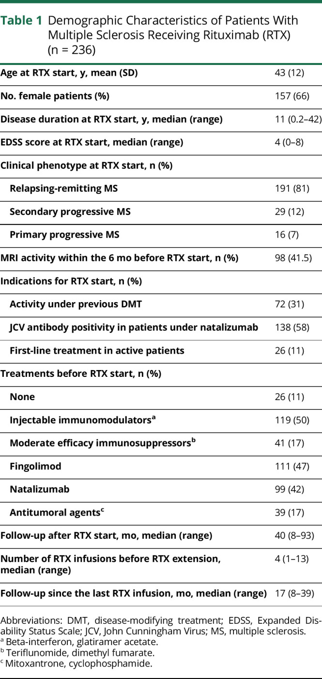
At the time of the first RTX infusion, the mean (SD) age of patients was 43 (12) years, 157 female patients (66%), median (range) disease duration 11 years (0.2–42), and median EDSS score 4 (0–8). The median follow-up after the first RTX infusion was 40 months (8–93). During RTX, the mean (SD) time between 2 clinical evaluations was 190 (44.8) days. The median number of RTX infusions was 4 (1–13). The median follow-up since the last RTX infusion, corresponding to the first interval of the “extended dosing” period analyzed here, was 17 months (8–39). In total, 22 of 236 (9.3%) patients were extended just after their first RTX infusion. RTX redosing was not performed in 6 patients within the predefined 24-month maximal interval for safety concerns (infection or hypogammaglobinemia). In that case, immunomodulatory treatment was introduced in one patient (glatiramer acetate), and no disease-modifying treatment in the remaining 5 patients. Moreover, in 3 patients because of disease stability and after discussion with their treating neurologist, RTX was postponed for an additional several months (26, 27, and 31 months).
The results related to safety were previously published.12
Evolution of MS During “Extended Dosing” Period
Whole Cohort Analysis
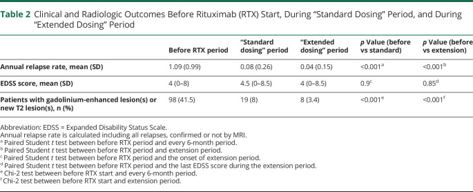
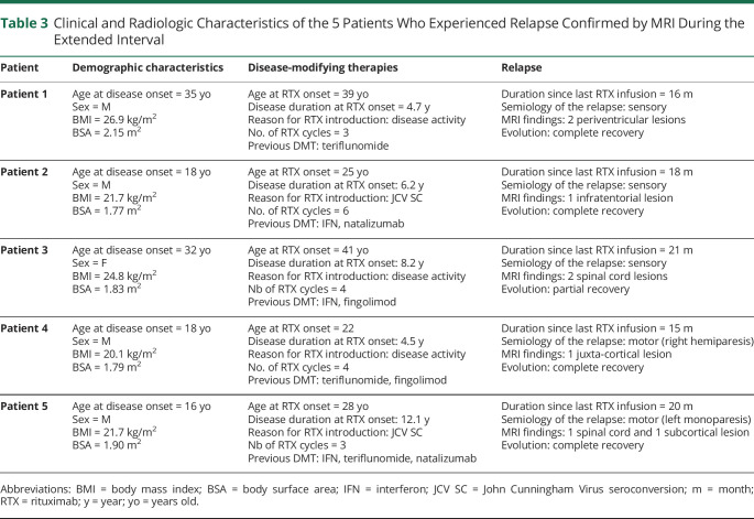
The mean ARR was lower after RTX start (including the “standard dosing” and “extended dosing” periods) than during the year before RTX start: 0.06 (0.15) vs 1.09 (0.99) (repeated ANOVA, p < 0.001) (Table 2). The mean ARR was higher but not significantly during the “standard dosing” than “extended dosing” period: 0.08 vs 0.04 (post hoc analysis, p = 0.64). Nonetheless, the mean ARR did not differ between the “standard dosing” and “extended dosing” periods when excluding relapses occurring during the first 6 months after RTX start: 0.03 and 0.04 (post hoc analysis, p = 0.99). During the “standard dosing” period, 31 relapses occurred in 27 patients (13 during the first 6 months and 18 after 6 months). Brain and spinal cord MRI performed during the 3-month period after these relapses revealed new T2 lesions in 10 of these patients. During the “extended dosing” period, 15 relapses occurred in 15 patients. Brain and spinal cord MRI performed during the 3-month period after these relapses revealed new T2 lesions in 5 of these patients. Table 3 presents the characteristics of the 5 patients with relapse confirmed by MRI during the “extended dosing” period.
Table 2.
Clinical and Radiologic Outcomes Before Rituximab (RTX) Start, During “Standard Dosing” Period, and During “Extended Dosing” Period
Table 3.
Clinical and Radiologic Characteristics of the 5 Patients Who Experienced Relapse Confirmed by MRI During the Extended Interval
During the “standard dosing” period, 19 of 236 (8%) patients showed at least one new T2 lesion or contrast-enhancing lesion on brain or spinal cord MRI as compared with the MRI performed before RTX onset. Of note, 14 of 19 (74%) cases reported during the “standard dosing” period occurred in the first 6 months after RTX start. During the “extended dosing” period, 8 of 236 (3.5%) patients showed at least one new T2 lesion or contrast-enhancing lesion on brain or spinal cord MRI as compared with the last MRI performed during the “standard dosing” period.
The median EDSS score did not differ between RTX start, the start of the “extended dosing” period, and the end of follow-up: 4 (0–8), 4.5 (0–8.5), and 4 (0–8.5) (repeated ANOVA, p = 0.98, pairwise post hoc p = 0.99 between RTX start and end of follow-up; p = 0.99 between the start of “extended dosing” period and the end of follow-up).
Subgroup Analysis in Patients With EDSS Score ≤3 at RTX Start
For 79 patients, the EDSS score was ≤3 when they started RTX (39 patients with disease duration ≤5 years). In this population, the mean (SD) ARR was lower after RTX start (including the “standard dosing” and “extended dosing” periods) than during the year before RTX start: 0.08 (0.21) vs 1 (1) (repeated ANOVA, p < 0.001). The mean (SD) ARR was higher but not significantly during the “standard dosing” than “extended dosing” period: 0.12 (0.39) vs 0.05 (0.17) (post hoc analysis, p = 0.71). Nonetheless, the mean ARR did not differ between the “standard dosing” and “extended dosing” periods when excluding relapses occurring during the first 6 months after RTX start: 0.05 and 0.05 (post hoc analysis, p = 0.99). During the “standard dosing” period, 12 relapses occurred in 11 patients (6 during the first 6 months and 6 after 6 months). Brain and spinal cord MRI performed during the 3-month period after these relapses revealed new T2 lesions in 6 of these patients. During the “extended dosing” period, 7 relapses occurred in 7 patients. Brain and spinal cord MRI performed during the 3-month period after these relapses revealed new T2 lesions in 4 of these patients.
The median EDSS score did not differ between RTX start, the start of the “extended dosing” period, and the end of follow-up: 1.5 (0–3), 1 (0–4), and 1 (0–5) (repeated ANOVA, p = 0.61, pairwise post hoc p = 0.66 between RTX start and end of follow-up; p = 1 between the start of “extended dosing” period and the end of follow-up).
Subgroup Analysis of Patients With Disease Duration ≤5 Years at RTX Start
In total, 71 patients started RTX in the first 5 years after clinical disease onset (39 patients with EDSS score ≤3). In this population, the mean (SD) ARR was lower after RTX start (including the “standard dosing” and “extended dosing” periods) than during the year before RTX start: 0.09 (0.22) vs 1.07 (1) (repeated ANOVA, p < 0.001). The mean (SD) ARR was higher but not significantly during the “standard dosing” than “extended dosing” period: 0.12 (0.39) vs 0.05 (0.21) (post hoc analysis, p = 0.63). Nonetheless, the mean ARR did not differ between the “standard dosing” and “extended dosing” periods when excluding relapses occurring during the first 6 months after RTX start: 0.03 and 0.05 (post hoc analysis, p = 0.98). During the “standard dosing” period, 12 relapses occurred in 10 patients (5 during the first 6 months and 5 after 6 months). Brain and spinal cord MRI performed during the 3-month period after these relapses revealed new T2 lesions in 6 of these patients. During the “extended dosing” period, 15 relapses occurred in 15 patients. Brain and spinal cord MRI performed during the 3-month period after these relapses revealed new T2 lesions in 5 of these patients.
The median EDSS score did not differ between RTX start, the start of the “extended dosing” period, and the end of follow-up: 3 (0–7), 3 (0–7.5), and 3 (0–7) (repeated ANOVA, p = 0.23, pairwise post hoc p = 0.26 between RTX start and end of follow-up; p = 0.98 between the start of “extended dosing” period and the end of follow-up).
B-Cell Repopulation
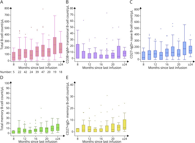
Figure 1 presents the dynamics of B-cell subset repopulation after the last RTX infusion. The median (range) proportion of IgM++CD27–CD38++ transitional B cells was 0% (0–26.5), IgD+CD27– naive B cells 92.4% (0–98.2), CD27+ memory B cells 4.6% (0–17.6) and IgM-IgD-CD27+ commuted memory B cells 1.5% (0–14.6) of total B cells.
Figure 1. Distribution of CD19+ B Cells (A), IgM++CD27−CD38++ Transitional B Cells (B), IgD+CD27− Naive B Cells (C), CD27+ Memory B Cells (D), and IgM−IgD−CD27+ Commuted Memory B Cells (E).
Samples were grouped into 2-month intervals since the last rituximab infusion.
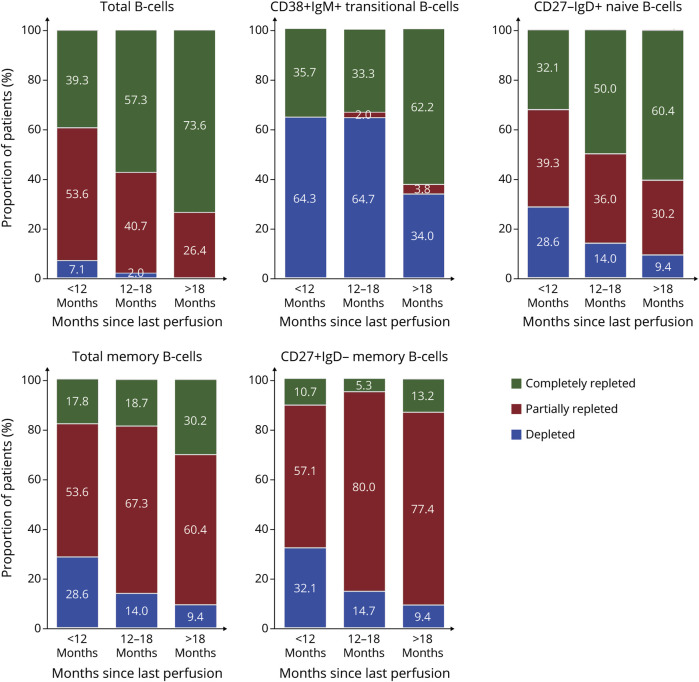
Figure 2 presents the proportion of patients with complete or partial repletion of B cells and subsets according to the delay from their last RTX infusion. It is noteworthy that after 12 months, more than 98% of PwMS showed partial or complete repletion of B cells. In opposite, CD27+ memory B cells are completely repleted in less than 20% of patients, even after 18 months.
Figure 2. Relative Proportion of Depleted (Under Level of Detection), Partially Repleted (Lower Level of Normality), and Completely Repleted B-Cell Subsets Counts at <12 Months, ≥12 to 18, and ≥18 Months since the Last Rituximab Infusion.
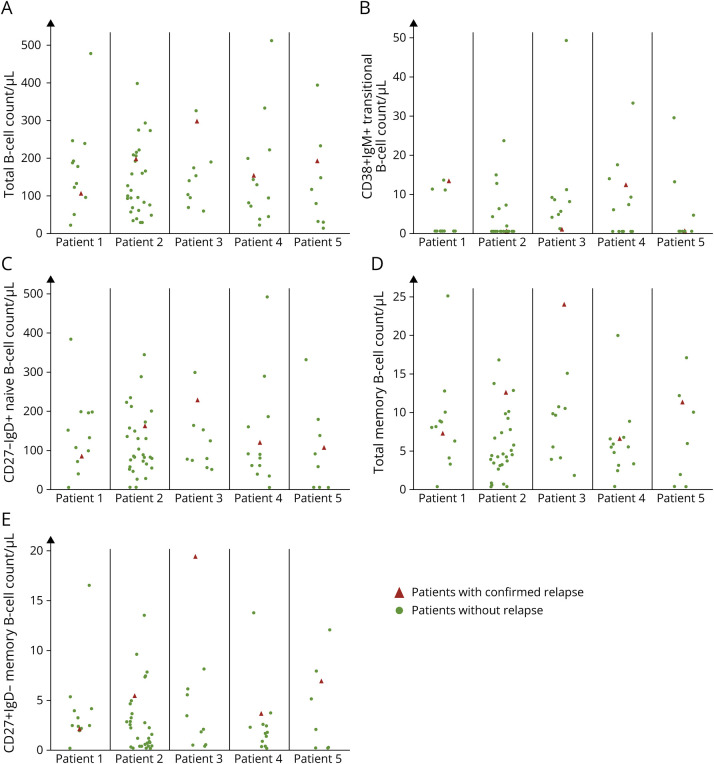
At the time of the confirmed relapses that occurred during the “extended dosing” period for 5 patients (Table 3), the median (range) proportion of B-cell repopulation was 8.8% (6.8–12.1) CD19+ B cells among total lymphocytes and 0.2% (0–14.7) IgM++CD27−CD38++ transitional B cells, 90.8% (89.6–93.1) IgD+CD27− naive B cells, 8.4% (5–9.7) of CD27+ memory B cells, and 3% (2.5–7.7) of IgM−IgD−CD27+ commuted memory B cells among total CD19+ B cells. The proportions of the different B-cell subsets at the time of the relapse did not differ from those for patients with a same extension window (n = 69; repeated-measures ANOVA, p = 0.75, 0.15, 0.17, 0.24, and 0.26, respectively) (Figure 3).
Figure 3. Frequency of CD19+ B Cells (A), IgM++CD27−CD38++ Transitional B Cells (B), IgD+CD27− Naive B Cells (C), CD27+ Memory B Cells (D), and IgM−IgD−CD27+ Commuted Memory B Cells (E) at the Time of the 5 Relapses Confirmed by MRI Occurring in 5 Distinct Patients During the “Extended Dosing” Period (Red Triangles).
The authors matched each patient at the time of confirmed relapse to all those without active disease and identical extended interval dosing time in months (green dots).
Finally, we determined factors associated with the repopulation proportion, considering the time since the last infusion. Slow CD19+ B-cell repopulation was associated with younger age (p = 0.001), male sex (p = 0.01), and treatment with cyclophosphamide and/or mitoxantrone before RTX (p = 0.04). Slow CD27+ memory B-cell repopulation was associated with the number of RTX infusions before extension (p = 0.03) and treatment with cyclophosphamide and/or mitoxantrone before RTX (p = 0.05).
Discussion
This study reports the evolution of all consecutive PwMS who received RTX in the MS center of the University Hospital of Marseille and crossing 2 distinct periods: when the dosing interval was 6 months and after our collegial decision to systematically extend the interval between 2 infusions up to 24 months. Patients mainly had a relapsing form of MS (81%), and many had high disease activity as demonstrated by MRI before RTX start (41%). Here, we report the evolution of these patients during their first extended interval and showed that disease activity remained very low after a median extension period of 17 months and did not differ from that in the period when RTX was infused every 6 months.
Unlike for ocrelizumab, for RTX infusions, there is no standard protocol for PwMS, and different dosing intervals have been used. In the preliminary open-label study conducted by Bar-Or et al. in 2008,23 2 infusions of RTX were administered at a 2-week interval at baseline and after 6 months, and patients were followed for 12 months after the last infusion. No patient exhibited disease activity on MRI during the year after the last infusion. In the pivotal phase II study conducted by Hauser et al.16 in 2008, 2 infusions of RTX were administered at a 2-week interval, no infusion was given at 6 months, and patients were followed for 1 year. Again, no patients exhibited disease activity on MRI during the year after the last RTX infusion. Of note, despite these previous data demonstrating the efficacy of anti-CD20 agents on disease activity for at least 12 months, a 6-month interval scheme was selected for the phase III OCR study in RRMS.27 This choice was probably motivated by the median time needed for B cells to repopulate being 32 weeks after the infusion.28 However, the apparent lack of correlation between B-cell repopulation and the return to disease activity evidenced in PwMS during the 12 months after the last RTX infusion reported above has not been questioned.
In the last few years, several observational studies have confirmed the results for the prolonged effect of RTX on disease activity in RRMS obtained in the pivotal studies. Juto et al.18 did not find any rebound of disease activity in patients interrupting RTX for different reasons and followed for a mean of 30 months. For 2 years, De Flon et al. prospectively followed patients who previously received 2 RTX infusions24: At 12 months after RTX infusions, MRI activity was extremely low; at 24 months, MRI activity remained low but tended to increase, thereby suggesting a waning effect of RTX. A Swedish retrospective study including a large sample of patients receiving RTX reported high efficacy of this treatment in patients whatever the dosing regimen, including 1,000 mg every 6 months to 500 mg every year.25 Recently, Boremalm et al. reported that a dose reduction of RTX was not associated with a return to disease activity even in patients with “super low dose,” defined as <500 mg annually.21
Several studies reported the disease evolution of PwMS receiving anti-CD20 therapy with B-cell or memory B-cell monitoring to tailor infusion.29-33 Using thresholds classically used in other pathologies to detect subtle re-emergence of B-cell repopulations, these studies found no disease reactivation. However, using these thresholds, the mean extended interval was limited, generally lower than 12 months. Recently, a large sample study of patients with RRMS treated with RTX and large extended interval dosing, frequently higher than 18 months, found no significant return to disease activity despite B-cell repopulation in most patients during the interval.22 However, B-cell count was not available for all patients presenting disease activity during the extended interval, which limits interpretation of the findings.
According to these previous studies, we found that the efficacy of RTX largely outlasted 6 months and was maintained, with substantial B-cell repopulation in most patients. During the “extended dosing” period, no changes in disease activity occurred despite a median CD19+ B-cell repopulation of 7% of total lymphocytes and a median CD27+ memory B-cell repopulation of 4% of total B cells. These levels of repopulation are considered as a complete or partial repopulation of CD19+ B cells for more than 90% of the patients in this study, with maintenance of therapeutic response. Importantly, the few patients (n = 5) experiencing renewed disease activity during the extended interval showed a similar level of repopulation of CD19+ B cells, IgM++CD27−CD38++ transitional B cells, IgD+CD27− naive B cells, CD27+ memory B cells, and IgM−IgD−CD27+ commuted memory B cells at the time of the relapse as patients without disease activity and with the same extension window. All these findings suggest that the level of circulating B-cell subsets does not represent an accurate biomarker to monitor the efficacy of anti-CD20 agents and that potential mechanisms underlying the efficacy of B-cell–depleting therapies in MS are different from those involved in AQP4 antibody neuromyelitis optica spectrum disorders, in which a slight repopulation of circulating memory B cells was found associated with relapse in several patients.32,33 In MS, renewal of the immune system probably mainly underlies the prolonged effect of B-cell–depleting therapies.34,35 Although normative values for B-cell subsets are not consensual, in the light of previous work performed in a large population of healthy subjects,26 it is noteworthy that CD27+ memory B cells are completely repleted in less than 20% of patients, even after 18 months. These results suggest a modified reconstitution of B-cell subsets after B-cell–depletion therapy in PwMS. In that way, previous studies demonstrated a less proinflammatory pattern of repopulated B cells in PwMS after B-cell depletion therapy.36,37 These changes are associated with a decrease in T-cell proinflammatory responses36,38 and a gain in regulatory functions characterized by an increase in blood T regulatory cell frequencies.39 Moreover, reconfiguration of CD27+ memory B cells by sustained reduction in autoreactive expansions as reported in another autoimmune disease could also participate to the prolonged effect of B-cell depletion therapy in PwMS.40 B-cell–depleting therapy may not reconstitute a fully healthy immune system in all PwMS, which explains why renewed disease activity can occur in some patients.34 This situation could be related to the existence of B cells in secondary lymphoid organs not accessible to anti-CD20 therapies, which could contribute to the re-emergence of pathogenic B cells.41 Better characterization of reconstituting B cells in patients with renewed disease activity is needed.
In this study, we were also interested in the potential factors that drive the kinetics of B-cell repopulation. We found slow CD19+ B-cell repopulation associated with young age, male sex, and prior treatment with cyclophosphamide and/or mitoxantrone. Moreover, slow CD27+ memory B-cell repopulation was associated with prior immunosuppression with cyclophosphamide and/or mitoxantrone. These observations agree with previous results in a rheumatology cohort42 and could underlie the increased risk of RTX-induced hypogammaglobulinemia in patients with previous immunosuppression.11 In addition, the kinetics of CD27+ memory B-cell repopulation was associated with the number of previous RTX infusions, as previously reported in patients with neuromyelitis optica spectrum disorders treated with RTX.43 All these factors affecting memory B-cell repopulation should be considered to optimize mitigating the risk associated with B-cell–depleting therapies.
Although the biomarker best tailoring B-cell–depleting therapy administration in MS is still not determined, this study found no disease reactivation in PwMS receiving RTX after a median expanded dosing interval of 17 months. Crucially, no relapse confirmed by MRI occurred before 15 months after the last RTX infusion. This extended interval dosing that leads to significant B-cell repopulation could significantly reduce the risk of infection related to hypogammaglobulinemia and could improve vaccine efficacy, as recently demonstrated for COVID-19 vaccination.6
Glossary
- ARR
annual relapse rate
- EDSS
Expanded Disability Status Scale
- LLN
lower limit of normality
- OCR
ocrelizumab
- PwMS
patients with multiple sclerosis
- RRMS
relapse-remitting MS
- RTX
rituximab
Appendix. Authors
Study Funding
The authors report no targeted funding.
Disclosure
The authors report no relevant disclosures. Go to Neurology.org/NN for full disclosures.
References
- 1.Luna G, Alping P, Burman J, et al. Infection risks among patients with multiple sclerosis treated with fingolimod, natalizumab, rituximab, and injectable therapies. JAMA Neurol. 2020;77(2):184. doi: 10.1001/jamaneurol.2019.3365 [DOI] [PMC free article] [PubMed] [Google Scholar]
- 2.van der Kolk LE, Baars JW, Prins MH, van Oers MHJ. Rituximab treatment results in impaired secondary humoral immune responsiveness. Blood. 2002;100(6):2257-2259. doi: 10.1182/blood.v100.6.2257.h81802002257_2257_2259 [DOI] [PubMed] [Google Scholar]
- 3.van Assen S, Holvast A, Benne CA, et al. Humoral responses after influenza vaccination are severely reduced in patients with rheumatoid arthritis treated with rituximab. Arthritis Rheum. 2010;62(1):75-81. doi: 10.1002/art.25033 [DOI] [PubMed] [Google Scholar]
- 4.Sormani MP, Rossi ND, Schiavetti I, et al. Disease modifying therapies and Covid-19 severity in multiple sclerosis. Ann Neurol. 2021;89(4):780–789. doi: 10.1002/ana.26028 [DOI] [PMC free article] [PubMed] [Google Scholar]
- 5.Sormani MP, Inglese M, Schiavetti I, et al. Effect of SARS-CoV-2 mRNA vaccination in MS patients treated with disease modifying therapies. EBioMedicine. 2021;72:103581. doi: 10.1016/j.ebiom.2021.103581 [DOI] [PMC free article] [PubMed] [Google Scholar]
- 6.Rico A, Ninove L, Maarouf A, et al. Determining the best window for BNT162b2 mRNA vaccination for SARS-CoV-2 in patients with multiple sclerosis receiving anti-CD20 therapy. Mult Scler J Exp Transl Clin. 2021;7(4):205521732110621. doi: 10.1177/20552173211062142 [DOI] [PMC free article] [PubMed] [Google Scholar]
- 7.Schiavetti I, Cordioli C, Stromillo ML, et al. Breakthrough SARS-CoV-2 infections in MS patients on disease-modifying therapies. Mult Scler. 2022;28(13):2106-2111. doi: 10.1177/13524585221102918 [DOI] [PubMed] [Google Scholar]
- 8.Levit E, Longbrake EE, Stoll SS. Seroconversion after COVID-19 vaccination for multiple sclerosis patients on high efficacy disease modifying medications. Mult Scler Relat Disord. 2022;60:103719. doi: 10.1016/j.msard.2022.103719 [DOI] [PMC free article] [PubMed] [Google Scholar]
- 9.Md Yusof MY, Vital EM, McElvenny DM, et al. Predicting severe infection and effects of hypogammaglobulinemia during therapy with rituximab in rheumatic and musculoskeletal diseases. Arthritis Rheumatol. 2019;71(11):1812-1823. doi: 10.1002/art.40937 [DOI] [PubMed] [Google Scholar]
- 10.Barmettler S, Ong M-S, Farmer JR, Choi H, Walter J. Association of immunoglobulin levels, infectious risk, and mortality with rituximab and hypogammaglobulinemia. JAMA Netw Open. 2018;1(7):e184169. doi: 10.1001/jamanetworkopen.2018.4169 [DOI] [PMC free article] [PubMed] [Google Scholar]
- 11.Avouac A, Maarouf A, Stellmann J-P, et al. Rituximab-induced hypogammaglobulinemia and infections in AQP4 and MOG antibody-associated diseases. Neurol Neuroimmunol Neuroinflamm. 2021;8(3):e977. doi: 10.1212/nxi.0000000000000977 [DOI] [PMC free article] [PubMed] [Google Scholar]
- 12.Perriguey M, Maarouf A, Stellmann J-P, et al. Hypogammaglobulinemia and infections in patients with multiple sclerosis treated with rituximab. Neurol Neuroimmunol Neuroinflamm. 2022;9(1):e1115. doi: 10.1212/nxi.0000000000001115 [DOI] [PMC free article] [PubMed] [Google Scholar]
- 13.Mizuhara K, Fujii N, Meguri Y, et al. Persistent hypogammaglobulinemia due to immunoglobulin class switch impairment by peri-transplant rituximab therapy. Int J Hematol. 2020;112(3):422-426. doi: 10.1007/s12185-020-02886-x [DOI] [PubMed] [Google Scholar]
- 14.Luterbacher F, Bernard F, Baleydier F, Ranza E, Jandus P, Blanchard-Rohner G. Case report: persistent hypogammaglobulinemia more than 10 years after rituximab given post-HSCT. Front Immunol. 2021;12:773853. doi: 10.3389/fimmu.2021.773853 [DOI] [PMC free article] [PubMed] [Google Scholar]
- 15.Etemadifar M, Nouri H, Pitzalis M, et al. Multiple sclerosis disease-modifying therapies and COVID-19 vaccines: a practical review and meta-analysis. J Neurol Neurosurg Psychiatry. 2022;93(9):986-994. doi: 10.1136/jnnp-2022-329123 [DOI] [PubMed] [Google Scholar]
- 16.Hauser SL, Waubant E, Arnold DL, et al. B-cell depletion with rituximab in relapsing–remitting multiple sclerosis. N Engl J Med. 2008;358(7):676-688. doi: 10.1056/nejmoa0706383 [DOI] [PubMed] [Google Scholar]
- 17.Baker D, Pryce G, James LK, Marta M, Schmierer K. The ocrelizumab phase II extension trial suggests the potential to improve the risk: benefit balance in multiple sclerosis. Mult Scler Relat Disord. 2020;44:102279. doi: 10.1016/j.msard.2020.102279 [DOI] [PubMed] [Google Scholar]
- 18.Juto A, Fink K, Al Nimer F, Piehl F. Interrupting rituximab treatment in relapsing-remitting multiple sclerosis; no evidence of rebound disease activity. Mult Scler Relat Disord. 2020;37:101468. doi: 10.1016/j.msard.2019.101468 [DOI] [PubMed] [Google Scholar]
- 19.Maarouf A, Rico A, Boutiere C, et al. Extending rituximab dosing intervals in patients with MS during the COVID-19 pandemic and beyond?. Neurol Neuroimmunol Neuroinflamm. 2020;7(5):e825. doi: 10.1212/nxi.0000000000000825 [DOI] [PMC free article] [PubMed] [Google Scholar]
- 20.Rolfes L, Pawlitzki M, Pfeuffer S, et al. Ocrelizumab extended interval dosing in multiple sclerosis in times of COVID-19. Neurol Neuroimmunol Neuroinflamm. 2021;8(5):e1035. doi: 10.1212/nxi.0000000000001035 [DOI] [PMC free article] [PubMed] [Google Scholar]
- 21.Boremalm M, Sundström P, Salzer J. Discontinuation and dose reduction of rituximab in relapsing-remitting multiple sclerosis. J Neurol. 2021;268(6):2161-2168. doi: 10.1007/s00415-021-10399-8 [DOI] [PMC free article] [PubMed] [Google Scholar]
- 22.Starvaggi Cucuzza C, Longinetti E, Ruffin N, et al. Sustained low relapse rate with highly variable B-cell repopulation dynamics with extended rituximab dosing intervals in multiple sclerosis. Neurol Neuroimmunol Neuroinflamm. 2023;10(1):e200056. doi: 10.1212/nxi.0000000000200056 [DOI] [PMC free article] [PubMed] [Google Scholar]
- 23.Bar-Or A, Calabresi PAJ, Arnold D, et al. Rituximab in relapsing-remitting multiple sclerosis: a 72-week, open-label, phase I trial. Ann Neurol. 2008;63(3):395-400. doi: 10.1002/ana.21363 [DOI] [PubMed] [Google Scholar]
- 24.de Flon P, Gunnarsson M, Laurell K, et al. Reduced inflammation in relapsing-remitting multiple sclerosis after therapy switch to rituximab. Neurology. 2016;87(2):141-147. doi: 10.1212/wnl.0000000000002832 [DOI] [PubMed] [Google Scholar]
- 25.Salzer J, Svenningsson R, Alping P, et al. Rituximab in multiple sclerosis. Neurology. 2016;87(20):2074-2081. doi: 10.1212/wnl.0000000000003331 [DOI] [PMC free article] [PubMed] [Google Scholar]
- 26.Morbach H, Eichhorn EM, Liese JG, Girschick HJ. Reference values for B cell subpopulations from infancy to adulthood. Clin Exp Immunol. 2010;162(2):271-279. doi: 10.1111/j.1365-2249.2010.04206.x [DOI] [PMC free article] [PubMed] [Google Scholar]
- 27.Hauser SL, Bar-Or A, Comi G, et al. Ocrelizumab versus interferon beta-1a in relapsing multiple sclerosis. N Engl J Med. 2017;376(3):221-234. doi: 10.1056/nejmoa1601277 [DOI] [PubMed] [Google Scholar]
- 28.Roll P, Palanichamy A, Kneitz C, Dorner T, Tony H-P. Regeneration of B cell subsets after transient B cell depletion using anti-CD20 antibodies in rheumatoid arthritis. Arthritis Rheum. 2006;54(8):2377-2386. doi: 10.1002/art.22019 [DOI] [PubMed] [Google Scholar]
- 29.Ellrichmann G, Bolz J, Peschke M, et al. Peripheral CD19+ B-cell counts and infusion intervals as a surrogate for long-term B-cell depleting therapy in multiple sclerosis and neuromyelitis optica/neuromyelitis optica spectrum disorders. J Neurol. 2019;266(1):57-67. doi: 10.1007/s00415-018-9092-4 [DOI] [PMC free article] [PubMed] [Google Scholar]
- 30.van Lierop ZY, Toorop AA, van Ballegoij WJ, et al. Personalized B-cell tailored dosing of ocrelizumab in patients with multiple sclerosis during the COVID-19 pandemic. Mult Scler. 2022;28(7):1121-1125. doi: 10.1177/13524585211028833 [DOI] [PMC free article] [PubMed] [Google Scholar]
- 31.Novi G, Bovis F, Fabbri S, et al. Tailoring B cell depletion therapy in MS according to memory B cell monitoring. Neurol Neuroimmunol Neuroinflamm. 2020;7(5):e845. doi: 10.1212/nxi.0000000000000845 [DOI] [PMC free article] [PubMed] [Google Scholar]
- 32.Kim S, Huh S, Lee S, Joung A, Kim H. A 5-year follow-up of rituximab treatment in patients with neuromyelitis optica spectrum disorder. JAMA Neurol. 2013;70(9):1110-1117. doi: 10.1001/jamaneurol.2013.3071 [DOI] [PubMed] [Google Scholar]
- 33.Durozard P, Rico A, Boutiere C, et al. Comparison of the response to rituximab between myelin oligodendrocyte glycoprotein and aquaporin‐4 antibody diseases. Ann Neurol. 2020;87(2):256-266. doi: 10.1002/ana.25648 [DOI] [PubMed] [Google Scholar]
- 34.Lünemann JD, Ruck T, Muraro PA, Bar’Or A, Wiendl H. Immune reconstitution therapies: concepts for durable remission in multiple sclerosis. Nat Rev Neurol. 2020;16(1):56-62. doi: 10.1038/s41582-019-0268-z [DOI] [PubMed] [Google Scholar]
- 35.Cencioni MT, Mattoscio M, Magliozzi R, Bar-Or A, Muraro PA. B cells in multiple sclerosis—from targeted depletion to immune reconstitution therapies. Nat Rev Neurol. 2021;17(7):399-414. doi: 10.1038/s41582-021-00498-5 [DOI] [PubMed] [Google Scholar]
- 36.Barr TA, Shen P, Brown S, et al. B cell depletion therapy ameliorates autoimmune disease through ablation of IL-6-producing B cells. J Exp Med. 2012;209(5):1001-1010. doi: 10.1084/jem.20111675 [DOI] [PMC free article] [PubMed] [Google Scholar]
- 37.Li R, Rezk A, Miyazaki Y, et al. Proinflammatory GM-CSF–producing B cells in multiple sclerosis and B cell depletion therapy. Sci Transl Med. 2015;7(310):310ra166. doi: 10.1126/scitranslmed.aab4176 [DOI] [PubMed] [Google Scholar]
- 38.Bar-Or A, Fawaz L, Fan B, et al. Abnormal B-cell cytokine responses a trigger of T-cell-mediated disease in MS? Ann Neurol. 2010;67(4):452-461. doi: 10.1002/ana.21939 [DOI] [PubMed] [Google Scholar]
- 39.Stasi R, Cooper N, Del Poeta G, et al. Analysis of regulatory T-cell changes in patients with idiopathic thrombocytopenic purpura receiving B cell-depleting therapy with rituximab. Blood. 2008;112(4):1147-1150. doi: 10.1182/blood-2007-12-129262 [DOI] [PubMed] [Google Scholar]
- 40.Maurer MA, Rakocevic G, Leung CS, et al. Rituximab induces sustained reduction of pathogenic B cells in patients with peripheral nervous system autoimmunity. J Clin Invest. 2012;122(4):1393-1402. doi: 10.1172/jci58743 [DOI] [PMC free article] [PubMed] [Google Scholar]
- 41.Häusler D, Häusser-Kinzel S, Feldmann L, et al. Functional characterization of reappearing B cells after anti-CD20 treatment of CNS autoimmune disease. Proc Natl Acad Sci USA. 2018;115(39):9773-9778. doi: 10.1073/pnas.1810470115 [DOI] [PMC free article] [PubMed] [Google Scholar]
- 42.Mitchell C, Crayne CB, Cron RQ. Patterns of B cell repletion following rituximab therapy in a pediatric rheumatology cohort. ACR Open Rheumatol. 2019;1(8):527-532. doi: 10.1002/acr2.11074 [DOI] [PMC free article] [PubMed] [Google Scholar]
- 43.Kim S-H, Kim Y, Kim G, et al. Less frequent rituximab retreatment maintains remission of neuromyelitis optica spectrum disorder, following long-term rituximab treatment. J Neurol Neurosurg Psychiatry. 2019;90(4):486-487. doi: 10.1136/jnnp-2018-318465 [DOI] [PubMed] [Google Scholar]
Associated Data
This section collects any data citations, data availability statements, or supplementary materials included in this article.
Data Availability Statement
All data analyzed during this study will be shared anonymized by reasonable request of a qualified investigator to the corresponding author.


