

ABSTRACT
Splenosis describe a clinical entity of autotransplantation after removal of the spleen secon-dary to a traumatic rupture or surgery. A 39-year-old female was referred to thoracic surgery department with complaints of severe chest pain. She had left thoracic and abdominal gun-shot injury that occurred 19 years earlier. Thorax computed tomograhy and thorax magnetic resonance imaging revealed pleural lesions. A video thoracoscopic biopsy disclosed splenosis in the patient. Splenic implants did not change in 6 years. The patient has mild thoracic pain. Thoracic splenosis can occur in patients who underwent abdominothoracic gunshot injury. The implants did not seem to change in long-term follow-up. Thoracic splenosis may occur, persist for years and it mimics pleural tumor after abdominal gun-shot injury and does not seem to necessitate any surgical intervention including diaphragmatic repair.
Keywords: Abdominal trauma, Fire-arm injury, long-term follow-up, pleural mass, splenosis, video-thoracoscopy
INTRODUCTION
Splenosis is defined as heterotopic autotransplantation of splenic tissue after splenic surgery, trauma or rupture. Splenosis frequently occurs in the peritoneal cavity. Intrathoracic location is rare. Most of splenosis patients are clinically asymptomatic and male.[1] Splenosis can be easily misdiagnosed because it may mimic pleural tumor/mesothelioma.[1]
CASE REPORT
A 39-year-old female patient was admitted to our unit because of continuous back and chest pain. There was a history of gunshot wound 21 years before, requiring splenectomy, partial gastrectomy, and pancreatectomy. Her past medical history was significant for tuberculosis. She had anti-tuberculosis therapy for 6 months. She was a current cigarette smoker with a 10 pack year smoking history. She had no remarkable family history. Systemic examination revealed no abnormality with the healed surgical scar at the abdomen.
A blood test demonstrates normal values except mild anemia (Hb: 9.1g/dL). Computed tomography (CT) revealed lobulated, solid nodule (38×16 mm) at subcutaneous fatty tissue of posterolateral thoracic wall and multiple nodular pleural thickening (3 cm) on left posterior costal pleura and diaphragmatic pleura (Fig. 1a and b). Patient was referred to our clinic due to the presence of the pleural nodules suggesting pleural tumor. Tc-99m labeled denatured red blood cell (RBC) scintigraphy showed multiple diaphragmatic pleural activities. A thorax magnetic resonance imaging (MRI) revealed multiple intrathoracic nodular lesions (Fig. 2a and b). For histopathological diagnosis, video-assisted thoracoscopic surgery biopsy was performed. The pathological examination reveals splenic tissue for all biopsies. The final diagnosis was thoracic splenosis. The patient’s postoperative recovery was uneventful. She was discharged on the 3rd postoperative day. She has been follow-up for 6 years. There was not any significant difference on her CT scans compared to the first postop CT scan during yearly follow-up scans (Fig. 3).
Figure 1.

Contrast enhanced computed tomography revealed pleural nodules (a and b; arrows) on left hemidiaphragm.
Figure 2.

Thorax magnetic resonance imaging disclosed intrathoracic pleural nodules (a and b; arrows) that are hyperintense on T2-weighted images.
Figure 3.
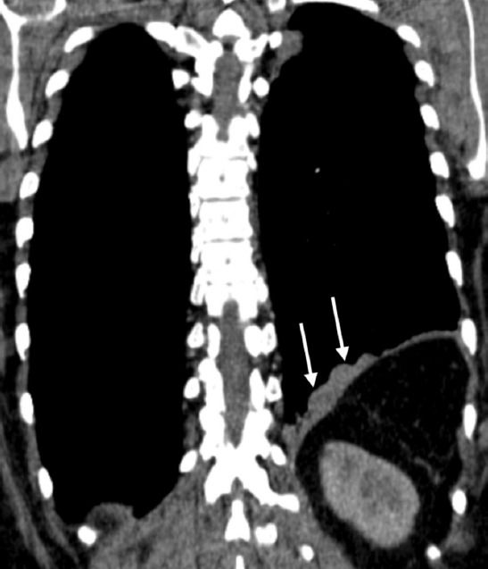
Coronary plane of computed tomography of the chest taken 6 years after the trauma shows splenosis(arrows) that has not been changed during follow-up.
DISCUSSION
Thoracic splenosis is autotransplantation of splenic tissue into the thoracic cavity. Diaphragmatic rupture following to traumatic disruption of the spleen leads to dissemination and auto implantation of splenic tissue.[2] Splenosis can occur within the abdominopelvic cavity, thoracic cavity, subcutaneous tissue, liver, or brain.[2] Splenosis is most commonly (65% of splenic rupture cases) seen in abdominal or pelvic cavities.[3] Intrathoracic splenosis is observed only in 18% of the splenic rupture cases.[3]
Thoracic splenosis can cause cough, chest pain, dyspnea, or hemoptysis. However, most pa-tients are asymptomatic.[1] Therefore, the time interval between the development of splenosis and trauma is not known exactly. Diagnosis is made ranging from 1 to 45 years with a mean delay of up to 21 years.[4] Our patient had had gunshot injury 19 years before her admission. Thus the frequency of thoracic splenosis might be underestimated.
Thoracic splenosis is generally seen as an incidental finding during imaging studies. Thorax CT can reveal multiple or solitary pleural-based nodules variable in size and usually on the left side. On MRI appearance of nodules are similar to normal splenic tissue.[4] Tc-99m scintigraphy using sulfur colloid or Tc-99m-tagged heat-damaged erythrocytes can be used to confirm diagnosis.[5]
Differantial diagnostic considerations based on imaging for splenosis including, primary lung carcinoma, lymphoma, infectious lesions, pleural metastases, asbestos-related pleural disease, thymoma, localized fibrous tumor of the pleura, empyema, sarcoidosis neurogenic tumor, or autoimmune diseases. If malignancy cannot be ruled out even after CT-guided biopsy, video-thoracoscopic biopsy or biopsy through thoracotomy should be performed to rule out any malignancy. Excision of nodules is not recommended and futile unless patient is not symptomatic or risk of malignancy eliminated. Besides, splenic autotransplantation might have a protective role against post-splenectomy infection or sepsis. The patient did not complain any higher incidence of infection. Written informed consent has been obtained from the patient for publication of this case.
Conclusion
Throcic splenosis is clinically asymptomatic in majority of the patients. Therefore, it should be taken in consideration in patients with a history of penetrating trauma when CT reveals pleural nodule and thickening. 99mTC-labeled RBC scintigraphy and thorax CT are appropri-ate radiologic examinations. Biopsy can be made to confirm a diagnosis and exclude malig-nancy. After a definitive diagnosis, patients do need any surgery. Thoracic splenic tissues do not seem to change in long-term (6 years) follow-up in addition to 19 years after trauma. There is no intervention necessary in these rare entities although mild thoracic pain ensues.
Footnotes
Informed Consent: Written informed consent was obtained from the patient for the publication of the case report and the accompanying images.
Peer-review: Internally peer-reviewed.
Authorship Contributions: Concept: Ö.S., B.K., A.T.; Design: Ö.S., B.K., A.T.; Supervision: Ö.S., B.K., A.T.; Resource: Ö.S., B.K., A.T.; Materials: A.T.; Data: A.T., Ö.S.; Literature search: Ö.S., A.T.; Writing: Ö.S., A.T.; Critical revision: A.T.
Conflict of Interest: None declared.
Financial Disclosure: The authors declared that this study has received no financial support.
REFERENCES
- 1.Tian Y, Jiang Q, Li K, Shen Q, Guo F, Fu F, et al. Diagnosis of multiple splenosis in right thorax and retroperitoneum:A case report and literature review. Int J Clin Exp Pathol. 2016;9:7498–502. [Google Scholar]
- 2.Khan AM, Manzoor K, Malik Z, Avsar Y, Shim C. Thoracic splenosis:Know it--avoid unnecessary investigations interventions and thoracotomy. Gen Thorac Cardiovasc Surg. 2011;59:245–53. doi: 10.1007/s11748-010-0706-8. [DOI] [PubMed] [Google Scholar]
- 3.Lake ST, Johnson PT, Kawamoto S, Hruban RH, Fishman EK. CT of splenosis:Pat-terns and pitfalls. Am J Roentgenol. 2012;199:W686–93. doi: 10.2214/AJR.11.7896. [DOI] [PubMed] [Google Scholar]
- 4.Yammine JN, Yatim A, Barbari A. Radionuclide imaging in thoracic splenosis and a review of the literature. Clin Nucl Med. 2003;28:121–3. doi: 10.1097/01.RLU.0000048681.29894.BA. [DOI] [PubMed] [Google Scholar]
- 5.Malik UF, Martin MR, Patel R, Mahmoud A. Parenchymal thoracic splenosis:History and nuclear imaging without invasive procedures may provide diagnosis. J Clin Med Res. 2010;2:180–4. doi: 10.4021/jocmr401w. [DOI] [PMC free article] [PubMed] [Google Scholar]