Abstract
BACKGROUND
A randomized clinical trial from 1984 to 1992 indicated that vitamin A supplementation had a beneficial effect on the progression of retinitis pigmentosa (RP), while vitamin E had an adverse effect.
METHODS
Sequencing of banked DNA samples from that trial provided the opportunity to determine whether certain genotypes responded preferentially to vitamin supplementation.
RESULTS
The genetic solution rate was 587 out of 765 (77%) of sequenced samples. Combining genetic solutions with electroretinogram outcomes showed that there were systematic differences in severity and progression seen among different genetic subtypes of RP, extending findings made for USH2A, RHO, RPGR, PRPF31, and EYS. Baseline electroretinogram 30-Hz flicker implicit time was an independent, strong predictor of progression rate. Using additional data and baseline implicit time as a predictor, the deleterious effect of vitamin E was still present. Surprisingly, the effect of vitamin A progression in the cohort as a whole was not detectable, with or without data from subsequent trials. Subgroup analyses are also discussed.
CONCLUSION
Overall, genetic subtype and implicit time have significant predictive power for a patient’s rate of progression, which is useful prognostically. While vitamin E supplementation should still be avoided, these data do not support a generalized neuroprotective effect of vitamin A for all types of RP.
TRIAL REGISTRATION
ClinicalTrials.gov NCT00000114, NCT00000116, and NCT00346333.
FUNDING
Foundation Fighting Blindness and the National Eye Institute: R01 EY012910, R01 EY031036, R01 EY026904, and P30 EY014104.
Keywords: Ophthalmology
Keywords: Retinopathy
Introduction
Retinitis pigmentosa (RP) is a slowly progressive, inherited, rod-cone retinal degeneration in which early rod photoreceptor death is typically followed by cone photoreceptor death, causing significant visual disability in most patients (1). Multiple strategies have been tested for treating RP, spanning many decades (2–4). Some studies focused on the potential role of certain nutritional supplements in slowing disease progression (5–8), including 3 clinical trials that were conducted at our institution from 1984 to 2008 (9–11). The investigators from those 3 trials not only maintained and preserved complete databases of clinical trial data from all participants but also created a comprehensive biobank for successful long-term storage of participant DNA samples. Now, 30 years later, this unique combination of clinical and genetic resources provides a rare opportunity to apply modern genetic and analytical techniques to a large cohort of RP study participants followed longitudinally.
The concept of the first trial (1984–1991), testing vitamin A and vitamin E supplementation to slow the progression of RP, was initially inspired by positive reports from patients who independently started taking one or both supplements (12). Formal dietary intake studies, combined with 3-year clinical data about progression rates, supported the hypothesis that vitamin A and/or E could be protective against the progression of RP (12). The roles of vitamin A and vitamin E in maintaining photoreceptor function were appreciated at the time as well. A 2 × 2 factorial design (“trace,” “A,” “E,” and “A plus E”) was selected to efficiently test the role of 2 supplements, and their combination, in the setting of a masked, randomized clinical trial. The dose of vitamin A used was 15,000 IU of vitamin A palmitate per day. This trial was notably large (n = 601 participants) and lengthy (4–6 years of follow-up) for a rare disease (9). The primary outcome was the 30-Hz cone flicker electroretinogram (ERG) amplitude, measured using signal averaging, bandpass filtering, and artifact rejection, to allow recording of smaller response amplitudes (9, 13). This cone flicker response amplitude shows a remarkably orderly exponential decay over a certain range of disease severity and correlates with clinically relevant outcomes, such as the ability to drive during the day or at night, walk alone at night, or be employed (14, 15).
The original results of the trial, that vitamin A supplementation slowed the progression of RP by 1.7% per year, and that vitamin E caused faster progression by 1.8% per year, were not met with uniform agreement (16–21). One criticism was that the largest effects of vitamins A and E were mostly seen in the last 2 years of the study (see Figure 5 of the original study in ref. 9), where the sample size was smaller. Regardless of this complexity, the addition of comprehensive genotyping to this data set provided a relatively clean opportunity to test the hypothesis that vitamin A’s effect could differ depending on the genetic cause of RP. One hypothesis is that some genetic subtype(s) benefited greatly, while others did not, resulting in the observed results seen in the ungenotyped RP cohort. For example, it could have been the case that RP associated with certain mutations in the rhodopsin gene (RHO) would benefit from vitamin A treatment, while other genetic types do not. This hypothesis was motivated by the biochemical observations that vitamin A, which is a covalently bound cofactor of RHO, helps certain class II RHO mutants to fold (22), and that vitamin A supplementation in mice expressing the p.Thr17Met but not the p.Pro347Ser mutant of RHO slowed progression of disease (23). In a different study, vitamin A supplementation was found to be adverse in an RHO p.Asp190Asn mutant mouse line. In a different study, an RHO p.Asp190Asn patient was shown to have a high fundus autofluorescence level, and treatment of an RHO p.Asp190Asn mouse model cause faster progression of disease (24). A recent study showed a correlation between serum vitamin A concentrations and disease severity in RHO p.G90D patients (25). Therefore, in this study, we tested the hypothesis that different genetic causes of RP might influence the responses to vitamin A and E supplementation. In the process of exploring this hypothesis, we had the opportunity to (a) determine genetic causes of disease in this well-characterized cohort, learning about the natural history of genetic subtypes of RP; and (b) reexamine details of the results of the original vitamin A/E study.
The original investigators did look for potential differential treatment responses among the genetic subtypes, which at the time were defined as recessive, dominant, X-linked, or unknown, based on pedigree analysis and limited molecular genetic solutions. No differential treatment responses were identified using these categories (9). With the intervening revolution in genetic sequencing, this study applied modern targeted DNA resequencing of all known inherited retinal disease (IRD) genes combined with expert variant annotation to reveal genetic diagnoses for a large fraction of previously unsolved participants in the study (26, 27). We also incorporated a recently defined biomarker for the progression of 30-Hz cone flicker ERG amplitudes, namely the baseline cone flicker implicit time, as a predictive variable (28, 29). Because the results from reanalyzing the original vitamin A/E trial were different than expected (see Results), additional participants were added for increased statistical power from the vitamin A–only arms of 2 later clinical trials that tested the effect of docosahexaenoic acid (DHA) supplementation or lutein supplementation (10, 11). Using this approach, we reevaluated the conclusions of the original study and produced a coherent data set describing the natural history of RP among different molecularly defined genotypes.
Results
Genetic solutions.
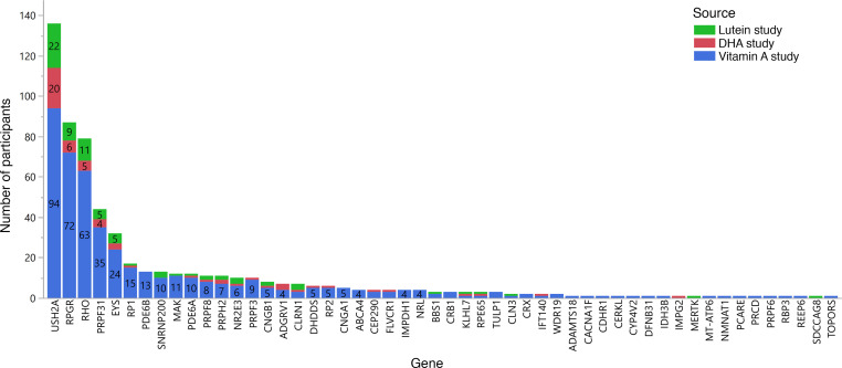
Preexisting genetic solutions from prior studies were known for 211 of the 799 participants included in these analyses. Of the remaining 588 participants, sequencing of banked DNA samples, which generally were in storage for 15–30 years, was highly successful. Of the 588 participants without existing solutions, 554 out of 588 (94%) had usable DNA samples available, which were used for next-generation sequencing (n = 550) or targeted Sanger sequencing when a familial solution was known (n = 4). Including the preexisting solutions and including participants whose samples could not be sequenced, the solution rate was 587 out of 799 (73%). Excluding participants with no remaining usable DNA sample, genetic solutions for 587 out of 765 (77%) sequenced participants were found. The genetic solutions spanned 53 different genes (Figure 1 and Supplemental Table 1; supplemental material available online with this article; https://doi.org/10.1172/jci.insight.167546DS1). The genetic solution rate (solved samples/sequenced samples) was similar between the 3 different study sources analyzed: vitamin A, 77%; DHA, 72%; and lutein, 77%. The most common causative gene was USH2A, found in 136 out of 587 (23%) of the participants with solutions. There were 19 genes identified as the cause of disease in only a single participant.
Figure 1. Histogram showing the number of study participants with a genetic solution in each gene.
Bars are labeled with the number of participants. Participant counts are reported separately for each study because the participants from the vitamin A study were recruited before any genetic solutions for RP were known, and therefore the distribution of genes in that study should be the most unbiased. Participants without a genetic solution (n = 178) are not shown.
Natural history.
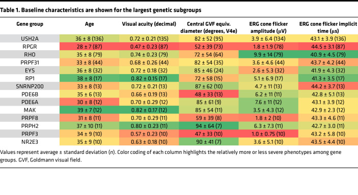
Baseline characteristics were analyzed by gene for participants with mutations in gene groups with 10 or more participants (n = 487 participants, 14 genes; Table 1). Data from less common gene groups (n < 10 participants) or unsolved samples are not shown in Table 1. Participants with RPGR mutations had their first clinical visit at the youngest age, reflecting a more severe phenotype, while participants with mutations in MAK were oldest, mirroring their good visual acuity and visual fields. Participants with PRPH2 and NR2E3 mutations had the best visual fields, whereas participants with mutations in PRPF3, PDE6B, and RPGR had the worst visual fields. Participants with RPGR mutations also had the lowest visual acuity. While a higher ERG amplitude is generally correlated with a shorter implicit time, participants with mutations in EYS had particularly short implicit times, paired with some of the lowest 30-Hz ERG amplitudes. Conversely, SNRNP200 participants had long implicit times with relatively high 30-Hz ERG amplitudes (based on n = 13 participants only). Although the severity of visual field and 30-Hz ERG amplitudes showed correspondence for many groups, there were also examples of one of these measures being disproportionately large relative to the other, for example in PRPF31. (These trends are comparisons between the gene group mean values and are not intended to represent all individual patient values that are spread within each gene group).
Table 1. Baseline characteristics are shown for the largest genetic subgroups.
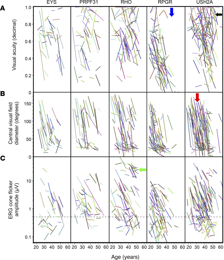
Figure 2 shows how visual acuity, central visual field diameter, and ERG cone flicker amplitude are related to age in the 5 largest genotype groups. These graphs demonstrate the heterogeneity between different participants even within gene groups. In general, all groups tend to lose visual field at a younger age, and acuity at a later age, especially the USH2A group; this is reflected in the cluster of lines appearing further to the left for the visual field row, and further to the right/center in the acuity row. Further gene-specific observations are described in Figure 2.
Figure 2. Natural history of retinitis pigmentosa by age among major genotypes.
Visual acuity (A), central visual field equivalent diameter (B), and ERG cone flicker amplitude (C) are shown for the 5 largest genetic subgroups. A linear curve fit is shown for the data for each participant. The dashed line in C represents 0.5 μV, below which the decay of the response amplitude is less reliably estimated. The lack of points below the blue arrow demonstrates the absence of RPGR participants with normal visual acuity after age 40. USH2A participants have relatively steep visual field declines starting at a variety of ages (red arrow), but can maintain visual acuity into older ages (black arrow). A subset of RHO participants has particularly mild deficits in the ERG amplitudes even at older ages (green arrow).
Effects of vitamin A/E supplementation on ERG cone flicker progression rates.
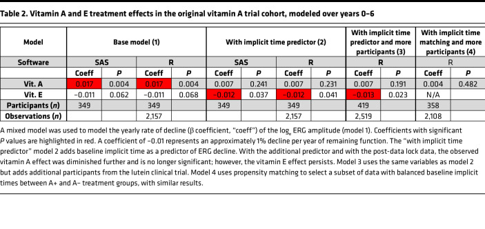
Modeling of the effects of vitamin A and vitamin E on the rate of ERG progression using the participants from the original vitamin A/E study initially produced different results than the original publication; vitamin A had a beneficial significant effect (Table 2; “base model,” P = 0.004), with a slightly smaller P value than that seen in the original paper (P < 0.001). Vitamin E, which had a significant negative influence in the original study (P = 0.04), also showed a negative trend in Table 2 (“base model,” P = 0.07). Reconciliation of available data sets revealed that the data in the “base model” includes additional year 5 and 6 data that were obtained after the data lock used in the original study, which excluded data mostly after September 1991. (Furthermore, a slightly different subset of participants met the minimum baseline ERG amplitude requirement when some minor data processing variabilities in the original data were strictly standardized, e.g., averaging right eye and left eye values before applying the minimum amplitude cutoff.) Using the additional post-data lock data (i.e., all available year 0–6 data) lowered the size of the vitamin A effect and removed the statistical significance of the vitamin E effect. Because the data at years 5 and 6 still had smaller numbers of participants than in prior years, we repeated the analysis on years 0–4 only using model 1. Vitamin A had no significant effect (P > 0.05) and vitamin E had a borderline negative effect (P = 0.046) when looking at the year 0–4 data. Further analyses were performed with all available year 0–6 data (Table 2).
Table 2. Vitamin A and E treatment effects in the original vitamin A trial cohort, modeled over years 0–6.
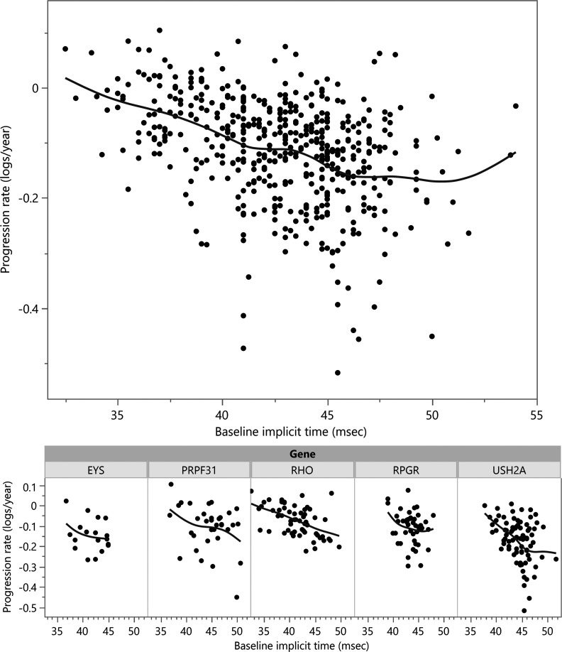
Next, the baseline implicit time was added to the regression model as a predictor of rate of progression (Table 2, model 2 “with implicit time predictor”). This biomarker was highly predictive, showing an additional ERG amplitude decline of –0.01 loge units (~1%) per millisecond of baseline implicit time compared with an overall –0.1 loge unit (~10%) yearly decline in the model without implicit time. A previous study showed implicit time was predictive for RHO RP patients (29). In this data set, the baseline implicit time was significantly predictive of rate of decline in general, and also within RHO, RPGR, and USH2A subgroups when analyzed separately (P < 0.05 for each). The relationship between ERG decline rate and baseline implicit time is shown graphically in Figure 3. Using a mixed model with implicit time as a predictor, there was no significant effect of vitamin A (P > 0.05) and a negative effect of vitamin E of –0.012 loge units (~1.2%, P = 0.04). Part of the vitamin A effect had already been lost by using additional post-data lock data from years 5–6 (see Table 2, model 1). When also using implicit time as a predictor (model 2), the remaining vitamin A effect was lost because, by coincidental imbalance at randomization, the vitamin A+ and A– arms were imbalanced for implicit time at baseline, with median baseline implicit times of 42.8 msec for A+/E± groups and 44.2 msec for A–/E± groups (P = 0.007). (The influence of implicit time as a predictive biomarker was not known at the time of the trial.)
Figure 3. Individual cone flicker ERG amplitude decay rates were calculated for each study participant (y axis), and plotted by baseline ERG cone flicker implicit time (x axis), for all participants (top).
A spline fit shows the trend toward worse progression rates with increasing baseline implicit time. Participants from the largest gene subgroups are shown below. (One outlier point beyond the y axis is not shown.)
Having noted that the original conclusions were not reproduced with these new analyses, we expanded the sample size by adding additional participants taking vitamin A only from the 2 control arms of the subsequent DHA and lutein trials (10, 11). The participants in the active arms of those trials, who took either DHA or lutein, were not included for sequencing or analysis to avoid adding additional treatment variables. The total data set consisted of 799 participants from all 3 trials. It was only possible to add participants to the A+/E– arm of the analysis, as participants in the subsequent trials had been advised to take vitamin A and avoid vitamin E supplementation based on the results of the original trial. Due to this imbalance, the analyses are presented both without (model 2) and with (model 3) these extra participants. The vitamin A–only participants from the DHA trial showed a marginally faster rate of decline (see Methods) than the participants in the original trial, and therefore were not used in this rate-of-change analysis (although including or excluding those participants made only trivial differences; data not shown). After adding additional participants from the lutein trial (Table 2, model 3 “with implicit time predictor and more participants”), there was no significant effect of vitamin A (P > 0.05), but the adverse effect of vitamin E persisted (–0.013 loge units, ~1.3%/year, P = 0.02). Interaction terms between vitamins A and E were not statistically significant for any of the above models.
To assess whether the choice of statistical packages and models contributed to these findings, the analyses were implemented in the R Lmer package (see Methods). Nearly identical results were obtained between SAS and R (Table 2). To assess for the possibility that there is hidden collinearity between baseline implicit time and assigned vitamin A treatment group, and to test for vitamin A effect in a smaller model with the minimal number of variables, propensity score matching was used to create a balanced data set where baseline implicit times are matched between groups. In other words, a subset of the data was used to create a balanced case-control type of analysis instead of using all participants and numerically controlling for the imbalance in implicit times between the groups. The matched data set (n = 358 participants, range 40–139 participants per group) showed homogeneity of baseline implicit time between groups (P = 0.97). With the balanced data set, there was still no beneficial effect of vitamin A treatment (Table 2, model 4 “with implicit time matching & more participants”).
In summary, adding additional year 5 and year 6 data, in combination with adding the predictive power of baseline implicit time to adjust for an imbalanced randomization at baseline, removed the evidence for the broad, beneficial effect of vitamin A that was seen in the original study. This lack of a significant vitamin A treatment effect persisted after adding additional participants from a later trial. The negative effect of vitamin E became smaller but remained statistically significant.
Rates of decline, by gene, and with vitamin A/E supplementation.
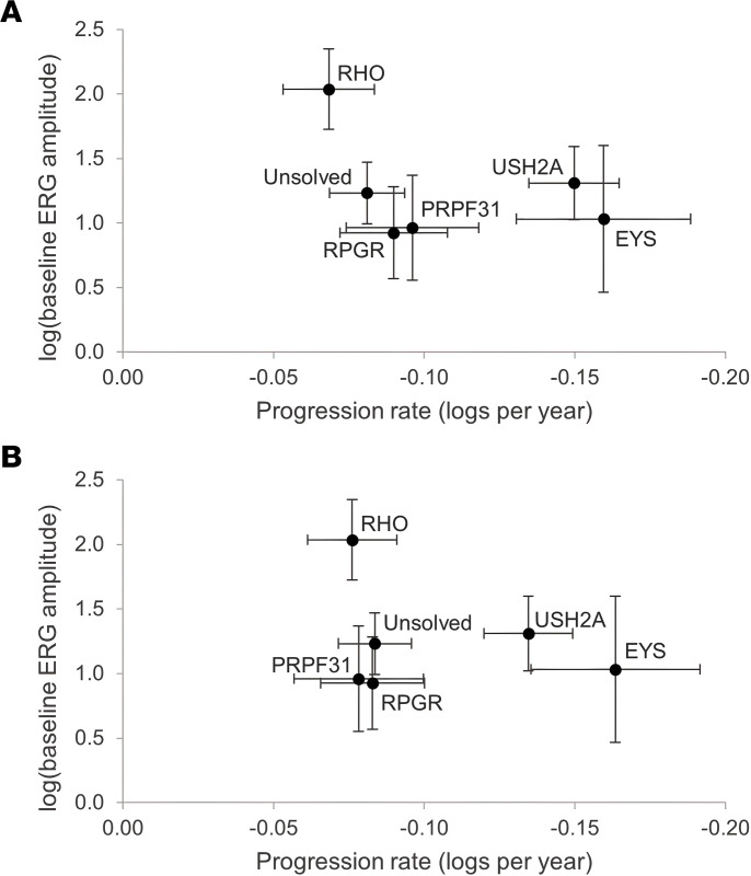
Next, genotype information was added to the progression models. For this purpose, “genotype-specific” is defined at the gene level, pooling together all participants with mutations in the same gene. Small gene groups were pooled into an “Other solutions” group to avoid spurious values from small subgroups (see Methods). A regression model was created estimating the cone flicker ERG amplitude and the cone ERG amplitude progression rate over time, with gene groups of EYS, Other, PRPF31, RHO, RPGR, Unsolved, and USH2A. The effect of the gene on amplitude and on the progression rate of amplitude was strong (P < 0.00001, P < 0.00001, respectively, type 3 test). Figure 4 and Supplemental Table 2 show the rates of progression across the 5 largest genotype groups. Among the participants who started the study with sufficient ERG amplitude to measure change over time (n = 419), participants with mutations in EYS and USH2A showed the highest progression rates, while RHO was the slowest. This observation was consistent whether or not baseline implicit time was controlled for. In order to determine the comparative importance of the different variables in influencing progression rate, calculation of standardized β-regression coefficients showed that there were similar effect sizes for gene effects and for the baseline implicit time effect (not shown). This indicates that gene effects and implicit time effects are of similar importance in predicting progression rate.
Figure 4. Gene-specific estimates of the baseline (time = 0) cone flicker ERG amplitude (y axis) and the yearly ERG amplitude progression rate (x axis).
Both x- and y-axis values are adjusted to remove any vitamin E treatment effect. The results are presented without (A) and with (B) adjustment for baseline implicit time. A negative value on the x axis represents a decrease in amplitude over time.
Note that because of measurement variability at very low ERG amplitudes, there is a “floor effect” beneath which progression cannot be assessed accurately. Therefore, these progression models only include participants (n = 419) whose baseline ERGs were sufficiently high to accurately determine rates of decline (see Methods). This selection of participants with a minimum starting amplitude causes all gene groups except RHO to artificially cluster on the y-axis “floor” at a starting loge(baseline ERG amplitude) of approximately 1–1.5; therefore, estimates of baseline disease severity should be obtained from the Natural history section above, in which there was no lower limit for inclusion in the analysis. The intrinsic progression rate, however, is shown on the x axis and demonstrates the differences in progression between gene groups.
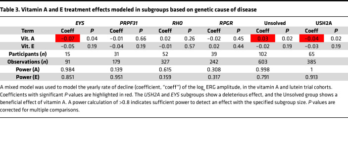
Table 3 shows the results of a regression model used to estimate the effects of vitamin A and vitamin E on progression rate within the genotype subgroups. When looking at the effect of treatment on progression rate, there was a significant interaction with the gene variable overall (P = 0.002). This justifies looking at the gene subgroups. There were no subgroups in which vitamin A showed a beneficial effect (except the “Unsolved” group, which does not have a specific biological meaning). In the USH2A group (n = 65 participants), an adverse effect of vitamin A treatment was observed (coefficient = –0.04, P = 0.02). A borderline adverse effect (P = 0.04) was seen in the EYS subgroup, based on a small sample size of 15. If the same analysis is repeated with minor changes, such as using the time variable rounded to the nearest integer year (as was done in the original study) or using outlier removal for baseline implicit time, the coefficients and P values showed only minor changes; the borderline vitamin A effect in the EYS subgroup had a P value of 0.044 with unrounded time variable data and 0.054 for the rounded time variable data. Because the EYS finding is based on a very small sample size (n = 15 participants split between all arms), the results from the EYS group should be viewed with additional caution. When outlier removal for baseline implicit time is performed while using the unrounded time data, only 4 participants were removed, and no significance level categories changed in Table 2. In Table 3, the vitamin A and E interaction term for the “Unsolved” group became significant, which again does not have a specific biological meaning.
Table 3. Vitamin A and E treatment effects modeled in subgroups based on genetic cause of disease.
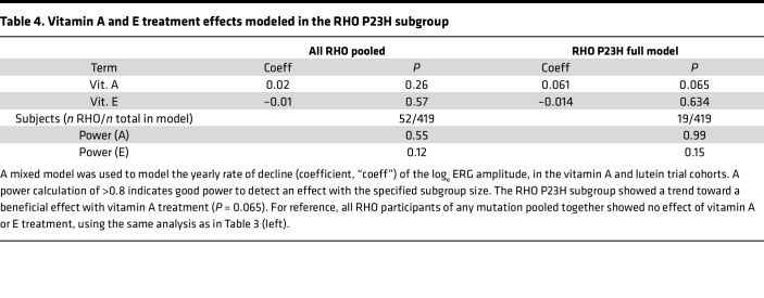
An additional hypothesis was made that certain RHO mutations might be particularly amenable to vitamin A chaperone therapy, while others might not be responsive (23). Among the 52 RHO participants, the largest groups were p.Pro23His (n = 19), p.Pro347Leu (n = 4), and p.Arg135Trp (n = 3), while all other mutations had n = 1 or 2. Therefore, only the p.Pro23His group (henceforth, “RHO P23H”) was considered further. A regression model was constructed with RHO P23H as a separate gene group (n = 19), along with the other gene groups shown in Table 3 (n = 419 total). The results in Table 4 show a trend toward a beneficial effect of vitamin A treatment in this subgroup (+0.061 loge units, ~6.1% per year, n = 19 participants), although with correction for multiple comparisons (n = 8 groups), this effect is not statistically significant (P = 0.065). Vitamin E was still adverse, though not significantly, in this model. While the power calculations are relatively high for this subgroup and the magnitude of the trend is relatively large, similar to the EYS subgroup finding above, the RHO P23H finding should be viewed with additional caution in this small subgroup (n = 19 participants across all arms).
Table 4. Vitamin A and E treatment effects modeled in the RHO P23H subgroup.
Discussion
This study clearly demonstrates the variety of severities and progression rates that occur in RP, based on a well-documented cohort of patients with RP with longitudinal data. Having determined a molecular cause of disease for 73% of all participants (587 out of 799) evaluated, we also demonstrate the success of what may be the oldest DNA biobanking effort for this rare disease. Combining this high-quality phenotype and genotype data improves our understanding of the severity and progression of various RP genetic subtypes. These data can be useful both for prognostic information for patients, as well as for planning patient selection and endpoints for interventional clinical trials. We also found that in contrast to the original reports, updated analyses did not show a benefit for vitamin A supplementation in reducing progression of disease for patients with RP.
Natural history of RP among major genotypes.
As enrollment in the vitamin A study was performed without knowledge of the underlying genotypes, it provides an unbiased view of the genetic composition and natural history of RP. Some genetic causes of RP had been discovered by the time the additional participants were recruited for the lutein and DHA trials, but panel-based genetic testing had not yet been implemented. There were recruitment efforts for RHO families in the department during the time period of the vitamin A studies. Therefore, the distribution of genes in the DHA and lutein trials in Figure 1, which make up a small part of the overall data set, may be slightly skewed by these efforts. For this reason, the gene counts for each trial are reported separately. However, this potential bias may be limited by the study design to include only 1 participant from each family (10, 11).
Starting amplitudes and rates of decline are shown in Figures 3 and 4. While the natural histories for the 4 largest groups (USH2A, RPGR, RHO, and PRPF31) have been published using data from our institution (30–33), this data set, which uses some of the same participants, integrates the findings into a single model where cross-comparison between the groups and to the “average” case of RP is straightforward. We additionally provide comparable progression rates for RP caused by mutations in EYS, the next-largest group in this data set. Baseline severity data for the 14 largest genotype groups are listed in Table 1.
Specifically, it is worth noting that RHO-associated RP remains the mildest among the major genotypes, with a higher baseline ERG and a slower rate of progression. While there are differences in severity among different RHO mutations (32), the average severity of the group as a whole is quite mild in comparison with other genotypes. The RPGR subgroup also continues to stand out as the most severe genotype among the large groups (Table 1), across nearly every metric. Mutations in genes that encode RNA splicing factors, including PRPF31, SNRNP200, PRPF8, and PRPF3, are a common cause of IRD, accounting for 76 participants in total; PRPF3 and PRPF8 cause relatively severe disease (Table 1). EYS participants had comparatively low ERG amplitudes despite having a faster (protective) cone flicker implicit time. EYS participants also had a surprisingly fast average progression rate, which was the fastest among the top 5 gene subgroups. The progression rate showed a trend toward faster progression with vitamin A or E treatment, which complicates the estimation of the true rate without supplementation. It will be interesting to see whether this faster rate of decline is captured in the measures being assessed in the larger cohort of individuals studied in the ongoing natural history study of EYS participants (ClinicalTrials.gov NCT04127006), and in a similar prospective natural history study of USH2A (ClinicalTrials.gov NCT03146078). While a faster progression rate is a poor prognostic sign for affected EYS patients, it may also provide an opportunity to plan shorter clinical trials for interventions that have the potential to slow progression of full-field metrics like the full-field ERG.
One limitation of these progression models, such as those in Figures 3 and 4, is that they can only be used with data from participants with sufficiently high baseline ERG amplitudes so that a decline can be reliably measured over time. In the RPGR group, nearly half (48%) of the participants started with very low ERGs in which progression cannot be assessed. Therefore, the progression rates in the most severe groups are unavoidably based on unusually mild participants in such groups. Of note, this issue does not apply to the cross-sectional data in Figure 2, which includes all participants regardless of initial ERG amplitude. The clinical trials included only participants with “typical RP,” and therefore the RPGR findings do not include participants with cone-rod dystrophies or cone dystrophies.
It was also notable that participants with NR2E3-associated RP had comparably particularly good visual fields. Indeed, it is interesting that 10 NR2E3 participants were included in this cohort of “typical RP” patients, despite a history in our department of attempting to separate out the Enhanced S-cone syndrome/Goldmann-Favre phenotype into a separate category based on factors such as the appearance of clumped pigment and blue-on-yellow visual field testing (34). Similarly, 4 ABCA4 participants were included in the cohort. While ABCA4 defects can cause RP, they more typically cause an inherited macular degeneration (Stargardt disease), with varying degrees of full-field cone or cone-rod dystrophy. While vitamin A processing is known to be defective in this genotype (35), only 1 ABCA4 participant had sufficient starting ERG amplitude to measure a progression rate, so no comparisons of progression between vitamin A treatment groups is possible in ABCA4 participants. Further “unexpected” genetic findings in Supplemental Table 1, such as the presence of 2 manifesting RPGR female carriers, and “dominant” pedigrees that turned out to have X-linked disease, have been previously described in other cohorts (36, 37).
This study did not estimate comparable progression rates by genotype for other outcomes, including visual field, visual acuity, or the mixed-response full-field ERG; comparing group differences and progression rates in these outcomes could be an area for future research. Additional areas of study could be comparison of syndromic versus nonsyndromic disease (38–40) and expanding on genotype/phenotype relationships within the larger gene groups.
In general, it remains a remarkable biological observation that the very particular and localized disease phenotype seen in RP can be caused by defects in so many different genes. While studies such as this one can dissect various important differences between the genotype subgroups in RP, the different genetic forms of RP are, overall, similar enough that it supports the theory of a shared underlying pathophysiology (41–43).
Implicit time.
It has long been known that in normal individuals, the retina produces slower electrical responses to dimmer light stimuli (44). This timing from light onset to response peak is measured as the implicit time, which is recorded in milliseconds. In the context of pathology, a large, healed chorioretinal scar will reduce the amplitude of response without affecting the implicit time, whereas in the case of RP, a decreased amplitude is observed as well as a lengthening of the implicit time (45). In effect, the chorioretinal scar is a situation where some of the retina is simply missing from an otherwise normally timed response, while in the case of RP, there is an abnormality of the response timing of the entire retina, as if the entire retina is seeing a dimmer stimulus. More formally, the effect is not simply due to a shortening of cone outer segments and resulting decreased quantal catch of photons, but instead is caused by abnormally low sensitivity of cone phototransduction consistent with a reduction in the amplification of transduction, as well as a slowing of the responses of the inner retina (46, 47).
There have been hints that a slower (larger) implicit time corresponds to worse disease, for example in sector RP, in which some quadrants of the fundus appear to be normal. When the implicit time is normal or near normal, sector RP has a stationary or more slowly progressive phenotype. However, patients with very delayed cone flicker implicit time, even if the fundus appears to have “sector RP,” have the progressive form of RP (48–50). Berson et al. took this observation further by noting that a patient’s initial implicit time can numerically help predict the rate of decline of the residual cone response amplitude (28). Therefore, the cone flicker implicit time is a “biomarker” in the sense that is a biologically derived measurement that has predictive value.
This study validates the use of the cone flicker implicit time as an important biomarker in the rate of progression of RP (Figures 3 and 4). Longer implicit times are associated with faster rates of ERG amplitude progression, both across the data set as a whole, and also within the largest genetic subgroups. The relationship may be different in EYS, though more data are needed to evaluate this fully. It is interesting that the baseline implicit time is nearly as powerful as the gene name in predicting rate of progression, emphasizing the importance of this biomarker.
Some previous studies have investigated the cone flicker implicit time as an outcome measure, rather than a predictor of disease (38, 40, 51). One noted that patients with smaller fields may have shorter implicit times due to residual foveal cones, which are faster (38). The findings in this study were facilitated by the use of a particular 30-Hz cone flicker ERG protocol (see Methods) that allows recording of responses of lower amplitudes and provides an opportunity to obtain useful amplitude and implicit time data in settings where standard methods may provide more unrecordable signals (e.g., see ref. 52).
Potential effects of vitamin A and E supplementation in the whole RP cohort.
It was not our intention to reevaluate the conclusions of the original study regarding vitamin A and E supplementation in the broader group of all typical RP patients, since the initial goal was to evaluate which genetic subgroups might best respond to vitamin A. However, the data surprisingly indicated that there was no overall robust effect of vitamin A when additional data plus a predictive biomarker were included in the analyses. It is always possible that different data processing or statistical techniques could have uncovered a treatment effect of vitamin A. As described in the Methods, we intentionally maintained data processing procedures as close as possible to those used in the original analysis, including use of a minimum starting ERG amplitude when estimating progression rates, and use of an average of both eyes’ amplitudes. We cannot rule out that alternative processing would be more sensitive. An additional floor cutoff at non-baseline visits and explicit modeling of right versus left eye values have been used in later studies, but these techniques were not used in this study in order to most closely replicate the original data processing of the original vitamin A study. As this result was surprising, we invested additional resources into expanding the sample size using participants in the subsequent trials. These extra participants did not make a fundamental difference in estimating the vitamin A and E treatment effects, but did further enhance the natural history and genetic solutions data set.
The updated analyses in this study also do not change the structure of the original data. It continues to be the case that the last 2 years of the original analysis contained fewer participants than in the original 4 years, even with additional data from after the original data lock. It has already been noted that the emergence of the vitamin A and E effects occurred in those latter years, where those data were more dispersed (Figure 5 of Berson et al., ref. 9). The lack of a vitamin A treatment effect was seen only when both additional data were used from years 5–6 and when implicit time was used as a predictive biomarker. The additional year 5 and 6 data brought the data in those years closer to what it had been in years 1–4, in which there were only small differences between the groups. The use of implicit time as a predictor caused the effect to shrink further, because the trial arms were unbalanced with respect to baseline implicit time in a way that coincidentally had made vitamin A look more protective. (The influence of implicit time as a predictive biomarker was not known at the time of the trial.) In summary, there was no robust effect of vitamin A on the progression of RP in this cohort as a whole. The potential negative effect within the USH2A subgroup is discussed in Subjective clinical recommendations below. The modeled negative effect in the EYS group may very well be spurious because of the small sample size. The trend toward a potential beneficial effect of vitamin A in the RHO P23H group is intriguing, although only based on 19 participants and not statistically significant after multiple comparison correction. Most of the additional RHO mutations are only represented by only 1 or 2 participants. Future work could involve binning these mutations by their biochemical properties in order to use a larger fraction of RHO participants to make more robust conclusions.
Summary.
Overall, this study demonstrates the systematic differences in severity and progression seen among different genetic subtypes of RP. It further demonstrates how the genetic cause of disease, and the 30-Hz ERG implicit time, can have significant predictive power for a patient’s rate of progression. We hope that the lasting contribution of this historical data set will be in helping RP patients and their doctors better understand the severity and expected progression of their disease. Future work may include methods of providing these estimate and predictions in a more accessible format and with refined specificity. Additional endpoints (including visual acuity, visual field, rod-dominant ERG responses) and their progression rates could be evaluated among genetic subgroups as well and may complement data from ongoing prospective natural history studies with additional outcomes such as full-field stimulus threshold testing (40). Furthermore, the validation of implicit time as a biomarker of disease progression in this large cohort may help with participant selection, participant stratification, and endpoint selection in clinical trials for future experimental therapies (53). Broader adoption of the specialized 30-Hz cone flicker ERG amplitude and implicit time protocol used in this study would likely facilitate the use of the implicit time as a predictive biomarker.
Subjective clinical recommendations.
Patients with RP and their doctors may want to know what is recommended for nutritional supplementation in RP based on this study. When data are complex and do not directly answer all questions of clinical relevance, different investigators who are presented with the same data may draw different conclusions and make different recommendations. Therefore, the rest of this section should be considered opinion rather than direct inference.
In our practice, we have stopped recommending vitamin A supplementation for patients who present with new diagnoses of RP. For patients already on vitamin A, there have been a range of approaches in our practice. One physician’s approach has been to recommend cession immediately or at least by the next visit for all patients, while another physician’s approach has been to allow for continuation under certain circumstances. For example, for patients who have been on vitamin A for many years and feel they are doing well, we have noted a natural tendency for them to want to continue. This seems reasonable as long as yearly liver function tests are performed, but becomes more concerning in the setting of osteopenia or osteoporosis since there is some evidence that high vitamin A intake can worsen bone density (54). A history of renal transplantation can also create additional risk (55). Conversely, we are also concerned that long-time patients who then stop vitamin A supplementation, after having had an experience of slow progression subjectively on vitamin A, may later regret their decision if their disease enters a worse stage. This willingness, by some of our physicians, to allow long-term patients to continue vitamin A supplements when deferring to the patient’s preference, is influenced by our experience that the safety record has been very good (56).
Regarding the potential negative effect of vitamin A supplementation in the subgroup of RP associated with USH2A mutations, and to a lesser extent those with EYS mutations, it was our impression that the results of the gene-specific subgroup analyses varied widely based on small additions or changes in input data. We speculate that if a large study was conducted in any specific subgroup, then the potential adverse effects would be unlikely to be replicated. However, notwithstanding the many limitations of any statistical test, the final statistical calculation was well powered to detect an effect of vitamin A in the USH2A subgroup, and the observed effect was adverse. Therefore, for patients with RP associated with USH2A mutations who are on vitamin A supplements, we make a recommendation to stop supplementation. The only genetic group for which vitamin A had a trend toward a beneficial effect was for those participants with the p.Pro23His mutation in RHO. The effect size was large, although based on a group of only 19 patients, and was not statistically significant after multiple-test correction. While vitamin A supplementation could be considered in this subgroup, there is not high confidence in any recommendation based on this modest amount of data.
For children, retrospective nonrandomized data suggest a significant benefit on progression rate in children with typical RP (28); on the other hand, the current study, which was masked and randomized, showed no benefit in adults. These 2 studies used completely independent data sets on different participants. The study in children used a small sample size and analyzed a variety of lengths of follow-ups. We speculate that it is more likely that the retrospective study in children could not adequately control for known or unknown confounding factors, compared with the possibility that there is a different effect in children than adults, or that this study encountered a type II error (failing to identify a true effect).
While this speculation is admittedly subjective and not at all certain, we have stopped recommending vitamin A supplementation for children with RP as well.
For vitamin E, we still recommend avoiding high-dose supplementation (>30 IU/day). Such supplements are typically marketed as strong antioxidants.
This study does not have any direct impact on the potential effects of DHA or lutein supplementation (5, 10, 11, 51).
In summary, we currently do not recommend vitamin A or E supplementation for patients with RP. It is possible that further research in subsets of individuals with biochemically distinct mutations would provide additional data. For context, it should be emphasized that other gene-independent, neuroprotective strategies for treatment of patients with IRDs are under study, including rod-derived cone viability factor (RdCVF) (57), N-acetyl cysteine (NAC) (57, 58), and NFE2-like bZIP transcription factor 2 (NFE2L2, previously known as NRF2) (59), which hopefully will provide benefit for RP patients. Gene-specific therapies are of course making great progress as well (60), along with strategies for end-stage disease in which there are no rods or cones (e.g., optogenetics) (61).
Methods
Cohorts.
From 1984 to 1991, 601 patients participated in the vitamin A/E trial (ClinicalTrials.gov NCT00000114) (12). Patients with certain forms of “atypical RP” were excluded, such as Usher syndrome type I, Bardet-Biedl syndrome, pericentral RP, sector RP, and X-linked RP carriers. Usher syndrome type II patients were included, and syndromic versus nonsyndromic presentations were not distinguished for purposes of analysis. Patients in all 4 treatment groups (“trace,” “A,” “E,” and “A plus E”) were included in the current analyses. Preliminary analyses of the effect of vitamin A and E on longitudinal progression rates were different than the findings of the original study (see Results), and therefore additional participants were added from later clinical trials to increase the sample size. Specifically, participants were added from the vitamin A–only controls arms of the subsequent, separate DHA trial (n = 91 unique additional participants, NCT00000116) and lutein trial (n = 107 unique additional participants, NCT00346333) (10, 11). The participants who received DHA or lutein were not included, neither in the DNA resequencing efforts nor in the data analysis.
We assessed for homogeneity of the 3 data sets (see Supplemental Methods). This model showed that, compared with the vitamin A trial, participants in the vitamin A–only arm of the lutein trial had a similar progression rate to that of the original trial (β = 0.01, P = 0.59), but participants in the vitamin A–only arm of the DHA trial showed a faster progression rate (β = –0.02, P = 0.039) using rounded time data, i.e., coincidentally the participants in the DHA trial showed faster progression despite being on the same treatment arm (vitamin A only). Therefore, the participants’ data from the DHA trial were not used for the purposes of regression modeling of rates of decline. Similar homogeneity results were obtained with unrounded time data, with (P = 0.57) or without (P = 0.075) implicit time outliers removed. Also, the mean baseline implicit values were lower overall in the DHA trial (42.94 msec for the original trial, 42.97 msec for the lutein trails, and 42.03 msec for the DHA trial; P < 0.001 by 1-way ANOVA), supporting the rationale that the patients in the vitamin A–only arm of the DHA trial should not be mixed with the data from the other 2 trials for the purposes of regression modeling of rates of decline. However, the data from all 3 trials were used elsewhere in this manuscript, where the rate of decline is not explicitly being modeled (see flow chart in Supplemental Figure 1).
Clinical data.
The methods for data collection for all 3 trials can be found in the original publications (9–11). We note that the ERG data acquisition methods and equipment were carefully maintained over the years to ensure consistent measurements (and remain in clinical use). These methods were developed before International Society of Clinical Electrophysiology of Vision (ISCEV) protocols were established and use a xenon flash of 0.2 cd•s/m2 instead of the now-standard 3 cd•s/m2 flashes for recording the 30-Hz flicker responses. The recording acquisition and processing also use signal averaging, bandpass filtering, and artifact rejection, to allow recording of smaller response amplitudes (13). Goldmann central visual field areas were measured by planimetry of the V4e response area that was contiguous with the center, and converted to an equivalent circular diameter using the formula diameter = 2 × sqrt(area/π).
Genotyping.
Preexisting genetic solutions were available for 211 samples. Five hundred fifty-four unsolved cases, for whom DNA samples were available, were analyzed with the Genetic Eye Disease (GEDi) targeted sequencing panel of all known IRD genes, as described previously (26), or by Sanger sequencing in 4 cases. Sequence data were aligned to the hg38 genome build and the subsequent variant calling, annotation, and analyses were performed as described previously (27). Copy number variation (CNV) predictions were produced using gCNV (62, 63), and the known MAK-Alu structural variant was identified using a custom script (64). Screening of mutations in the RPGR ORF15 was performed by PCR amplification of the target region and Sanger sequencing using established methods (65). Variants were classified according to the American College of Medical Genetics and Genomics (ACMG) guidelines (66, 67) and adjudicated by the authors. Recessive solutions were required to have 2 mutations identified, but segregation testing was not available in many cases. Samples were marked as either solved or unsolved, with the solved samples being pooled by gene into gene-specific groups for further analysis. The small number of samples without sequencing data available (n = 34) were also marked as unsolved. Some solutions have been previously published (see Supplemental Table 1). Sixty-one of the 765 solutions (8%) were obtained from whole-exome sequencing, as reported previously (27, 63).
Longitudinal analysis of ERG data.
Data processing of ERG parameters was performed as closely as possible to the original study in consultation with the original data manager of the study (C Weigel DiFranco) (9). For additional description of ERG data processing and statistical methods, please see Supplemental Methods.
Over the course of several iterations of data processing (e.g., as more participants were solved genetically and added), it was noted that when looking at small subsets of participants, there were unstable estimates of the effects of vitamin A and E on progression rates, with small changes in the input data set (data not shown). Power calculations further supported the notion that very small subgroups should not be analyzed. Therefore, analyses for single-gene subgroups were only performed for the larger gene groups: USH2A, RHO, RPGR, PRPF31, and EYS. Genetic solutions for smaller gene groups were pooled into an “Other solutions” group for the regression models. The “Unsolved” group represents the group of participants who had no genetic solution identified after sequencing and analysis. In general, all mutations in the same gene were pooled together; however, an additional hypothesis was made that certain mutations or mutation classes of RHO mutations might be differentially responsive to vitamin A (23). There was only 1 subgroup, RHO P23H, with enough patients (n = 19) to attempt a subgroup analysis.
There are some participants with additional long-term follow-up data that are now available, including those who participated in multiple trials and accumulated very long total follow-up time. Initial regression modeling using imbalanced, very-long-term data (e.g., 23 years) from some participants and only year 4–6 data from the rest of the participants produced results that were overly weighted by the outcomes of the very-long-term participants (data not shown). It also would have required additional variables to model participants who switched vitamin A treatment status during these longer follow-up time periods. Therefore, for each participant, only the first 6 years of data were used for regression models.
Subsequent analyses were performed in R; see Supplemental Methods.
Study approval.
The research was conducted in compliance with Mass Eye and Ear Institutional Review Board (IRB) approval. Written informed consent had been obtained from the participants after explanation of the nature and possible consequences of the study.
Author contributions
JC and CWD designed the study, processed and analyzed phenotype and genotype data, performed statistical analyses, and wrote the manuscript. KS and RMH analyzed data and wrote the manuscript. E Place, EZ, and KMB processed genotype data and analyzed variants. MM processed phenotype and genotype data. YZ performed statistical analyses. E Pierce designed the study, analyzed phenotype and genotype data, wrote the manuscript, and obtained funding. Co–first author order was determined by the authors’ relative contributions to the content, analysis, and writing of the manuscript.
Supplementary Material
Acknowledgments
Funding for this study was provided by the Foundation Fighting Blindness and the National Eye Institute R01 EY012910 (to E Pierce), R01 EY031036 (to JC) R01 EY026904 (to KB and E Pierce), and P30 EY014104 (MEEI core support). The original vitamin A/E study was funded by the National Eye Institute and the Foundation Fighting Blindness. Center grants from the Foundation Fighting Blindness over the past 4 decades allowed for the creation and maintenance of the resources used in this study, including the DNA biobank and multiple generations of genetic testing infrastructure. The authors would like to thank the research participants who contributed to the original clinical trials in our department, most of whom traveled across the country every year for extensive testing as trial participants. We would like to thank Eliot Berson, Ted Dryja, Bernard Rosner, their research groups, and all the original investigators and data managers, including Carol Weigel DiFranco and Shyana Harper, who over the last 30+ years contributed to the collection and maintenance of the large data sets and the large DNA sample repository that were used for this study. We thank Michael Sandberg for his contributions to implicit time as a predictive biomarker and to the software and hardware used for accurate measurement of low-amplitude ERG recordings. We thank the clinical staff of the Inherited Disorders Service (formerly, ERG Service) at Mass Eye and Ear. While we know that Eliot Berson (1937–2017) would be less than pleased with certain findings of this study, we hope that he would be encouraged by the recent progress in the field that he helped create, and by the efforts of the doctors and scientists he trained who share his commitment toward developing better therapies for patients with inherited retinal diseases.
Version 1. 06/01/2023
In-Press Preview
Version 2. 08/08/2023
Electronic publication
Footnotes
Conflict of interest: RMH, JC, and EP disclose payments to their institution for clinical trial agreements not directly related to this study. RMH and JC disclose receipt of consulting fees or other services from Biogen, Gensight, and Spark Therapeutics. RMH discloses receipt of consulting fees or equity from AGTC, Annexon, BlueRock Therapeutics, ProQR, Regeneron, Sunovion, Vida Ventures, and Intergalatic therapeutics.
Copyright: © 2023, Comander et al. This is an open access article published under the terms of the Creative Commons Attribution 4.0 International License.
Reference information: JCI Insight. 2023;8(13):e167546.https://doi.org/10.1172/jci.insight.167546.
Contributor Information
Jason Comander, Email: jason_comander_public@meei.harvard.edu.
Matthew Maher, Email: MMAHER@MGH.HARVARD.EDU.
Erin Zampaglione, Email: erzampag@gmail.com.
Yan Zhao, Email: yzhao38@meei.harvard.edu.
Rachel M. Huckfeldt, Email: Rachel_Huckfeldt@MEEI.HARVARD.EDU.
Kinga M. Bujakowska, Email: kinga_bujakowska@meei.harvard.edu.
References
- 1.Hartong DT, et al. Retinitis pigmentosa. Lancet. 2006;368(9549):1795–1809. doi: 10.1016/S0140-6736(06)69740-7. [DOI] [PubMed] [Google Scholar]
- 2.Brooks BP, Jeffrey B. Methodological insights for randomized clinical trials of retinitis pigmentosa: lessons learned from a trial of valproic acid. JAMA Ophthalmol. 2018;136(8):857–858. doi: 10.1001/jamaophthalmol.2018.1167. [DOI] [PMC free article] [PubMed] [Google Scholar]
- 3.Newell FW. Clinical trials and retinitis pigmentosa. Am J Ophthalmol. 1978;85(5 pt 1):720–721. doi: 10.1016/S0002-9394(14)77115-6. [DOI] [PubMed] [Google Scholar]
- 4.Macdonald AE, McKenzie KG. Sympathectomy for retinitis pigmentosa. Trans Am Ophthalmol Soc. 1934;32:172–190. [PMC free article] [PubMed] [Google Scholar]
- 5.Hoffman DR, et al. Docosahexaenoic acid slows visual field progression in X-linked retinitis pigmentosa: ancillary outcomes of the DHAX trial. Invest Ophthalmol Vis Sci. 2015;56(11):6646–6653. doi: 10.1167/iovs.15-17786. [DOI] [PMC free article] [PubMed] [Google Scholar]
- 6.Chatzinoff A, et al. Eleven-CIS vitamin A in the treatment of retinitis pigmentosa. A negative study. Arch Ophthalmol. 1968;80(4):417–419. doi: 10.1001/archopht.1968.00980050419001. [DOI] [PubMed] [Google Scholar]
- 7.Dagnelie G, et al. Lutein improves visual function in some patients with retinal degeneration: a pilot study via the internet. Optometry. 2000;71(3):147–164. [PubMed] [Google Scholar]
- 8.Brito-Garcia N, et al. Effectiveness and safety of nutritional supplements in the treatment of hereditary retinal dystrophies: a systematic review. Eye (Lond) 2017;31(2):273–285. doi: 10.1038/eye.2016.286. [DOI] [PMC free article] [PubMed] [Google Scholar]
- 9.Berson EL, et al. A randomized trial of vitamin A and vitamin E supplementation for retinitis pigmentosa. Arch Ophthalmol. 1993;111(6):761–772. doi: 10.1001/archopht.1993.01090060049022. [DOI] [PubMed] [Google Scholar]
- 10.Berson EL, et al. Clinical trial of docosahexaenoic acid in patients with retinitis pigmentosa receiving vitamin A treatment. Arch Ophthalmol. 2004;122(9):1297–1305. doi: 10.1001/archopht.122.9.1297. [DOI] [PubMed] [Google Scholar]
- 11.Berson EL, et al. Clinical trial of lutein in patients with retinitis pigmentosa receiving vitamin A. Arch Ophthalmol. 2010;128(4):403–411. doi: 10.1001/archophthalmol.2010.32. [DOI] [PMC free article] [PubMed] [Google Scholar]
- 12.Berson EL, et al. Natural course of retinitis pigmentosa over a three-year interval. Am J Ophthalmol. 1985;99(3):240–251. doi: 10.1016/0002-9394(85)90351-4. [DOI] [PubMed] [Google Scholar]
- 13.Andreasson SO, et al. Narrow-band filtering for monitoring low-amplitude cone electroretinograms in retinitis pigmentosa. Am J Ophthalmol. 1988;105(5):500–503. doi: 10.1016/0002-9394(88)90241-3. [DOI] [PubMed] [Google Scholar]
- 14.Berson EL. Long-term visual prognoses in patients with retinitis pigmentosa: the Ludwig von Sallmann lecture. Exp Eye Res. 2007;85(1):7–14. doi: 10.1016/j.exer.2007.03.001. [DOI] [PMC free article] [PubMed] [Google Scholar]
- 15.Berson EL. Retinitis pigmentosa. The Friedenwald lecture. Invest Ophthalmol Vis Sci. 1993;34(5):1659–1676. [PubMed] [Google Scholar]
- 16.Norton EW. A randomized trial of vitamin A and vitamin E supplementation for retinitis pigmentosa. Arch Ophthalmol. 1993;111(11):1460–1465. doi: 10.1001/archopht.1993.01090110018003. [DOI] [PubMed] [Google Scholar]
- 17.Marmor MF. A randomized trial of vitamin A and vitamin E supplementation for retinitis pigmentosa. Arch Ophthalmol. 1993;111(11):1460–1465. doi: 10.1001/archopht.1993.01090110018004. [DOI] [PubMed] [Google Scholar]
- 18.Clowes DD. A randomized trial of vitamin A and vitamin E supplementation for retinitis pigmentosa. Arch Ophthalmol. 1993;111(11):1461–1465. doi: 10.1001/archopht.1993.01090110018005. [DOI] [PubMed] [Google Scholar]
- 19.Massof RW, Finkelstein D. Supplemental vitamin A retards loss of ERG amplitude in retinitis pigmentosa. Arch Ophthalmol. 1993;111(6):751–754. doi: 10.1001/archopht.1993.01090060039019. [DOI] [PubMed] [Google Scholar]
- 20.Massof RW, Fishman GA. How strong is the evidence that nutritional supplements slow the progression of retinitis pigmentosa? Arch Ophthalmol. 2010;128(4):493–495. doi: 10.1001/archophthalmol.2010.46. [DOI] [PubMed] [Google Scholar]
- 21.Gamel JW, Barr CC. A randomized trial of vitamin A and vitamin E supplementation for retinitis pigmentosa. Arch Ophthalmol. 1993;111(11):1462–1463. doi: 10.1001/archopht.1993.01090110018006. [DOI] [PubMed] [Google Scholar]
- 22.Wan A, et al. Characterizing variants of unknown significance in rhodopsin: A functional genomics approach. Hum Mutat. 2019;40(8):1127–1144. doi: 10.1002/humu.23762. [DOI] [PMC free article] [PubMed] [Google Scholar]
- 23.Li T, et al. Effect of vitamin A supplementation on rhodopsin mutants threonine-17 --> methionine and proline-347 --> serine in transgenic mice and in cell cultures. Proc Natl Acad Sci U S A. 1998;95(20):11933–11938. doi: 10.1073/pnas.95.20.11933. [DOI] [PMC free article] [PubMed] [Google Scholar]
- 24.Cui X, et al. Long-term vitamin A supplementation in a preclinical mouse model for RhoD190N-associated retinitis pigmentosa. Hum Mol Genet. 2022;31(14):2438–2451. doi: 10.1093/hmg/ddac032. [DOI] [PMC free article] [PubMed] [Google Scholar]
- 25.Krasovec T, et al. Correlation between the serum concentration of vitamin A and disease severity in patients carrying p.G90D in RHO, the most frequent gene associated with dominant retinitis pigmentosa: implications for therapy with vitamin A. Int J Mol Sci. 2023;24(1):780. doi: 10.3390/ijms24010780. [DOI] [PMC free article] [PubMed] [Google Scholar]
- 26.Consugar MB, et al. Panel-based genetic diagnostic testing for inherited eye diseases is highly accurate and reproducible, and more sensitive for variant detection, than exome sequencing. Genet Med. 2015;17(4):253–261. doi: 10.1038/gim.2014.172. [DOI] [PMC free article] [PubMed] [Google Scholar]
- 27.Zampaglione E, et al. The importance of automation in genetic diagnosis: lessons from analyzing an inherited retinal degeneration cohort with the Mendelian Analysis Toolkit (MATK) Genet Med. 2022;24(2):332–343. doi: 10.1016/j.gim.2021.09.015. [DOI] [PMC free article] [PubMed] [Google Scholar]
- 28.Berson EL, et al. Association of vitamin A supplementation with disease course in children with retinitis pigmentosa. JAMA Ophthalmol. 2018;136(5):490–495. doi: 10.1001/jamaophthalmol.2018.0590. [DOI] [PMC free article] [PubMed] [Google Scholar]
- 29.Rahmani S, et al. A biomarker for disease course in patients with autosomal dominant retinitis pigmentosa due to RHO mutations. Invest Ophthalmol Vis Sci. 2016;57(12):134 [Google Scholar]
- 30.Sandberg MA, et al. Disease course in patients with autosomal recessive retinitis pigmentosa due to the USH2A gene. Invest Ophthalmol Vis Sci. 2008;49(12):5532–5539. doi: 10.1167/iovs.08-2009. [DOI] [PMC free article] [PubMed] [Google Scholar]
- 31.Sandberg MA, et al. Disease course of patients with X-linked retinitis pigmentosa due to RPGR gene mutations. Invest Ophthalmol Vis Sci. 2007;48(3):1298–1304. doi: 10.1167/iovs.06-0971. [DOI] [PubMed] [Google Scholar]
- 32.Berson EL, et al. Disease progression in patients with dominant retinitis pigmentosa and rhodopsin mutations. Invest Ophthalmol Vis Sci. 2002;43(9):3027–3036. [PubMed] [Google Scholar]
- 33.Hafler BP, et al. Course of ocular function in PRPF31 retinitis pigmentosa. Semin Ophthalmol. 2016;31(1–2):49–52. doi: 10.3109/08820538.2015.1114856. [DOI] [PMC free article] [PubMed] [Google Scholar]
- 34.Jacobson SG, et al. SWS (blue) cone hypersensitivity in a newly identified retinal degeneration. Invest Ophthalmol Vis Sci. 1990;31(5):827–838. [PubMed] [Google Scholar]
- 35.Sajovic J, et al. The role of vitamin A in retinal diseases. Int J Mol Sci. 2022;23(3):1014. doi: 10.3390/ijms23031014. [DOI] [PMC free article] [PubMed] [Google Scholar]
- 36.Comander J, et al. Visual function in carriers of X-linked retinitis pigmentosa. Ophthalmology. 2015;122(9):1899–1906. doi: 10.1016/j.ophtha.2015.05.039. [DOI] [PMC free article] [PubMed] [Google Scholar]
- 37.Daiger SP, et al. Molecular findings in families with an initial diagnose of autosomal dominant retinitis pigmentosa (adRP) Adv Exp Med Biol. 2018;1074:237–245. doi: 10.1007/978-3-319-75402-4_29. [DOI] [PMC free article] [PubMed] [Google Scholar]
- 38.Sengillo JD, et al. Electroretinography reveals difference in cone function between syndromic and nonsyndromic USH2A patients. Sci Rep. 2017;7(1):11170. doi: 10.1038/s41598-017-11679-y. [DOI] [PMC free article] [PubMed] [Google Scholar]
- 39.Pierrache LH, et al. Visual prognosis in USH2A-associated retinitis pigmentosa is worse for patients with Usher syndrome type IIa than for those with nonsyndromic retinitis pigmentosa. Ophthalmology. 2016;123(5):1151–1160. doi: 10.1016/j.ophtha.2016.01.021. [DOI] [PubMed] [Google Scholar]
- 40.Birch DG, et al. The RUSH2A study: best-corrected visual acuity, full-field electroretinography amplitudes, and full-field stimulus thresholds at baseline. Transl Vis Sci Technol. 2020;9(11):9. doi: 10.1167/tvst.9.11.9. [DOI] [PMC free article] [PubMed] [Google Scholar]
- 41.Punzo C, Cepko C. Cellular responses to photoreceptor death in the rd1 mouse model of retinal degeneration. Invest Ophthalmol Vis Sci. 2007;48(2):849–857. doi: 10.1167/iovs.05-1555. [DOI] [PubMed] [Google Scholar]
- 42.Cepko C, Punzo C. Cell metabolism: sugar for sight. Nature. 2015;522(7557):428–429. doi: 10.1038/522428a. [DOI] [PubMed] [Google Scholar]
- 43.Amamoto R, et al. Retinoic acid signaling mediates peripheral cone photoreceptor survival in a mouse model of retina degeneration. Elife. 2022;11:e76389. doi: 10.7554/eLife.76389. [DOI] [PMC free article] [PubMed] [Google Scholar]
- 44.Bornschein H, et al. Temporal aspects of the human electroretinogram; a study of the implicit time-amplitude relationship of the B-wave. AMA Arch Ophthalmol. 1957;57(3):386–392. doi: 10.1001/archopht.1957.00930050398010. [DOI] [PubMed] [Google Scholar]
- 45.Berson EL, et al. Temporal aspects of the electroretinogram. Arch Ophthalmol. 1969;81(2):207–214. doi: 10.1001/archopht.1969.00990010209011. [DOI] [PubMed] [Google Scholar]
- 46.Hood DC, Birch DG. Abnormalities of the retinal cone system in retinitis pigmentosa. Vision Res. 1996;36(11):1699–1709. doi: 10.1016/0042-6989(95)00246-4. [DOI] [PubMed] [Google Scholar]
- 47.Birch DG, Sandberg MA. Dependence of cone b-wave implicit time on rod amplitude in retinitis pigmentosa. Vision Res. 1987;27(7):1105–1112. doi: 10.1016/0042-6989(87)90025-3. [DOI] [PubMed] [Google Scholar]
- 48.Berson EL, Howard J. Temporal aspects of the electroretinogram in sector retinitis pigmentosa. Arch Ophthalmol. 1971;86(6):653–665. doi: 10.1001/archopht.1971.01000010655008. [DOI] [PubMed] [Google Scholar]
- 49.Ballios BG, et al. Beyond sector retinitis pigmentosa: expanding the phenotype and natural history of the rhodopsin gene codon 106 mutation (Gly-to-Arg) in autosomal dominant retinitis pigmentosa. Genes (Basel) 2021;12(12):1853. doi: 10.3390/genes12121853. [DOI] [PMC free article] [PubMed] [Google Scholar]
- 50.Georgiou M, et al. Sector retinitis pigmentosa: extending the molecular genetics basis and elucidating the natural history. Am J Ophthalmol. 2021;221:299–310. doi: 10.1016/j.ajo.2020.08.004. [DOI] [PMC free article] [PubMed] [Google Scholar]
- 51.Hoffman DR, et al. Four-year placebo-controlled trial of docosahexaenoic acid in X-linked retinitis pigmentosa (DHAX trial): a randomized clinical trial. JAMA Ophthalmol. 2014;132(7):866–873. doi: 10.1001/jamaophthalmol.2014.1634. [DOI] [PMC free article] [PubMed] [Google Scholar]
- 52.Testa F, et al. Clinical presentation and disease course of Usher syndrome because of mutations in MYO7A OR USH2A. Retina. 2017;37(8):1581–1590. doi: 10.1097/IAE.0000000000001389. [DOI] [PubMed] [Google Scholar]
- 53. Thompson DA, et al. Advancing clinical trials for inherited retinal diseases: recommendations from the second Monaciano Symposium. Transl Vis Sci Technol. 2020;9(7):2. [DOI] [PMC free article] [PubMed] [Google Scholar]
- 54.Tanumihardjo SA. Vitamin A and bone health: the balancing act. J Clin Densitom. 2013;16(4):414–419. doi: 10.1016/j.jocd.2013.08.016. [DOI] [PubMed] [Google Scholar]
- 55.Makoff R. Vitamin replacement therapy in renal failure patients. Miner Electrolyte Metab. 1999;25(4–6):349–351. doi: 10.1159/000057473. [DOI] [PubMed] [Google Scholar]
- 56.Sibulesky L, et al. Safety of <7500 RE (<25000 IU) vitamin A daily in adults with retinitis pigmentosa. Am J Clin Nutr. 1999;69(4):656–663. doi: 10.1093/ajcn/69.4.656. [DOI] [PubMed] [Google Scholar]
- 57.Byrne LC, et al. Viral-mediated RdCVF and RdCVFL expression protects cone and rod photoreceptors in retinal degeneration. J Clin Invest. 2015;125(1):105–116. doi: 10.1172/JCI65654. [DOI] [PMC free article] [PubMed] [Google Scholar]
- 58.Campochiaro PA, et al. Oral N-acetylcysteine improves cone function in retinitis pigmentosa patients in phase I trial. J Clin Invest. 2020;130(3):1527–1541. doi: 10.1172/JCI132990. [DOI] [PMC free article] [PubMed] [Google Scholar]
- 59.Wu DM, et al. Nrf2 overexpression rescues the RPE in mouse models of retinitis pigmentosa. JCI Insight. 2021;6(2):e145029. doi: 10.1172/jci.insight.145029. [DOI] [PMC free article] [PubMed] [Google Scholar]
- 60.Russell S, et al. Efficacy and safety of voretigene neparvovec (AAV2-hRPE65v2) in patients with RPE65-mediated inherited retinal dystrophy: a randomised, controlled, open-label, phase 3 trial. Lancet. 2017;390(10097):849–860. doi: 10.1016/S0140-6736(17)31868-8. [DOI] [PMC free article] [PubMed] [Google Scholar]
- 61.Fortuny C, Flannery JG. Mutation-independent gene therapies for rod-cone dystrophies. Adv Exp Med Biol. 2018;1074:75–81. doi: 10.1007/978-3-319-75402-4_10. [DOI] [PubMed] [Google Scholar]
- 62.McKenna A, et al. The genome analysis toolkit: a MapReduce framework for analyzing next-generation DNA sequencing data. Genome Res. 2010;20(9):1297–1303. doi: 10.1101/gr.107524.110. [DOI] [PMC free article] [PubMed] [Google Scholar]
- 63.Zampaglione E, et al. Copy-number variation contributes 9% of pathogenicity in the inherited retinal degenerations. Genet Med. 2020;22(6):1079–1087. doi: 10.1038/s41436-020-0759-8. [DOI] [PMC free article] [PubMed] [Google Scholar]
- 64.Bujakowska KM, et al. Efficient in silico identification of a common insertion in the MAK gene which causes retinitis pigmentosa. PLoS One. 2015;10(11):e0142614. doi: 10.1371/journal.pone.0142614. [DOI] [PMC free article] [PubMed] [Google Scholar]
- 65.Nassisi M, et al. Retrospective natural history study of RPGR-related cone- and cone-rod dystrophies while expanding the mutation spectrum of the disease. Int J Mol Sci. 2022;23(13):7189. doi: 10.3390/ijms23137189. [DOI] [PMC free article] [PubMed] [Google Scholar]
- 66.Richards S, et al. Standards and guidelines for the interpretation of sequence variants: a joint consensus recommendation of the American College of Medical Genetics and Genomics and the Association for Molecular Pathology. Genet Med. 2015;17(5):405–424. doi: 10.1038/gim.2015.30. [DOI] [PMC free article] [PubMed] [Google Scholar]
- 67.Riggs ER, et al. Technical standards for the interpretation and reporting of constitutional copy-number variants: a joint consensus recommendation of the American College of Medical Genetics and Genomics (ACMG) and the Clinical Genome Resource (ClinGen) Genet Med. 2020;22(2):245–257. doi: 10.1038/s41436-019-0686-8. [DOI] [PMC free article] [PubMed] [Google Scholar]
Associated Data
This section collects any data citations, data availability statements, or supplementary materials included in this article.


