Abstract
The biological properties of wild-type A75/17 and cell culture-adapted Onderstepoort canine distemper virus differ markedly. To learn more about the molecular basis for these differences, we have isolated and sequenced the protein-coding regions of the attachment and fusion proteins of wild-type canine distemper virus strain A75/17. In the attachment protein, a total of 57 amino acid differences were observed between the Onderstepoort strain and strain A75/17, and these were distributed evenly over the entire protein. Interestingly, the attachment protein of strain A75/17 contained an extension of three amino acids at the C terminus. Expression studies showed that the attachment protein of strain A75/17 had a higher apparent molecular mass than the attachment protein of the Onderstepoort strain, in both the presence and absence of tunicamycin. In the fusion protein, 60 amino acid differences were observed between the two strains, of which 44 were clustered in the much smaller F2 portion of the molecule. Significantly, the AUG that has been proposed as a translation initiation codon in the Onderstepoort strain is an AUA codon in strain A75/17. Detailed mutation analyses showed that both the first and second AUGs of strain A75/17 are the major translation initiation sites of the fusion protein. Similar analyses demonstrated that, also in the Onderstepoort strain, the first two AUGs are the translation initiation codons which contribute most to the generation of precursor molecules yielding the mature form of the fusion protein.
Canine distemper virus (CDV) is a negative-strand RNA virus which belongs to the morbillivirus group of the paramyxovirus family. CDV is closely related to measles virus and causes, in dogs and other carnivores, a demyelinating disease which is considered to be an animal model for multiple sclerosis in humans (22). In the infected host, the virus establishes a persistent infection in the central nervous system which is poorly understood (6) but which appears to be associated with defects in virus assembly (described below).
Field isolates of CDV readily replicate in dog or ferret macrophages (8, 19) as well as in primary dog brain cell cultures (DBCC) (26). In contrast, cell lines such as Vero cells do not allow the propagation of field isolates (12). This is very different from the situation observed with cell culture-adapted CDV, such as the Onderstepoort (OP-CDV) vaccine strain, which is able to replicate in all cell lines tested.
Virulent CDV may be adapted to multiply in cell lines, which, however, requires several passages and is associated with major changes in its biological properties. Perhaps the most striking difference from wild-type virus is the failure of cell culture-adapted CDV to cause distemper in experimental animals (12).
Strain A75/17 may be considered as the prototype virulent CDV. In DBCC, this virus produces a noncytolytic, persistent infection with very selective virus spread via microfusions between cell processes (25), characteristic of the in vivo situation in the central nervous system (23). In sharp contrast, OP-CDV produces a highly cytolytic infection in DBCC and other cells, characterized by the formation of giant syncytia. Furthermore, whereas in OP-CDV massive amounts of progeny virus are released from infected cells by budding from the cell membrane, budding appears to be a rare event in strain A75/17, the infectious progeny virions being barely detectable in the culture medium (25). This is consistent with electron microscopic studies which showed major differences in spike formation and alignment between the two CDV strains (24a). In A75/17 CDV, spike formation is very rare, whereas in OP-CDV, large amounts of spikes are seen in the infected cell membrane.
At least some of the biological differences between virulent CDV field isolates and cell culture-adapted CDV may be attributed to differences in the two envelope glycoproteins, the fusion (F) and attachment (H) proteins, due to mutations that have occurred upon serial passage of the virus in cell culture. Thus, loss of host cell specificity in OP-CDV is likely to be associated with changes in the attachment protein, whereas increased formation of syncytia in this virus is likely to be due to changes in the fusion protein.
The fusion proteins of morbilliviruses are essential for virus penetration and cell-to-cell spread (18). The mature protein is formed by cleavage of a large F0 precursor molecule into the F1 and F2 subunits, which are linked by disulfide bridges (11). The precise cleavage site in CDV F0 has been determined by amino acid sequence analysis of the N terminus of the F1 subunit (24). In measles virus, N-linked oligosaccharides are important for proteolytic cleavage, stability, and biological activity of the F protein (1, 2), which, most likely, is also the case for CDV F.
In OP-CDV, the F open reading frame potentially encodes a protein of 662 amino acids (4). However, translation initiation does not appear to occur at the first in-frame AUG. Based on deletion analysis and in analogy to other morbilliviruses, the fourth AUG, which in fact is the third in-frame AUG, has been proposed as a translation initiation codon (10). Translation from this AUG would reduce the size of the resulting F2 subunit to half of the size it would be if the first AUG was used for translation initiation.
The CDV attachment protein H is the equivalent of the measles virus hemagglutinin, but has no hemagglutinating activity. The protein is extensively glycosylated, but does not undergo proteolytic cleavage. After synthesis, the H protein forms disulfide-bound dimers (13). Tetramers and larger structures have also been seen in cross-linking studies (17).
The OP-CDV H protein consists of 604 amino acids (9). The H proteins of field isolates of CDV have 607 amino acids, which is due to three additional residues at the C terminus of the protein (7, 14). By sodium dodecyl sulfate-polyacrylamide gel electrophoresis, the H proteins of CDV field isolates have a higher apparent molecular mass, which is due to differences in glycosylation (14).
In this study, we have investigated the fine structure and expression of the H and F proteins of CDV strain A75/17.
MATERIALS AND METHODS
Cell cultures and viruses.
CV1 African green monkey kidney cells were grown in Dulbecco’s modified Eagle’s medium with 10% fetal bovine serum at 37°C in the presence of 5% CO2. Virulent CDV strain A75/17 was obtained from M. Appel, Cornell University, Ithaca, N.Y. The OP-CDV strain was derived from the so-called Green’s distemperoid virus, which had been isolated from a natural case of distemper and serially passaged in ferrets. The ferret-passaged virus was then adapted to chicken eggs, after which it was called OP-CDV (3). The strain was obtained from the Swiss Federal Vaccine Institute, where it had been propagated in Vero cells. The vaccinia virus strain Western Reserve (WR) was used for transient expression studies (see below) and was obtained from B. Moss, National Institutes of Health, Bethesda, Md.
Isolation of H and F genes of A75/17 CDV and OP-CDV.
Total RNA was extracted from the thymus of a dog infected with A75/17 CDV or from Vero cells infected with OP-CDV by using the RNeasy Midi kit from Qiagen. For first-strand cDNA synthesis, the Advantage RT-for-PCR kit from Clontech was used according to the instructions of the supplier. Reactions were performed with 10 μg of total RNA and oligo(dT) at a 20 μM final concentration as a primer.
PCR amplification of the desired genes was performed with the appropriate primers (Table 1). PCR amplifications were performed with 30 cycles of denaturation at 95°C for 1 min, annealing at 60°C for 1 min, and extension at 72°C for 2 min.
TABLE 1.
Primers used for reverse transcription-PCR amplification of CDV genes
| Strain (reference) | Gene | Primer (position of underlined nucleotide)a |
|---|---|---|
| A75/17-CDV | H | GGGGTACCGCTCAGGTAGTCCAACAATGCT (4) |
| H | GCGTCGACCTAAAAGGATCCTGGATCCTTAG (1969) | |
| OP-CDV (9) | H | CGGGATCCAGGGCTCAGGTAGTCCAGCAA (1) |
| H | CGGGATCCGTGTATCATCATACTGTCAGGGA (1860) | |
| A75/17-CDV | F | CGGAATTCAGGGTCCAGGACGTAGCAAGC (1) |
| F | CGGAATTCAAGACGTGTGACCAGAGTGCTTTAG (2095) | |
| OP-CDV (4) | F | CGGGATCCAGGGTCCAGGACATAGCAAGC (1) |
| F | CGGGATCCAATCACGTAATCATGGTCAGTC (2197) |
Position in the relevant OP-CDV gene.
Cloning and sequencing of H and F genes.
The PCR-amplified strain A75/17 H gene was cleaved with KpnI and SalI, gel purified, and cloned into the KpnI and SalI sites of plasmid pCI (Promega, Madison, Wis.). The insert was then excised, the ends were rendered blunt, and the fragment was cloned into the blunt-ended BamHI site of plasmid pHGS-1 (5), which contains a strong vaccinia virus late promoter. The PCR-amplified H gene of OP-CDV was cleaved with BamHI, gel purified, and cloned into the BamHI site of pHGS-1. The PCR-amplified F gene of strain A75/17 was cleaved with EcoRI, gel purified, and cloned into the EcoRI site of pHGS-1. The PCR-amplified F gene of OP-CDV was cleaved with BamHI, gel purified, and cloned into the BamHI site of pHGS-1. All inserts were sequenced by the dideoxynucleotide chain-termination method (20).
Antibodies.
The complete H gene of A75/17 CDV was cloned into the pQE16 bacterial expression plasmid (Qiagen). Expression from this plasmid results in a recombinant protein containing a six-histidine tag, allowing purification on an Ni-nitrilotriacetic acid agarose column (Qiagen). The purified, denatured protein was injected into New Zealand White rabbits. A total of five injections were performed with 300 μg of purified protein. The sera were used at a 1/1,000 dilution for the Western blotting.
The same procedure was used to obtain polyclonal antiserum against A75/17 F protein, except that the sequences encoding the hydrophobic domains of the protein were deleted by recombinant PCR before cloning of the gene into the pQE16 plasmid. The protein which was expressed in bacteria thus contained the amino acid sequence between Gln 136 and Arg 220, which was fused to the region between Glu 267 and Asn 605. This serum was used at a 1/500 final dilution for Western blotting.
Both antisera were also used in Western blots to detect the corresponding proteins of OP-CDV.
Mutagenesis.
Site-directed mutagenesis of AUG codons was performed with the Quick Change Site Direct mutagenesis kit (Stratagene). Briefly, plasmids were amplified by PCR using complementary primers containing the desired mutation (UUG instead of AUG). Then, parental, naturally Dam-methylated plasmids were digested with DpnI restriction enzyme, and Escherichia coli cells were transformed with the newly synthesized, nonmethylated and thus DpnI-resistant plasmids. The appropriate regions of all plasmids were sequenced to confirm that the desired mutation had been introduced.
Protein expression and tunicamycin treatment.
H and F proteins of A75/17 and OP-CDV were expressed by using the vaccinia virus transient expression system. Briefly, CV1 cells grown in 3.5-cm-diameter dishes were infected with vaccinia virus at a multiplicity of infection of 5 in 400 μl of medium and left for 1 h at room temperature. Then, 2 ml of medium was added, and the cells were incubated for another hour at 37°C. The cells were then transfected with 2.5 μg of vaccinia virus expression plasmids by using the SuperFect reagent (Qiagen). Medium was changed 3 h after transfection, and the cells were incubated for 15 h at 37°C with or without 20 μg of tunicamycin per ml (Fluka). The cells were then lysed in a buffer containing 20% glycerol, 2% sodium dodecyl sulfate, 0.2% bromophenol blue, 100 mM Tris-HCl (pH 6.8), and 5% β-mercaptoethanol.
Western blotting.
Protein samples were fractionated on sodium dodecyl sulfate–8% polyacrylamide gels under denaturing conditions. Separated proteins were transferred to nitrocellulose membranes by electroblotting, and the membranes were then soaked in Tris-buffered saline (TBS)-Tween (25 mM Tris-HCl [pH 7.5], 137 mM NaCl, 3 mM KCl, 0.1% Tween 20) containing 5% nonfat dry milk. The membranes were then incubated with either of the polyclonal rabbit antisera at the appropriate dilution. After one wash for 20 min and two washes for 5 min with TBS-Tween, the membranes were incubated for 1 h at room temperature with a 1:3,000 dilution in blocking buffer of a rabbit anti-immunoglobulin G antibody conjugated with horseradish peroxidase (Sigma). After three washes with TBS-Tween, the bound antibodies were detected with the enhanced chemiluminescence kit (Amersham) according to the manufacturer’s instructions.
Nucleotide sequence accession numbers. The F and H gene sequences have been submitted to GenBank (accession no. AF112188 and AF112189, respectively).
RESULTS
Sequence analysis of the H and F protein-coding regions.
To avoid introducing mutations by cell culture adaptation of the virus, the protein-coding sequences of the two envelope glycoproteins of CDV strain A75/17 were isolated directly from infected dog lymphoid tissue and sequenced. For comparison, we also isolated and sequenced the H and F protein-coding sequences of OP-CDV propagated in cell culture.
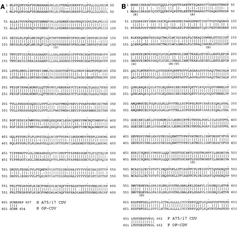
The nucleotide sequence corresponding to the OP-CDV H protein obtained in our laboratory (data not shown) showed only one difference with respect to the published sequence of OP-CDV (9), which changes a Phe residue to a Ser residue (Fig. 1A). However, a total of 139 nucleotide differences were observed within the region encoding the A75/17 H protein, compared to the sequence of the corresponding OP-CDV open reading frame (data not shown). The deduced amino acid sequences of the H proteins for OP-CDV and A75/17 CDV (Fig. 1A) differ in 57 amino acids which are distributed evenly over the entire length of the H protein. The most striking difference is that the AUU translation termination codon in OP-CDV is UCA, coding for a Ser residue in A75/17. Translation termination occurs at a UGA, three codons further downstream. The A75/17 H protein thus has 607 amino acid residues, compared to 604 in OP-CDV.
FIG. 1.
Comparison of the H (A) and F (B) proteins of A75/17 (top line) and OP-CDV (bottom line). Differences in the OP-CDV genes with respect to the published sequences (4, 9) are indicated in parentheses.
The nucleotide sequence of the OP-CDV F protein obtained in our laboratory (data not shown), starting with the first in-frame AUG, differed at nine positions with respect to the published sequence (4). Eight of these nucleotide differences resulted in amino acid changes (Fig. 1B). Comparison of our OP-CDV and A75/17 CDV F protein-coding sequences showed a total of 142 nucleotide differences, of which 67 are located in the F2 portion, and 75 are located in the F1 part of the protein. Interestingly, these differences result in 60 amino acid changes, of which 44 are found in the much smaller F2 portion (224 residues) of the molecule, and only 16 are found in the F1 portion (438 residues [Fig. 1B]). The most striking difference, however, was found at the position corresponding to the AUG, which in OP-CDV had been proposed as the translation initiation codon (10). In the A75/17 strain, the codon at this position is AUA, coding for Ile. To rule out the possibility that this codon was a cloning or PCR artifact, we isolated four additional F protein-coding sequences from independent PCR amplifications and sequenced the corresponding regions. In each case, we found an AUA codon at that position instead of AUG. We therefore believe that this is a true difference between the two CDV strains, and this finding led us to reexamine the utilization of various AUGs for translation initiation of the CDV F protein (see below).
Expression of the H protein.
The H proteins of CDV field isolates were reported to have a reduced electrophoretic mobility in polyacryamide gels compared to that of OP-CDV H, which is due to differences in glycosylation (14). We therefore tested whether this is also the case for the H protein of the A75/17 strain. To facilitate detection of the proteins by Western blotting, we used a vaccinia virus transient expression system in which the corresponding genes are contained in a plasmid under the control of a strong vaccinia virus promoter. These plasmids were then transfected into cells previously infected with vaccinia virus, providing the transcription machinery. An additional advantage of this system is that the RNAs are transcribed in the cytoplasm of the cell, as in the case of a CDV infection. The risk of aberrant splicing of RNAs normally not in contact with the RNA processing machinery can thus be avoided by using this system, in contrast to expression from transfected plasmids in the nucleus. On the other hand, proteins expressed in the transient expression system may not necessarily represent functional proteins.
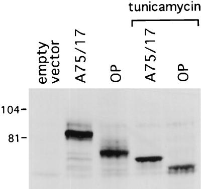
Figure 2 shows a Western blot analysis of the A75/17 and OP-CDV H proteins expressed from transfected plasmids in the cytoplasm. A prominent band with an apparent molecular mass of about 80 kDa is observed in strain A75/17. A faster-migrating species is observed with the transfected OP-CDV H gene.
FIG. 2.
Expression of H proteins of A75/17 CDV and OP-CDV. Extracts from vaccinia virus-infected CV1 cells transfected with empty vector or plasmids containing the respective genes were analyzed by Western blotting. H proteins were also expressed in the presence of tunicamycin, as indicated. The positions and molecular masses (kilodaltons) of markers are shown to the left.
We considered the possibility that the different electrophoretic mobility was due to differences in glycosylation of the H proteins of the two viruses. Indeed, treatment of cells with tunicamycin reduced the apparent molecular masses of both proteins, albeit to different extents, suggesting that A75/17 H contains more N glycosylations than OP-CDV H. Interestingly, in the presence of tunicamycin, the A75/17 protein still had a higher apparent molecular mass.
Expression of A75/17 and OP-CDV F proteins.
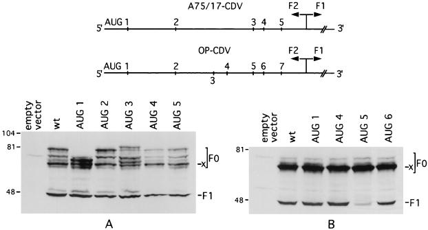
As noted above, the AUG of the OP-CDV F protein, which has been proposed to be the translation initiation codon, is an AUA codon in strain A75/17. We therefore investigated which AUG is used for translation initiation in strain A75/17. Site-directed mutagenesis was used to change all five in-frame AUGs in the F2 portion of the F protein into UUG codons. The mutated plasmids were transfected into vaccinia virus-infected CV1 cells. Cell extracts were analyzed by Western blotting with a polyclonal rabbit antiserum directed against the F0 precursor (Fig. 3A).
FIG. 3.
Expression of A75/17 CDV and OP-CDV F genes containing individually mutated AUGs. (Top) Positions of AUGs in the 5′ regions of the RNAs encoding the F2 portion of the F protein of A75/17 CDV and OP-CDV. Note that, in A75/17 CDV, only in-frame AUGs are shown, whereas in OP-CDV, the numbering of AUGs is according to that of Evans et al. (10), which takes into account the third AUG, which is not in frame. (A and B) Effect of mutations of individual AUGs on expression of A75/17 (A) and OP-CDV (B) F proteins. Plasmids containing the wild-type (wt) F coding sequences or genes carrying mutations in individual AUGs as indicated at the top were transfected into vaccinia virus-infected cells. Cell extracts were analyzed by Western blotting. Extracts from cells transfected with empty vector are shown as controls. The positions of F0 precursors and of the F1 proteins are indicated. A band of unknown origin (x) is marked (see text). The positions of molecular mass markers and their sizes in kilodaltons are shown to the left.
In extracts of cells expressing the A75/17 F wild-type gene, a series of high-molecular-mass bands were observed. These most likely represent F0 precursors translated from different AUGs. In addition, a band of about 46 kDa was also detected by the antiserum. From its size, we concluded that this protein was the F1 cleavage product.
When the first in-frame AUG was mutated, the highest-molecular-mass band disappeared, and a smaller F0 precursor, barely visible in extracts of cells transfected with the wild-type gene, became the most prominent high-molecular-mass band. This band was lost when the second AUG was mutated, and the highest-molecular-mass band reappeared instead. Mutation of the other AUGs also affected the band pattern of F0 precursors and the relative abundance of individual bands. However, it was not possible to assign all high-molecular-mass bands to particular AUGs. Interestingly, a prominent protein species (designated x) was not affected by any of the mutations, and its origin remains to be investigated (see Discussion).
The intensity of the F1 band was not significantly affected by any of the AUGs mutated individually. This suggests that the processing of the F0 precursor, giving rise to the C-terminal F1 cleavage product, is not affected by translation of F0 from alternative AUGs. Mutations in individual AUGs in strain A75/17 showed that the first AUG in the F open reading frame can be used as a translation initiation codon, yielding an F0 precursor. In OP-CDV, the first AUG is located at the same position. In addition, the nucleotides at positions −3 and +4, which are particularly important for translation initiation (16), are identical in both strains. It is therefore very likely that the first AUG is also used as a translation initiation codon in OP-CDV. In order to test this possibility, we performed site-directed mutagenesis of relevant AUGs in the OP-CDV gene (Fig. 3B). Surprisingly, the banding pattern of high-molecular-mass precursors in cells expressing the wild-type gene was different from that obtained with strain A75/17. Particularly the highest-molecular-mass species represented as an intense band with A75/17 was not visible with OP-CDV in this experiment. However, in some experiments (Fig. 4B), a faint band corresponding to an F0 precursor initiated at the first AUG was seen. Thus, in OP-CDV, the first AUG is either not a major translation initiation site, or, alternatively, precursors initiated at the first AUG are more efficiently cleaved into the mature proteins in OP-CDV than in A75/17. Additional experiments showed that the latter hypothesis is correct (described below). As with A75/17 F, a high-molecular-mass band referred to as band x was also seen at a similar position in the gel in OP-CDV F, and this band was also not affected by mutation of any AUG.
FIG. 4.
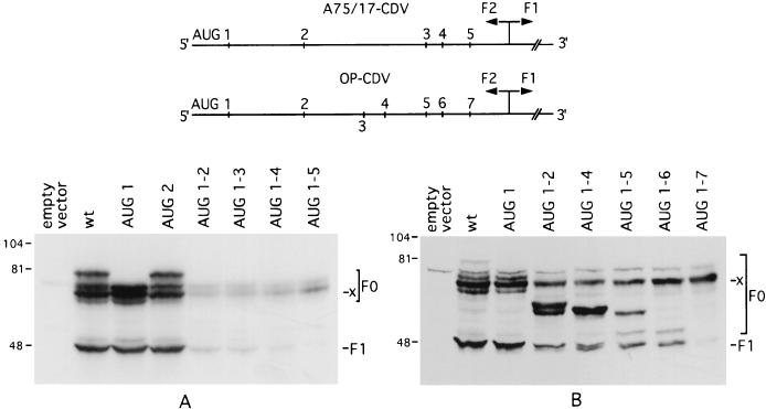
Expression of A75/17 CDV and OP-CDV F genes containing multiple mutations of in-frame AUGs. Plasmids containing the wild-type F genes from A75/17 CDV (A) or from OP-CDV (B) or genes carrying mutations in increasing numbers of AUGs, as indicated at the top, were transfected into vaccinia virus-infected cells. Cell extracts were analyzed by Western blotting. The positions of F0 precursors and of the F1 proteins are indicated. A band of unknown origin (x) is marked (see text). The positions of molecular mass markers and their sizes in kilodaltons are shown to the left.
Similar to what was observed for A75/17 F, mutations of individual AUGs had little effect on the intensity of the F1 band. Significantly, this was also true for the fourth AUG, which has been proposed as translation initiation codon (10) and which is absent in strain A75/17. The exception was mutation of AUG 5, which greatly reduced the amount of F1 (see Discussion).
The idea that for both strain A75/17 and OP-CDV F proteins, different AUGs may be used for translation initiation to generate precursors which can be cleaved into the mature proteins was tested by analyzing the expression of F genes in which increasing numbers of AUGs in the same construct had been mutated (Fig. 4). As seen before, individual mutations of the first or second AUG in A75/17 F had no effect on the intensity of the F1 band (Fig. 3A). In contrast, when both AUGs were mutated, a drastic reduction of F1 occurred (Fig. 4A). We therefore conclude that the main precursors giving rise to the F1 cleavage product in CDV strain A75/17 are translated from the first or second AUG. Mutation of additional AUGs further decreased the intensity of the F1 band, which disappeared when all in-frame AUGs were mutated. Surprisingly, band x was present in all constructs and even increased slightly in the construct where all AUGs had been mutated. This indicates that band x is not a precursor of F1.
Similar results were obtained for the OP-CDV F protein (Fig. 4B). Again, upon mutation of the first AUG, the highest-molecular-mass precursor was lost, which, in this experiment, was more clearly visible in cells expressing the wild-type gene. Whereas mutation of the first AUG had little effect on the intensity of the F1 band, mutation of both the first and second AUGs reduced the amount of F1. This effect was less drastic than that with A75/17 F, but it still indicated that, also in OP-CDV F, the first two AUGs are the major translation initiation sites for the synthesis of precursors yielding the mature proteins. In contrast to A75/17 F, mutation of AUGs located further downstream resulted in the synthesis of shorter precursors that do not appear to be readily cleaved, since, although present in large amounts, they do not contribute significantly to the production of F1. Interestingly again, upon mutation of all AUGs, band x did not disappear and rather increased in intensity. Thus, as in A75/17, this polypeptide of unknown origin is not a precursor for F1.
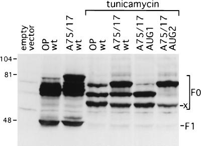
F0 precursors are not cleaved in the presence of tunicamycin (21). Therefore, the intensity of a particular F0 precursor band should directly reflect the frequency of translation initiation from the corresponding AUG and not be masked by processing events. The above observation and the availability of plasmids with mutations in various AUGs, allowing identification of the initiation codon from which individual F0 precursors are translated, provided an additional possibility of estimating the usage of the first and second AUGs for translation initiation of A75/17 and OP-CDV F proteins. A transient expression experiment was therefore performed with wild-type and mutated F genes in cells treated with tunicamycin (Fig. 5).
FIG. 5.
Expression of OP-CDV or A75/17 F proteins in the absence or presence of tunicamycin. Plasmids carrying wild-type (wt) F genes or plasmids in which the first or second AUG of the F gene had been mutated were transfected into vaccinia virus-infected cells. After incubation in the absence or presence of tunicamycin, cell extracts were analyzed by Western blotting. As a control (shown in lane 1), empty plasmid was transfected. A band of unknown origin (x) is indicated (see text). The positions of molecular mass markers and their sizes in kilodaltons are shown to the left.
With the wild-type genes of both A75/17 and OP-CDV, three major bands were observed in the presence of the glycosylation inhibitor. Mutation of the first or second AUG in the A75/17 F resulted in the disappearance of the first and second bands, respectively. Therefore, the first and second bands represent unglycosylated F0 precursors translated from the first and second AUGs. The third band most likely represents the unglycosylated protein x.
The largest F0 precursor in the A75/17 wild-type construct was clearly the most intense band, indicating that the first AUG is the major translation initiation site. In the OP-CDV wild-type construct, the second band representing the unglycosylated F0 precursor starting at the second AUG was somewhat more intense than the first band, suggesting that, in OP-CDV, there is a slight preference for the use of the second AUG as a translation initiation codon.
DISCUSSION
In this study, we have investigated the fine structure and expression of the CDV H and F surface glycoproteins. We chose these two proteins because they are likely to be involved in determining some of the biological differences between highly attenuated, cell culture-adapted vaccine strain Onderstepoort and strain A75/17, the prototype virulent CDV field isolate.
In the attachment protein H, we found 57 amino acid differences between OP-CDV and strain A75/17. The A75/17 protein has six potential N-linked glycosylation sites in the extraviral domain, three more than OP-CDV H (9). Other field isolates have either seven (7) or eight potential N-linked glycosylation sites in the extraviral domain (14).
With respect to OP-CDV, strain A75/17 H contains three additional amino acids at the C terminus of the protein, like the H protein of the Convac strain (15) and many field isolates (7, 14). Interestingly, these amino acids create an additional potential N glycosylation site.
When OP-CDV and A75/17 H were expressed in cells treated with tunicamycin, significant reductions in the apparent molecular masses of both proteins were observed, indicating that many of the potential glycosylation sites are used. In Japanese field isolates (14), the H proteins comigrated in polyacryamide gels with OP-CDV H following treatment with the glycosylation inhibitor. Significantly, in our study, the A75/17 protein still had a higher apparent molecular mass than OP-CDV H when expressed in the presence of tunicamycin. We are unable to explain this difference at the present time.
The most significant difference between the fusion proteins of CDV strain A75/17 and OP-CDV was the absence in A75/17 of the AUG that has been proposed as the translation initiation codon in OP-CDV (10). This has been designated the fourth AUG, since a third AUG, which is not in frame, has also been counted. The absence of this AUG in strain A75/17 led us to examine which AUG was used for translation initiation in this strain. To our surprise, site-directed mutagenesis of individual AUGs had little effect on the F1 protein, although, as expected, F0 precursors initiated at the first or second AUG disappeared when the corresponding AUG was mutated. This clearly indicated that, in A75/17, at least the first and second AUGs are used for translation initiation and that mutation of single AUGs is compensated for by more extensive use of the remaining ones. Therefore, to identify which AUGs contribute most to the production of F1, we mutated increasing numbers of AUGs in the same construct. This analysis clearly showed that the first two AUGs in strain A75/17 are by far the most important translation initiation codons of the F protein.
These results led us to reexamine the utilization of various AUGs in OP-CDV. As in A75/17, mutations of individual AUGs, including the fourth one, had little effect on the amount of F1 produced, except for a drastic reduction when the fifth AUG was mutated. Thus, this AUG is either the major translation initiation site in OP-CDV F, or, alternatively, precursors initiated from AUGs located further upstream are not readily cleaved when they contain a Leu instead of a Met residue at the position corresponding to the fifth AUG in the mRNA. The results obtained by mutation of several AUGs in the same construct are in favor of the latter hypothesis. Indeed, mutation of both the first and second AUGs in the same construct greatly reduced the amount of F1, albeit not to the extent as in A75/17. We therefore conclude that, in OP-CDV, the first two AUGs in the F open reading frame are also the major translation initiation sites. The observation that mutation of both of these two AUGs still allowed production of some F1 is best explained by assuming that in OP-CDV, AUGs located further downstream can be more readily used for translation initiation than those in A75/17. Indeed, such small F0 molecules initiated at these AUGs were present in substantial amounts (Fig. 4B), but are apparently not efficiently cleaved, since in these constructs, the F1 band is rather faint. In contrast, F0 precursor molecules initiated at the first and second AUGs appear to be more easily cleaved in OP-CDV F than in strain A75/17 F, since these molecules are either not detectable (Fig. 3B) or are present in small amounts (Fig. 4B), despite the fact that they contribute most to the generation of F1.
The efficiency with which individual F0 precursors are cleaved is most likely determined by their secondary structure, which depends on the amino acid sequence and the AUG used for translation initiation. In this context, it is interesting to note that we have isolated from infected dog lymphoid tissue an A75/17 strain F protein coding sequence which yielded an F protein which was not cleaved (data not shown). Sequence analysis revealed only two amino acid differences with respect to the cleavable version, both of which were located far away from the F1-F2 cleavage site. In view of the large number of amino acid differences between the F proteins of strain A75/17 and OP-CDV, it is not surprising that their F0 precursors are cleaved with different efficiencies. Structural differences between individual F0 precursors initiated at different AUGs presumably also account for the fact that some of them are not readily cleaved into the mature proteins.
An intriguing observation in the mutation analysis was the presence of a band, designated x, which was more intense in OP-CDV than in strain A75/17 and which did not disappear even when all in-frame AUGs were mutated. We therefore also mutated all AUGs which are not in frame (data not shown); however, this band was still present. It is possible that this protein, which does not appear to be a precursor of the mature F proteins, is initiated at another codon than an AUG. Work is in progress to determine the origin of this protein. Preliminary deletion analysis has identified a 75-nucleotide sequence which is required for its synthesis.
Mutation analysis clearly showed that both in strain A75/17 and in OP-CDV, the first and second AUGs are the most important translation initiation sites (described above). To determine their relative usage, we performed expression studies in the presence of tunicamycin. In the presence of this glycosylation inhibitor, F0 precursors are not cleaved, and their abundance should therefore directly reflect the usage of the corresponding AUGs. This analysis showed that in A75/17, the first AUG is the major translation initiation site which is used about three times more efficiently than the second one (Fig. 5). In OP-CDV, the opposite is true. Here, the second AUG appears to be used about three times more frequently than the first one. These results were confirmed with recombinant vaccinia viruses expressing the F proteins under control of the 7.5-kDa early promoter. With this expression system, the same differences in the usage of the AUGs were observed, but the preferences were much more pronounced (data not shown). Interestingly, the first AUG (underlined) is in the same, not ideal, sequence context in both viruses (CCCAUGC). The second (underlined), however, is in a more favorable context in OP-CDV (ACCAUGA) than in A75/17 CDV (AUCAUGA), which may explain why translation initiation occurs more frequently at this AUG in OP-CDV. (Residue differences are in boldface.)
Our study has revealed major differences in the structure and expression of the H and F proteins between strain A75/17 and OP-CDV. These differences are likely to account for at least some of the biological differences between the two CDV strains, one of which establishes a persistent infection and one of which produces a lytic infection. Transport to the cell surface, spike formation, fusion activity, and ability to interact with other viral structural elements in the virus assembly process all depend on structural and biological properties of the viral surface glycoproteins. By studying these properties at a more functional level, we believe that we will learn more about why CDV strain A75/17, but not OP-CDV, establishes a persistent infection in the host.
ACKNOWLEDGMENTS
We thank Lorenz Rindisbacher and Marc Vandevelde for critically reading the manuscript and Jacqueline Goenaga for technical assistance.
This study was supported by grants 31-43434.95 (R.W.) and 31-45609.95 (A.Z.) from the Swiss National Science Foundation and by a grant from the European Biotechnology Program (contract no. BIO 4 CT 960473).
REFERENCES
- 1.Alkhatib G, Roder J, Richardson C, Briedis D, Weinberg R, Smith D, Taylor J, Paoletti E, Shen S-H. Characterization of a cleavage mutant of the measles virus fusion protein defective in syncytium formation. J Virol. 1994;68:6770–6774. doi: 10.1128/jvi.68.10.6770-6774.1994. [DOI] [PMC free article] [PubMed] [Google Scholar]
- 2.Alkhatib G, Shen S-H, Briedis D, Richardson C, Massie B, Weinberg R, Smith D, Taylor J, Paoletti E, Roder J. Functional analysis of N-linked glycosylation mutants of the measles virus fusion protein synthesized by recombinant vaccinia virus vectors. J Virol. 1994;68:1522–1531. doi: 10.1128/jvi.68.3.1522-1531.1994. [DOI] [PMC free article] [PubMed] [Google Scholar]
- 3.Appel M J G, Gillespie J H. Anonymous virology monographs. Vienna, Austria: Springer-Verlag; 1972. Canine distemper virus; pp. 1–96. [Google Scholar]
- 4.Barrett T, Clarke D K, Evans S A, Rima B K. The nucleotide sequence of the gene encoding the F protein of canine distemper virus: a comparison of the deduced amino acid sequence with other paramyxoviruses. Virus Res. 1987;8:373–386. doi: 10.1016/0168-1702(87)90009-8. [DOI] [PubMed] [Google Scholar]
- 5.Bertholet C, Stocco P, Van Meir E, Wittek R. Functional analysis of the 5′ flanking sequence of a vaccinia virus late gene. EMBO J. 1986;5:1951–1957. doi: 10.1002/j.1460-2075.1986.tb04449.x. [DOI] [PMC free article] [PubMed] [Google Scholar]
- 6.Bollo E, Zurbriggen A, Vandevelde M, Fankhauser R. Canine distemper virus clearance in chronic inflammatory demyelination. Acta Neuropathol. 1986;72:69–73. doi: 10.1007/BF00687949. [DOI] [PubMed] [Google Scholar]
- 7.Bolt G, Jensen T D, Gottschalck E, Arctander P, Appel M J G, Buckland R, Blixenkrone-Moller M. Genetic diversity of the attachment (H) protein gene of current field isolates of canine distemper virus. J Gen Virol. 1997;78:367–372. doi: 10.1099/0022-1317-78-2-367. [DOI] [PubMed] [Google Scholar]
- 8.Brügger M, Jungi T W, Zurbriggen A, Vandevelde M. Canine distemper virus increases procoagulant activity of macrophages. Virology. 1992;190:616–623. doi: 10.1016/0042-6822(92)90899-z. [DOI] [PubMed] [Google Scholar]
- 9.Curran M D, Clarke D K, Rima B K. The nucleotide sequence of the gene encoding the attachment protein H of canine distemper virus. J Gen Virol. 1991;72:443–447. doi: 10.1099/0022-1317-72-2-443. [DOI] [PubMed] [Google Scholar]
- 10.Evans S A, Belsham G J, Barrett T. The role of the 5′ nontranslated regions of the fusion protein mRNAs of canine distemper virus and rinderpest virus. Virology. 1990;177:317–323. doi: 10.1016/0042-6822(90)90486-b. [DOI] [PubMed] [Google Scholar]
- 11.Graves M C, Silver S M, Choppin P W. Measles virus polypeptides synthesis in infected cells. Virology. 1978;86:254–263. doi: 10.1016/0042-6822(78)90025-9. [DOI] [PubMed] [Google Scholar]
- 12.Hamburger D, Griot C, Zurbriggen A, Örvell C, Vandevelde M. Loss of virulence of canine distemper virus is associated with a structural change recognized by a monoclonal antibody. Experientia. 1991;47:842–845. doi: 10.1007/BF01922469. [DOI] [PubMed] [Google Scholar]
- 13.Hardwick J M, Bussell R H. Glycoproteins of measles virus under reducing and nonreducing conditions. J Virol. 1978;25:687–692. doi: 10.1128/jvi.25.2.687-692.1978. [DOI] [PMC free article] [PubMed] [Google Scholar]
- 14.Iwatsuki K, Miyashita N, Yoshida E, Gemma T, Shin Y S, Mori T, Hirayama N, Kai C, Mikami T. Molecular and phylogenetic analyses of the haemagglutinin (H) proteins of field isolates of canine distemper virus from naturally infected dogs. J Gen Virol. 1997;78:373–380. doi: 10.1099/0022-1317-78-2-373. [DOI] [PubMed] [Google Scholar]
- 15.Kovamees J, Blixenkrone Moller M, Norrby E. The nucleotide and predicted amino acid sequence of the attachment protein of canine distemper virus. Virus Res. 1991;19:223–233. doi: 10.1016/0168-1702(91)90048-z. [DOI] [PubMed] [Google Scholar]
- 16.Kozak M. Point mutations define a sequence flanking the AUG initiator codon that modulates translation by eukaryotic ribosomes. Cell. 1998;44:283–292. doi: 10.1016/0092-8674(86)90762-2. [DOI] [PubMed] [Google Scholar]
- 17.Malvoisin E, Wild T F. Measles virus glycoproteins: studies on the structure and interaction of the haemagglutinin and fusion proteins. J Gen Virol. 1993;74:2365–2372. doi: 10.1099/0022-1317-74-11-2365. [DOI] [PubMed] [Google Scholar]
- 18.Merz D C, Scheid A, Choppin P W. Importance of antibodies to the fusion glycoprotein of paramyxoviruses in the prevention of spread of infection. J Exp Med. 1980;15:275–288. doi: 10.1084/jem.151.2.275. [DOI] [PMC free article] [PubMed] [Google Scholar]
- 19.Metzler A E, Higgins R J, Krakowka S, Koestner A. Virulence of tissue culture-propagated canine distemper virus. Infect Immun. 1980;29:940–944. doi: 10.1128/iai.29.3.940-944.1980. [DOI] [PMC free article] [PubMed] [Google Scholar]
- 20.Sanger F, Nicklen S, Coulson A R. DNA sequencing with chain-terminating inhibitors. Proc Natl Acad Sci USA. 1977;74:5463–5467. doi: 10.1073/pnas.74.12.5463. [DOI] [PMC free article] [PubMed] [Google Scholar]
- 21.Sato T A, Kohama T, Sugiura A. Intracellular processing of measles virus fusion protein. Arch Virol. 1988;98:39–50. doi: 10.1007/BF01321004. [DOI] [PubMed] [Google Scholar]
- 22.Vandevelde M, Zurbriggen A. The neurobiology of canine distemper virus infection. Vet Microbiol. 1995;44:271–280. doi: 10.1016/0378-1135(95)00021-2. [DOI] [PMC free article] [PubMed] [Google Scholar]
- 23.Vandevelde M, Zurbriggen A, Higgins R J, Palmer D. Spread and distribution of viral antigen in nervous canine distemper. Acta Neuropathol. 1985;67:211–218. doi: 10.1007/BF00687803. [DOI] [PubMed] [Google Scholar]
- 24.Varsanyi T M, Jornvall H, Orvell C, Norrby E. F1 polypeptides of two canine distemper virus strains: variation in the conserved N-terminal hydrophobic region. Virology. 1987;157:241–244. doi: 10.1016/0042-6822(87)90335-7. [DOI] [PubMed] [Google Scholar]
- 24a.Zurbriggen, A. Unpublished observations.
- 25.Zurbriggen A, Graber H U, Wagner A, Vandevelde M. Canine distemper virus persistence in the nervous system is associated with noncytolytic selective virus spread. J Virol. 1995;69:1678–1686. doi: 10.1128/jvi.69.3.1678-1686.1995. [DOI] [PMC free article] [PubMed] [Google Scholar]
- 26.Zurbriggen A, Vandevelde M, Dumas M, Griot C, Bollo E. Oligodendroglial pathology in canine distemper virus infection in vitro. Acta Neuropathol. 1987;74:366–373. doi: 10.1007/BF00687214. [DOI] [PubMed] [Google Scholar]


