Abstract
Cervical teratomas are extremely rare germ cell tumours and it is much more common in newborn than adults, and in contrast to the paediatric cases adult teratomas have been highly malignant. Cervical teratoma incorporates lesions arising in the anterior and posterior triangles of the neck. This tumor can reach enormous size and cause airway obstruction and patients should be quickly treated. Surgery is the primary modality of treatment as malignant transformation can occur. Hereby, we present a case of benign teratoma of neck in adult which was completely misdiagnosed preoperatively due to its rare occurrence in adults.Even though cervical teratoma of adult is extremely rare, it should be considered as an important differential diagnosis in patient of midline cystic neck swelling. Preoperative radiological investigations requires high index of suspicion. Complete surgical resection is recommended. We believe that upper cervicotomy approach is a safe and effective method for the treatment of mature cervical teratoma with a few protruding into the superior mediastinum.
Supplementary Information
The online version contains supplementary material available at 10.1007/s12070-023-03748-8.
Keywords: Surgical resection, Cervical mature teratoma, Adult
Introduction
Teratomas are abnormal growths, benign or malignant, in which tissue derived from each of the three germinal layers is recognized. Cervical Teratoma (CT)are very uncommon [1, 2]. The malignancy potential of the lesion is determined by the degree of immaturity of the tissue [2, 3]. Germ cell tumors are extra-gonadal in 5–10% of all teratoma and the most commun extra-gonadal site is mediastinum [4]. The neck regions may be affected in rare cases [4–6]. CT occurred mainly in newborn, children and they are extremely rare in adult [1, 2] with high incidence of malignancy [1, 7, 8]. CT can reach enormous size causing airway obstruction and nearby structures compression, requiring surgery [2, 3]. We report the case of a 42-year-old woman operated for a benign CT, and we review CT adults in the literature.
Case Report
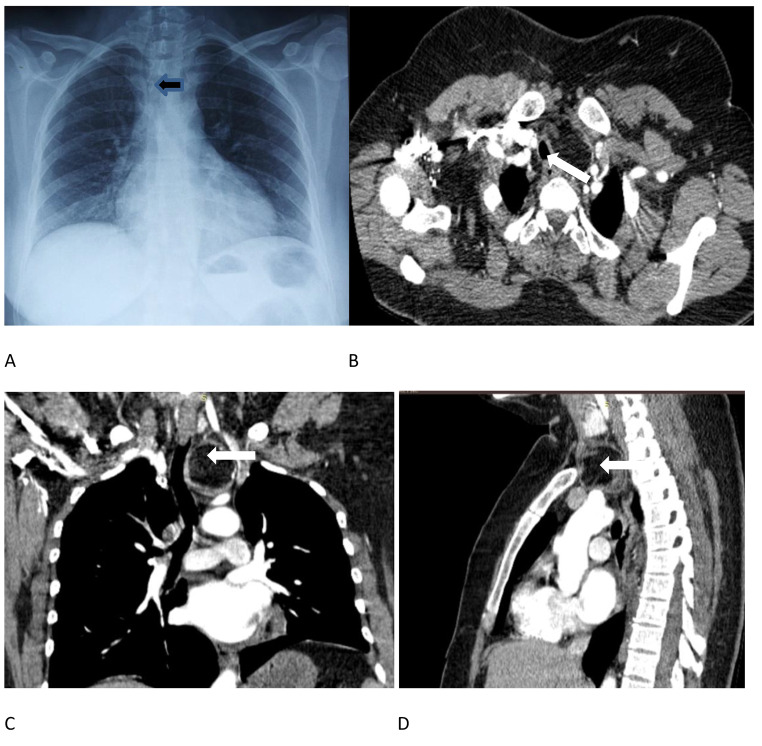
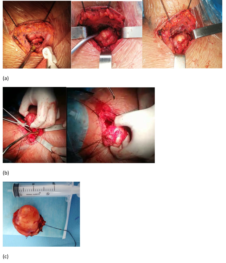
A previously healthy 42-year-old woman presented with a recurrent bronchopneumpathy associated to a slowly enlarging neck tumor noticed 2 months earlier. Clinical exam showed, an elastic soft nodule,4 × 5 cm in size,in the base of the left anterior neck. The tumor was not tender and was attached to the deeper portion of the neck. No palpable cervical lymph nodes were noted. A neck X-ray demonstrated a deviation of the trachea. Computerized tomography of the chest showed a well-defined, lobulated tumor mesuring 4 cm with predominant fat density, having smaller foci of soft tissue densities and calcifications, in the left paratracheal region of the lower neck, minimally extending to the upper thorax (Fig. 1). Surgery was performed through upper cervicotomy (Fig. 2), revealing a tumor separated from the thyroid. Intra-operatively,the tumor was well encapasuled. Adhesion between the capsule and the anterior neck mass and surrounding tissues was severe. The capsule wasn’t ruptured during dissection. The tumor was completely excised(Fig. 2). It measured 6 × 5 × 2 cm, showed many cystic lumina containing butter-like material and opaque fluid. The cyst wall of the tumor is lined with squamous epithelium, with smooth muscle tissue, sebaceous gland and sweat gland tissue visible in it, and fibrous tissue in the center. Histological exam of the specimen showed features that are in keeping with mature teratoma. The patient was discharged three days after operation.
Fig. 1.
A. Chest radiograph shows left neck swelling (arrow) with right tracheal deviation B. Axial CT image showing the cervical mass with tracheal deviation to the right. Septation within the mass is indicated by the arrow. C. Coronal CT image showing relationship of the mass to the great vessels. Dark area within the mass {arrow) corresponds to the lipomatous element and ….calcifications. D. Sagittal Axial views showing a well defined cystic and solid mass in upper mediastinum extending to the neck
Fig. 2.
Intraoperative appearance of cystic lesion (a) Initial appearance of neck mass adherent to trachea (b) Dissection of the well encapsulated cervical solid lesion (c) Specimen photograph of the excised cervical teratoma
Discussion
Teratomas are embryonal neoplasms composed of tissues foreign to the anatomic site of origin with all three blastodermic layers (ectoderm, endoderm, and mesoderm) [3, 9, 10]. The mediastinum is the second most common anatomic site of teratoma [8]. The Neck is one of the least common extra-gonadal sites [1, 9, 11]. CT constitutes only 1,5–5% of all teratomas [2–4, 6, 8, 10, 12]. CT in adulthood represents 10,6% of all CT with a high incidence of malignancy [3, 10]. They pre-dominate in females (75% of the cases) [3]. In the course of our review of the literature during 70 years we were able to find 19 documented cases of benign CT in adults, as listed in Table 1.The exact cause of CT is unknown [4, 5].Mediastinal teratomas are usually asymptomatic [6, 11]. If teratoma arises in the cervical region, it increase rapidly in the growth, so we note symptoms related to compression of surrounding structures,such as,dyspnea,dysphagia,or recurrent episodes of infection [6, 11]. Differential diagnosis of CT are a papillary carcinoma of thyroid with cystic formation, a metastasis from thyroid carcinoma,cystic squamous cell carcinoma,lymphangiomas,and bronchial cysts [3, 10, 11].
Table 1.
Documented cases of cervical teratoma in adults during 70 years (since 1954)
| First author (year) | Age Sex | Size of tumor | Outcome |
|---|---|---|---|
| Cavellero (1954) | 24 Female | - | - |
| Keyness (1959) | 24 Male | - | 15 months ; alive |
| Ohara (1959) | 39 Male | 10*7,8 cm | 7 years ;alive |
| Muto (1968) | 25 Female | 5*5*3 cm | 4 years ;alive |
| Woods(1978) | 40 Female | - | 12months ; alive |
| Mochizuki (1986) | 26 Female | 3*3*2 cm | 16 Months ; alive |
| Endo (1992) | 29 Male | - | - |
| Sawafuji (1993) | 21 Male | - | 19 Months; alive |
| Kuhel (1996) | 32 Female | 4,8*4*2 cm | 22 Months; alive |
| Abe (1997) | 21 Female | 7,5*4,5*2,7 | 7 months ;alive |
| KHazama(2003) (12) | 27 Female | 10 cm | 6 months ;alive |
| Omranipour (2007) | 36 Male | 5*5*8 cm | |
| Gaurav (2008) | 19 Male | 16*7*2 cm | 36 months ;alive |
| Alimehmeti (2013) (10) | 25 Female | 4 cm | |
| Siow (2015) (4) | 18 Male | 10*6,5 cm | 1 year ; alive |
| Ansari (2017) (3) | 24 Male | 3*4 cm | - |
| Lee (2018) | 38 Male | 6,5*5, 2*2, 2 cm | 5 years ;alive |
| Birla Roy (2020) | 18 Female | 6 cm | - |
| Liu (2020) | 33 Male | 4*4*5 cm | 9 months ; alive |
| Our Patient | 42 Female | 4*5 cm | 1 month; alive |
| Total | 20 cases | - | - |
Chest x-ray can evoke CT [11]. Ultrasonography should demonstrate relationships between masses with thyroid gland and great vessels. Neck CT scan with coverage of mediastinum is essential to evaluate intrathoracic extension and assists planning surgery [2, 6, 11]. In our patient CT scan established the continuity of mediastinal mass into the neck. Magentic resonance imaging can be also very useful in evaluating the relationship of the tumor to the great vessels and vital structures [12]. Calcified structures present in only 26% of patients with matured teratomas have been reported to present the typical shadow [12]. When a CT is encountered in an adult,the surgeon should anticipate the possibility of a tedious dissection due to adherence to the pretracheal fascia or the thyroid and the great vessels [4, 8].
Complete surgical removal of CT is the curative treatment. It allows establishing the diagnosis and preventing life threatening complications [11]. Anatomically, CT lie in the visceral space between the anterior strap muscles of the neck and the pretrachea fascia [4]. For approach of this tumor,we did not require a median sternotomy as the inferior edge was just at the level of the thoracic inlet and we were able to retract it superiorly [4]. If the mass extends to the thoracic inlet or the supraclavicular region, a manubrial or sternal osteotomy should be performed [8]. Some surgeons suggest that surgical removal of a CT may involve the removal of a portion of or the entire thyroid gland [3, 5]. Some researches conclude that radiation therapy may be used before surgery or after surgery as an adjuvant therapy [5]. Chemotherapy immediately after surgery has also been used to treat individuals with cervical teratoma [5] but adjuvant chemotherapy is indicated when malignancy is confirmed [4].
In conclusion, even though CT of adult is extremely rare, it should be considered as an important differential diagnosis in adult of cystic neck [3]. The preliminary diagnosis can be suggested on preoperative investigations [2, 4]. Complete surgical resection is recommended due to unpredictable compressive complication and the high potential of malignant transformation [3, 4].
Electronic Supplementary Material
Below is the link to the electronic supplementary material.
Acknowledgements
Not appplicable.
Abbreviations
- CT
Cervical teratoma
Author Contribution
Abdennadher Mahdi: conceived the cae report, contiubuted to writing, reviewing and finalization of the manuscript.
Ben Amara Kouathar: collected clinical details.
Abdelkebir Amina: approval of the final version.
Zribi Hazem: approval of the final version.
Ben Mansour Amani: Helped in data acquisition.
Sahnoun Imen: Helped in data acquisition.
Zairi Sarra: Evaluated of the final version.
Marghli Adel: Critical of the final version.
Funding
No.
Data Availability
Yes.
Declarations
Ethics Approval and Consent to Participate
Not applicable.
Consent for Publication
Yes.
Competing Interest
No.
Footnotes
Publisher’s Note
Springer Nature remains neutral with regard to jurisdictional claims in published maps and institutional affiliations.
Contributor Information
Mahdi Abdennadher, Email: abdennadhermahdi@gmail.com.
Kaouthar Ben Amara, Email: kawtherbenamara@gmail.com.
Amina Abdelkebir, Email: aminaabdelkbir@gmail.com.
Hazem Zribi, Email: zribihazem@yahoo.fr.
Amani Ben Mansour, Email: benmansour_amani@yahoo.fr.
Imen Sahnoun, Email: imensahnounj@gmail.com.
Sarra Zairi, Email: sarra.zairi@gmail.com.
Adel Marghli, Email: marghli_adel@yahoo.fr.
References
- 1.Woods RD, Pearson BW, Weiland LH. Benign cervical cystic teratoma. Otolaryngol May-Jun. 1978;86(3 Pt 1):ORL468–ORL472. doi: 10.1177/019459987808600317. [DOI] [PubMed] [Google Scholar]
- 2.Jiangjiang Liu X, Ang X, Guo Yu, Feng H Ma. Mature cystic teratoma of the Suprasternal Fossa in an adult: Report of Case. January 2020.Case Reports in Clinical Medicine09(12):385–391DOI: 10.4236/crcm.2020.912053
- 3.Humaam Ansari M, Balajirao Gujrathi A, Ambulgekar V. Benign teratoma of neck in adult: a rare case report. F Int J Otorhinolaryngol Head Neck Surg. 2017;3(4):1136–1139. doi: 10.18203/issn.2454-5929/ijohns20174351. [DOI] [Google Scholar]
- 4.Siow SL, Mahendran HA. Cervical teratoma: a rare Neck Swelling in an adult. Am J Med Case Rep. 2015;3(3):75–78. [Google Scholar]
- 5.Adzick NS, Koop CE (2007) Cervical Teratoma. NORD (National Organization for Rare Disorders). Available from: https://rarediseases.org/rarediseases
- 6.You Jin L, Yeon Joo J, Hee Bum S, Hak Jin K, Byung Ju L, Ho Seok I et al (2018 Sep) Cystic Neck Mass in an adult: unusual manifestation of a Mediastinal mature teratoma. J Korean Soc Radiol 79(3):171–174 English. Published online Aug 20, 2018. 10.3348/jksr.2018.79.3.171
- 7.Jordan RB, Gauderer MW (1988 Jun) Cervical teratomas: an analysis. Literature review and proposed classification J Pediatr Surg. 23:583–591. 10.1016/s0022-3468(88)80373-7. 6 [DOI] [PubMed]
- 8.Kuhel WI, Yagoda M, Peterson P (1996 Jul) Benign cervical teratoma in the adult: report of a rare case with dense fibrosis involving adjacent vital structures Otolaryngol Head Neck Surg. 115:152–155. 10.1016/S0194-5998(96)70154-7. 1 [DOI] [PubMed]
- 9.Abe H, Sako H, Tamura Y, Tango Y, Tani T, Kodama M. Benign cervical teratoma in an adult: report of a case. Surg Today. 1997;27(5):469–472. doi: 10.1007/BF02385717. [DOI] [PubMed] [Google Scholar]
- 10.Alimehmeti M, Alimehmeti R, Ikonomi M, Saraci M, Petrela M Cystic benign teratoma of the neck in adultWorldJ Clin Cases. 2013 Sep16; 1(6):202–204. Published online 2013 Sep 16. doi: 10.12998/wjcc.v1.i6.202 [DOI] [PMC free article] [PubMed]
- 11.Gaurav Agarwal, Dilip K, Kar Teratoma of the anterior mediastinum presenting as a cystic neck mass: a case report.J Med Case Rep. 2008 Jan28;2:23. doi: 10.1186/1752-1947-2-23 [DOI] [PMC free article] [PubMed]
- 12.Kenji H, Shinichiro M, Mitsunori O, Hikaru M (2003 Sep) Matured mediastinal teratoma extending into the cervical neck of an adult. Interact Cardiovasc Thorac Surg 2(3):265–267. 10.1016/S1569-9293(03)00053-7 [DOI] [PubMed]
Associated Data
This section collects any data citations, data availability statements, or supplementary materials included in this article.
Supplementary Materials
Data Availability Statement
Yes.


