Abstract
Benign soft tissue chondroma is a rare type of extraskeletal chondrocytic tumour. It usually can be found in skeletal system in extremities. Head and neck region is one of the most uncommon sites for extraskeletal chondroma .Most common site is tongue and there has been paucity of cases arising from the other subsites .We present a case of 56 years gentleman who came to our OPD with a right masticator space swelling. It was nonmalignant on FNAC. He underwent wide local excision through a transparotid approach. Final biopsy & IHC report showed presence of benign chondrocytic neoplasm- soft tissue chondroma (extraskeletal). No further therapy was used and he has been in follow up since then. To our knowledge ,this is the third reported case of masseteric space chondroma.
Supplementary Information
The online version contains supplementary material available at 10.1007/s12070-023-03705-5.
Keywords: Chondroma, Masseteric space, Extraskeletal, Transparotid approach
Introduction
Benign chondrocytic neoplasm or soft tissue chondroma is commonly referred to as chondroma of a soft part, due to its extraskeletal origin it poses a diagnostic challenge. It is a benign cartilage-forming tumor that occurs in extraarticular soft tissue. This type of benign tumor usually arises in extremities but very rarely is found in head and neck regions like the parotid gland, parapharyngeal space, masticator space, auricle, tongue, and neck region [1–3]. Here is described the third case to the author’s knowledge, of a benign chondrocytic neoplasm of masticatory space.
Case Report
A normotensive nondiabetic 56 yrs old male, presented with right facial swelling for 3 months. On examination 3 × 3 cm firm swelling right parotid region with restricted mobility. Overlying skin free. Neck: No lymph nodes palpable. FNAC showed sialadenosis. Biopsy: no evidence of malignancy. PET CT: Right masseter shows mildly metabolically active hypodense nodule with peripheral hyperdensities calcification (1.9 × 1.9 cm, SUV max 3.4), abutting anterior cortex of the right mandibular condyle. Adjacent stranding is seen. Few up to cm-sized lymph nodes are seen in the left axillary (SUV max 6.7) region with heterogenous increased tracer uptake (possibly unrelated). The right lung upper lobe shows a few sub cm metabolically inactive subpleural infiltrates, likely to be nonspecific. MRI (25.8.21): Right masticator space shows a well-circumscribed altered signal intensity (T1 intermediate and T2 intermediate to hyperintense) bilobed nodular lesion showing minimal contrast enhancement and no restriction on DWI/ADC with mild perilesional edema is seen between the ramus of the mandible and the masseter muscle however no erosion/altered marrow signal seen within ramus, measuring 2.2(AP)x1.9(TR)x3.2(CC)cm. laterally; the Lesion is abutting the superficial lobe of the right parotid gland on the anterior aspect with a distinct plane. Repeat USG guided FNAC from right masseteric space lesion showed occasional scattered serous acini, ductal epithelial cells, stromal fragment, scattered squamous and degenerated cells.no evidence of malignancy in the submitted smears.
So preoperatively no definitive diagnosis could be made. As per imaging mass seems to arise from the deep lobe of parotid .So after tumor board discussion the patient was planned for excision of lesion.
We did a wide local excision lesion through transparotid approach.
Surgical Technique.
All steps were performed under general anesthesia.
The surgical procedure can best be described in 4 main steps.
A modified Blair incision was made in the right facial region.
Cheek flap elevated.
Superficial parotidectomy was done after identifying the facial nerve trunk and its branches.
Facial nerve branches lifted from the masseter muscle. A well-capsulated lesion was seen between the masseter muscle and ramus of the mandible, with dense adhesion with the periosteum of the mandible (Fig. 1).
Mass was dissected out with cuff of masseter muscle en bloc (Fig. 2).
Haemostasis achieved and wound closed in 2 layers over suction drain.
Fig. 1.
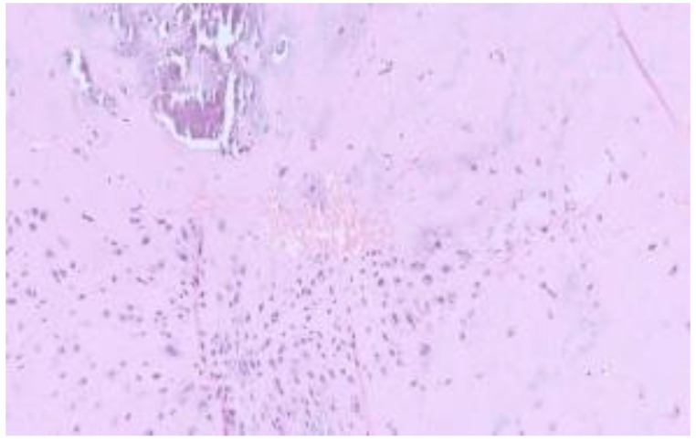
The tumour is cellular with well formed hyaline cartilaginous matrix with chondrocytes arranged in lacunae exhibiting minimal atypia & scant mitosis. Coarse Calcification & evidence of enchondral ossification is seen
Fig. 2.
On IHC, chondrocytes express s-100 (weak to moderate immunoexpression)
The histopathology report showed a well-circumscribed multilobular cartilaginous tumor. the tumor is cellular with a well-formed hyaline cartilaginous matrix with chondrocytes arranged in lacunae exhibiting minimal atypia & scant mitosis.Coarse calcification & evidence of endochondral ossification is seen. Foci of multinucleated giant cells were also identified along the interlobular vascular channels. No evidence of invasion into the adjacent salivary gland tissue was identified. there is no evidence of malignancy. On IHC, focal expression is seen with S100, while no expression of CK or p40 is seen (Figs. 3 and 4). ERG 1 highlights the interlobular vascular channels. Overall features suggestive of benign chondrocytic neoplasm- soft tissue chondroma (extraskeletal).
Fig. 3.
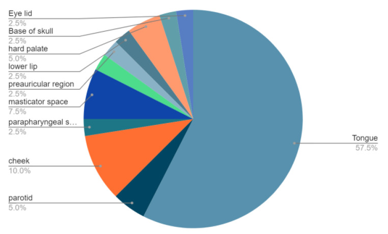
Frequency of distribution among subsites of head & neck region
Fig. 4.
Intraoperative: mass held up with help of Babcock forceps
Patient had mild facial nerve paresis in the post-op period for which he had complete recovery in 3 weeks.
Discussion
Chondroma was first described by Lichtenstein and Hall [1]. These are benign neoplasm predominantly composed of mature hyaline cartilage, which accounts for about 1.5% of all benign soft tissue tumors [2]. Extraskeletal chondroma usually is characterized by a slow-growing, enlarging nodule that may cause pain or tenderness, predominantly in hands and feet of male adult patients, with an excellent prognosis and sporadic malignant transformation [3]. According to a study of 104 cases reported by.
Chung and Enzinger [4], chondroma occurred in fingers in about 80% of the cases. Although chondromas occur in patients of all ages, extraskeletal chondroma typically affects adults between 30 and 60 years of age, and there is no obvious gender difference [5].Typically, the tumors are asymptomatic and manifest as slowly growing masses [6]. To best of authors knowledge, total 40 cases of extraskeletal chondromas have been reported in head and neck region and out of which 25 cases reported in tongue while only 2 cases were reported in masticator space (Table 1) .This is only 3rd case report in literature in masticator space [7].
Table 1.
Review of literature on soft tissue chondroma of the head and neck
| Author | site |
|---|---|
| Bruce, 1953 | Tongue |
| Rosen 1961 | Tongue |
| Yoel 1965 | Tongue |
| Ramachandran 1968 | Tongue |
| Hankey 1968 | Cheek |
| Huppertz 1969 | Cheek |
| Viglioglia 1970 | Tongue |
| Samant 1971 | Tongue |
| Gutmann 1974 | Tongue |
| Zegarelli 1977 | Tongue |
| del Rio 19,781 | Tongue |
| Jungueira 1982 | Tongue |
| Yasuoka 1984 | Tongue |
| Segal 1984 | Tongue |
| Sambo 1986 | Tongue |
| Ling 1986 | Tongue |
| Van der Wal 1987 | Tongue |
| Aguirre 1988 | Tongue |
| Tani 1989 | Tongue |
| Ishibashi 1989 | Tongue |
| Yamanaka 1989 | Tongue |
| Sanchez-Aniceto 1990 | Tongue |
| Munro 1990 | Tongue |
| Kostopoulos 1993 | Parotid gland |
| Blum 1993 | Cheek |
| Kamysz 1996 | Neck |
| Wang 1998 | Parapharyngeal space |
| Sera 2005 | Tongue |
| Onodera 2005 | Cheek |
| Aslam 2006 | Parotid gland |
| De Riu 2007 | Masticatory space |
| Vazquez Mahia2007 | Preauricular region |
| Scivetti 2008 | Tongue |
| J. Falleti 2009 | Masseter muscle |
| Yoome Kim 2009 | Lower lip |
| ToshihiroKawano 2011 | Hard palate |
| KirteeRaparia 2013 | Base of skull |
| Vescovi 2014 | hard palate |
| Aseem 2018 | Eye lid |
| This report 2023 | Masticator space |
Soft tissue chondromas may occur in people of any age; typically, The isolated tumors of masticator spaces are very rare. Usually it involves the tumor of adjacent subsite like oral cavity, oropharynx, nasopharynx and parotid [8, 9].
The pathological features of chondromas vary considerably from case to case. Some tumours show focal fibrosis, ossification or myxoid change. Chondromas can be very cellular, with mild-to-moderate nuclear pleomorphism and scattered mitosis [10].
Histopathologically, the tumor exhibits many lobular structures and some parts similar to hyaline cartilage containing mucous components [11]. Approximately 10% of soft-tissue chondromas contain osteoclast-like multinucleated giant cells or epithelioid histiocytes with granuloma-like proliferation. Regarding the mechanism of onset of a chondroma in the masticatory space, hypotheses include aberrant embryonic cartilage and metaplasia owing to chronic inflammation [12].
Clinically chondroma remain asymptomatic but if they increased in size may cause pressure/obstructive symptom depending on there site of origin. Like parapharyngeal space tumor may cause upper airway obstruction [13, 14].
In this case, the patient presented with right preauricular swelling. Owing to the high and deep location of tumor with superior extension up to zygoma – surgical excision of the tumour required a wide exposure and external access. An intraoral approach to the medial condylar region does not allow complete control of the posterior aspects of the neoplasia and is not without risk of injury to the internal maxillary artery, mandibular nerve and other surrounding structures. So we took the transparotid approach for wide excision.
At the 12-month follow-up, there were no signs of recurrence of the neoplasia. The patient showed no functional limitation or aesthetic defect, and there was no evidence of complications.
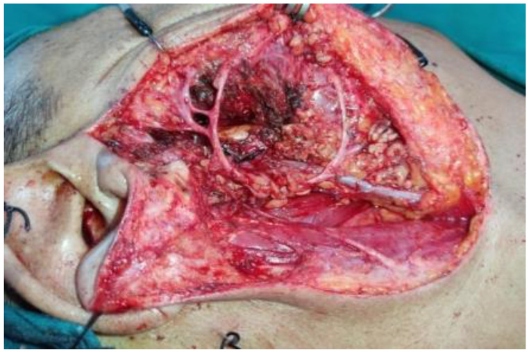
Fig. 5.
Post excision operative site
Electronic Supplementary Material
Below is the link to the electronic supplementary material.
Funding Information
None.
Declarations
Ethical implication
The study involves a human participant. Ethical committee clearance was taken.
Informed Consent
A proper informed consent was obtained from her before writing this report quoting the details.
Conflict of interest
The authors declare that they don’t have any conflict of interest.
Footnotes
Publisher’s Note
Springer Nature remains neutral with regard to jurisdictional claims in published maps and institutional affiliations.
References
- 1.Lichtenstein L, Hall JE. Periosteal chondroma; a distinctive benign cartilage tumor. J Bone Joint Surg Am. 1952;24–A–3:691–697. doi: 10.2106/00004623-195234030-00026. [DOI] [PubMed] [Google Scholar]
- 2.Kedhaier A, Maala R, Ennouri N, Regaya N. Soft tissue chondroma of the hand:a report of five cases. Acta Orthop Belg. 2007;73:458–461. [PubMed] [Google Scholar]
- 3.Dahlin DC, Salvador AH (1974) Cartilaginous tumors of the soft tissues of the hands and feet. Mayo Clin Proc. ;49:721–6. [PubMed] [Google Scholar] [Ref list] [PubMed]
- 4.Chung EB, Enzinger FM. Chondroma of soft parts. Cancer. 1978;41:1414–1424. doi: 10.1002/1097-0142(197804)41:4<1414::AID-CNCR2820410429>3.0.CO;2-O. [DOI] [PubMed] [Google Scholar]
- 5.Enzinger FM (1983) Cartilaginous tumors and tumor-like lesions of soft tissue; in Enzinger. In: Weiss FM SW (ed) Soft tissue tumors. St. Louis, Mosby Co, pp 698–719
- 6.Kamysz JW, Zawin JK, Gonzalez-Crussi F. Soft tissue chondroma of the neck: a case report and review of the literature. Pediatr Radiol. 1996;26:145–147. doi: 10.1007/BF01372095. [DOI] [PubMed] [Google Scholar]
- 7.Falleti J, et al. Extraskeletal Chondroma of the Masseter muscle: a Case Report with Review of the literature. Int J Oral Maxillofac Surg. 2009;38(8):895–899. doi: 10.1016/j.ijom.2009.02.020. [DOI] [PubMed] [Google Scholar]
- 8.Park Y, Kim HY, Yu IK, Jang DS, Kim JH (2022) Imaging characteristics of slow-growing soft tissue chondroma of the tongue: A case report. Medicine (Baltimore). May 13;101(19):e29276. doi: 10.1097/MD.0000000000029276. PMID: 35583538; PMCID: PMC9276242 [DOI] [PMC free article] [PubMed]
- 9.Sera H, Shimoda T, Ozeki S, Honda T. A case of chondroma of the tongue. Int J Oral Maxillofac Surg. 2005;34:99–100. doi: 10.1016/j.ijom.2004.03.011. [DOI] [PubMed] [Google Scholar]
- 10.Kim MJ, Cho KJ, Ayala AG, Ro JY. Chondrosarcoma: with updates on molecular genetics. Sarcoma. 2011;2011:405437. doi: 10.1155/2011/405437. [DOI] [PMC free article] [PubMed] [Google Scholar]
- 11.Saito M, Nishimoto K, Nakayama R, Kikuta K, Nakamura M, Matsumoto M, Morioka H. Extraskeletal Chondroma of the Index Finger: a Case Report. Case Rep Oncol. 2017;10:479–484. doi: 10.1159/000477237. [DOI] [PMC free article] [PubMed] [Google Scholar]
- 12.Vescovi P, Meleti M, Merigo E, Manfredi M, Corradi D, Giovannacci I, Poli T, Nammour S. Soft tissue chondroma of the oral cavity: an extremely rare tumour localized on the hard palate. Case Rep Med. 2014;2014:414861. doi: 10.1155/2014/414861. [DOI] [PMC free article] [PubMed] [Google Scholar]
- 13.Ferlito A, Devaney KO, Mäkitie AA (2019 Oct) Differing characteristics of cartilaginous lesions of the larynx. Eur Arch Otorhinolaryngol 276(10):2635–2647. 10.1007/s00405-019-05563-wEpub 2019 Jul 23. PMID: 31338576; PMCID: PMC6757023 [DOI] [PMC free article] [PubMed]
- 14.Alobida NW, Doubi AO, Alswayyed M, Dima Z, Jamjoom, Khalid Al-Qahtani Chondroid chordoma of the parapharyngeal space: a case report and review of literature. Acta Otolaryngol Case Rep. 2020;5(1):69–73. doi: 10.1080/23772484.2020.1827952. [DOI] [Google Scholar]
Associated Data
This section collects any data citations, data availability statements, or supplementary materials included in this article.


